Search results (124 results)
-
---thumb.jpg/image-square;max$300,300.ImageHandler) inferior peripheral snowbanking
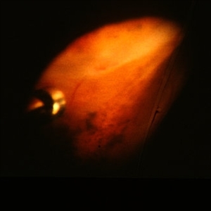
inferior peripheral snowbanking
Feb 14 2013 by From the Collections of Thomas M. Aaberg, MD and Thomas M. Aaberg Jr., MD
intraoperative indirect ophthalmoscopic photograph showing inferior peripheral snowbanking
Condition/keywords: pars planitis, snowbank
-
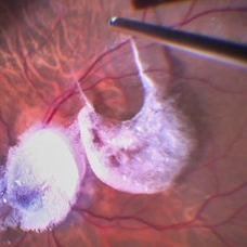
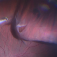
 peripheral pre-retinal fibrosis and neovascularization with vitreous cutter
Feb 14 2013 by From the Collections of Thomas M. Aaberg, MD and Thomas M. Aaberg Jr., MD
intraoperative photo of peripheral pre-retinal fibrosis and neovascularization with vitreous cutter
Condition/keywords: pars planitis, peripheral retinal neovascularization
-
 Intraocular Multiple Cysticercus
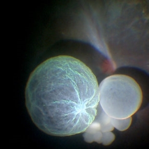
Oct 10 2018 by Vishal Agrawal, MD, FRCS,FACS,FASRS
Intraoperative fundus picture of right eye of a 18-year-old boy with complaints of DOV for the past 2 months. There were 12 intravitreal cysts in total with vitritis sclerosis retinal vessels and TRD. To note here, the largest cyst has a flimsy wall and no scolex (possibly ruptured) and the rest of the smaller cysts have a scolex and a taut wall.
Photographer: Vishal Agrawal MD,FRCS
Imaging device: SONY PMW-10 MD HD
Condition/keywords: cysticercosis, scolex
-
 Retinal Detachment with Giant Retinal Tear and Macular Hole
Jan 6 2020 by MATTEO FORLINI, MD
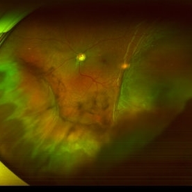
A 61-year-old-male patient presented with sudden diminution of vision in the right eye due to retinal detachment with giant retinal tear and macular hole. Best corrected visual acuity (BCVA) at presentation was 20/200. A 23 G vitrectomy was performed. The edges of the tear were unrolled and complete retinal re-attachment under PFCL was achieved. A 360 degree intraoperative endolaser was performed on the peripheral retina as well as around the edges of the tears. PFCL was exchanged with silicone oil 5000cs as final tamponade. At six-months follow-up retina was attached and macular hole was repaired. Best-corrected visual acuity is 20/125 at present.
Photographer: Matteo Forlini MD, San Marino Hospital, Republic of San Marino
Condition/keywords: full thickness macular hole, giant retinal tear, silicone oil
-
 Repair of Retinal Detachment
Feb 11 2015 by Darrell E. Baskin, MD
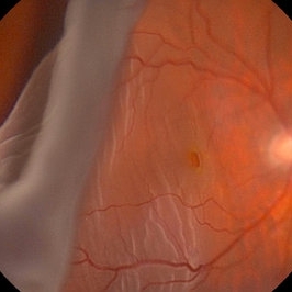
Intraoperative photo of a 75-year-old woman with a retinal detachment status post open globe repair.
Photographer: Darrell Baskin, Wilford Hall, Lackland Air Force Base, Texas
Imaging device: Zeiss Lumera ReSight 700
Condition/keywords: perfluorocarbon fluid, vitreoretinal surgery
-
 TA Stained Posterior Hyaloid Face
Apr 11 2014 by Subhendu Kumar Boral, MBBS, MD(AIIMS), DNB, FASRS (USA)
Intraoperative step of posterior hyaloid face staining by triamcinolone acetonide particles during PVD induction in a case of diabetic epiretinal membrane left eye in a 68-year-old gentleman.
Photographer: Subhendu Kumar Boral
Condition/keywords: hyaloid
-
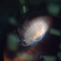 Pars Plana Cysts
Jan 29 2018 by Shani Pillar
During a pars plana vitrectomy for fixation of a dislocated IOL, this finding of pars plana cysts was seen, while performing indentation. Pars plana cysts are not uncommon, but rarely visualized so clearly, given their extremely peripheral location.
Photographer: Dr. Shani Pillar, Meir Medical Center, Kfar Saba, Israel
Imaging device: Intraoperative microscope
Condition/keywords: cyst of the pars plana, ora serrata, peripheral fundus lesion
-
 Chronical Submacular Hemorrhage in the Setting of Neovascular AMD
Mar 23 2015 by Rita Couceiro, MD, MS
An 80-year-old male, with a history of hypertension and high cholesterol, complained of acute and painless vision loss in his left eye (OS) in the previous 5 months. On observation best corrected visual acuity in OS was hand motion. A dense vitreous opacity in OS precluded fundus examination. Ocular ultrasound revealed vitreous hemorrhage and thickening of the macular area. The patient was submitted to pars plana vitrectomy, which disclosed a large submacular hemorrhage with chronical features and disciform scarring in the setting of neovascular AMD.
Imaging device: Intraoperative fundus photograph
Condition/keywords: neovascular age-related macular degeneration (AMD), submacular hemorrhage, wet age-related macular degeneration (wet AMD)
-
---thumb.jpg/image-square;max$300,300.ImageHandler) peripheral pre-retinal fibrosis and neovascularization with vitreous cutter
peripheral pre-retinal fibrosis and neovascularization with vitreous cutter
Feb 14 2013 by From the Collections of Thomas M. Aaberg, MD and Thomas M. Aaberg Jr., MD
intraoperative photo of peripheral pre-retinal fibrosis and neovascularization with vitreous cutter
Condition/keywords: pars planitis, peripheral retinal neovascularization
-
 Vitreomacular Traction
Sep 27 2012 by Virgilio Morales-Canton, MD
Intraoperative image of a patient with a macular traction syndrome. Brilliant blue was used. DORC 27 Ga system.
Photographer: Virgilio Morales-Canton
Imaging device: optronics surgical camera
Condition/keywords: brilliant blue, macular traction, vitreomacular interface disorders
-
 Endogenous Candida Endophthalmitis
Apr 17 2014 by Shlomit Schaal, MD, PhD, MHCM
Intraoperative photo of vitrectomy for endogenous endopthalmitis.
Photographer: Shlomit Schaal MD, PhD and Nathan Podoll MD, University of Louisville, Louisville, KY
Condition/keywords: endogenous endophthalmitis
-
---thumb.jpg/image-square;max$300,300.ImageHandler) peripheral pre-retinal fibrosis and neovascularization with vitreous cutter
peripheral pre-retinal fibrosis and neovascularization with vitreous cutter
Feb 14 2013 by From the Collections of Thomas M. Aaberg, MD and Thomas M. Aaberg Jr., MD
intraoperative photo of peripheral pre-retinal fibrosis and neovascularization with vitreous cutter
Condition/keywords: pars planitis, peripheral retinal neovascularization
-
 Suprachoroidal Hemorrhage
Sep 2 2020 by Rinal Pandit
Fundus photograph of left eye of a 56-year-old female with primary angle closure glaucoma showing massive hemorrhagic choroidal detachment that developed following trabeculectomy surgery. Suprachoroidal hemorrhage is defined as the accumulation of blood within the potential space between the choroid and sclera, with the source of the blood being the long or short posterior ciliary artery. Delayed suprachoroidal hemorrhage (DSHC) remains one of the most dreaded and sight threatening complications of glaucoma filtration surgery. The risk factors include old age, hypertension, high myopia, arteriosclerosis, chronically elevated IOP, sudden hypotony, trauma, aphakia/pseudophakia, prior vitrectomy, history of 5 FU injections and anti-platelet agents. The incidence of postoperative SCH after trabeculectomy varies between 0.6%- 1.4%. DSCH after surgery varies considerably in severity but is generally characterized by the sudden onset of severe pain, decreased vision, and a shallow anterior chamber usually associated with raised intraocular pressure. B-scan ultrasonography can help to distinguish serous from hemorrhagic choroidals.Suprachoroidal hemorrhages appear as dome-shaped elevations of the retina with increased echo densities that are often heterogeneous and within the suprachoroidal space. Choroidal effusions appear as dome-shaped elevations with hypoechoic suprachoroidal space. The first step in the management is the timely diagnosis. Medical management includes oral and topical antiglaucoma drugs to lower IOP, oral and topical steroids to control inflammation and topical cycloplegics and oral analgesics to tackle pain. Serial ultrasound B scans of the affected eye should be performed in order to monitor progression of the SCH and help determine apposition, height, and liquefaction of the SCH. Indications of surgical drainage include non resolution with medical management,concurrent retinal detachment, central retinal apposition (kissing choroidals) and incarceration of vitreous in the wound site. The ideal time of drainage is between 7-14 days depending upon clot lysis. The prognosis of both intraoperative and postoperative SCH is poor. An overwhelming majority of patients do not achieve pre-hemorrhage visual acuity and most do not recover to a visual acuity of 20/200 or better. The major determinants of good or bad visual outcomes of SCH’s are preoperative visual acuity and retinal detachment at the time of hemorrhage, respectively.
Imaging device: OPTOS,Ultra wide field retinal imaging system
Condition/keywords: suprachoroidal hemorrhage, trabeculectomy, ultra-wide field imaging
-
 Triamcinolone Stained Hyaloid
Feb 11 2017 by Manish Nagpal, MD, FRCS (UK), FASRS
Intraoperative photo of triamcinolone stained hyaloid being being lifted with vacuum generated by the cutter to detach it.
Photographer: Manish Nagpal
Imaging device: Still captured from a 3 chip HD camera on microscope
Condition/keywords: hyaloid, triamcinolone
-
 Intraoperative Photo During Vitrectomy
Jan 26 2017 by Manish Nagpal, MD, FRCS (UK), FASRS
Intraoperative photo while doing vitectomy for peripheral base shaving near the canula port enhanced by peripheral scleral indentation while using chandelier light.
Photographer: Manish Nagpal
Imaging device: Still captured from a 3 chip HD camera on microscope
Condition/keywords: canula, indentation, scleral depression
-
 peripheral pre-retinal fibrosis and neovascularization with vertical scissors
Feb 14 2013 by From the Collections of Thomas M. Aaberg, MD and Thomas M. Aaberg Jr., MD
intraoperative photo of peripheral pre-retinal fibrosis and neovascularization with vertical scissors
Condition/keywords: pars planitis, peripheral retinal neovascularization
-
 IOFB Over Disc BRAO Post Hyaloid Removal
Feb 25 2017 by Manish Nagpal, MD, FRCS (UK), FASRS
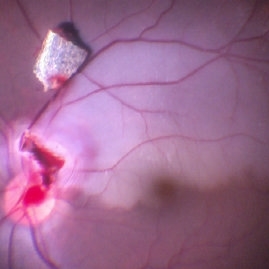
Intraoperative photo of a foreign body piercing the inferotemporal margin of disc revealing a infero temporal BRAO immediately after hyaloid removal. The foreign boy has just been removed from the disc and is freely lying on the retina.
Photographer: MANISH NAGPAL
Imaging device: STILL CAPTURED FROM A 3CHIP HD camera attached to microscope
Condition/keywords: intraocular foreign body
-
 iOCT of Dislocated IOL
Dec 20 2017 by Sidney A Schechet, MD
Intraoperative optical coherence tomography image of a dislocated IOL being safely grasped and lifted of the surface of the retina with microforceps.
Imaging device: Leica EnFocus intraoperative optical coherence tomography
Condition/keywords: dislocated posterior chamber intraocular lens (PCIOL), optical coherence tomography (OCT), pars plana vitrectomy (PPV)
-
 Triamcinolone Stained Hyaloid
Feb 11 2017 by Manish Nagpal, MD, FRCS (UK), FASRS
Intraoperative photo of triamcinolone stained hyaloid showing the whitish haloes of its attachments on macula and disc.
Photographer: manish nagpal
Imaging device: Still captured from 3 Chip HD camera on microscope
Condition/keywords: hyaloid, triamcinolone
-
 Unfolding of GRT flap
May 31 2017 by Manish Nagpal, MD, FRCS (UK), FASRS
Intraoperative photo of unfolding of flap of giant retinal tear.
Photographer: MANISH NAGPAL
Imaging device: SONY 3 CHIP HD CAMERA
Condition/keywords: giant retinal tear, perfluorocarbon fluid
-
 Intraoperative Photo Taken During Vitrectomy
Jan 26 2017 by Manish Nagpal, MD, FRCS (UK), FASRS
Intraoperative photo while doing vitectomy near a horseshoe tear to clear the adherent vitreous enhanced by peripheral scleral indentation while using chandelier light.
Photographer: Manish Nagpal
Imaging device: Still captured from a 3 chip HD camera on microscope
Condition/keywords: cutter, scleral indentation, vitrectomy, vitreous
-
 Triamcinilone Stained Posterior Hyaloid
Jan 25 2017 by Manish Nagpal, MD, FRCS (UK), FASRS
Intraoperative photo in a case of macular hole, the diffuse triamcinolone is removed and only the stained paramacular hyaloid is showing the staining. The cutter is in the process of pulling that hyaloid to release traction.
Photographer: Manish Nagpal
Imaging device: Still captured from a 3 chip HD camera on microscope
Condition/keywords: hyaloid, macular hole, stained posterior
-
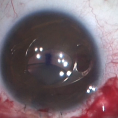 Artisan Lens
Aug 23 2018 by Jessica G Lee, MD
Intraoperative photo of a 13 -year-old boy with Marfan's syndrome with dislocated crystalline lens, undergoing procedure for placement of Artisan lens.
Condition/keywords: intraocular lens (IOL), Marfan's syndrome
 Loading…