Search results (124 results)
-
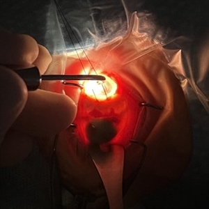
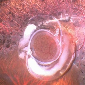
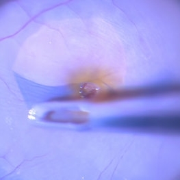
 Dislocated Cataractous Lens
Jun 19 2025 by Mrinali Gupta, MD, FASRS
Intraoperative image of a chronically dislocated cataractous lens. The patient underwent pars plana vitrectomy, lensectomy, and placement of an anterior chamber intraocular lens, with improvement in vision from Count Fingers to 20/20 without correction.
Photographer: Mrinali Gupta, MD
Imaging device: Intraoperative surgical video (Zeiss Lumera scope, Resight lens)
Condition/keywords: dislocated crystalline lens
-
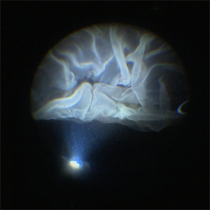
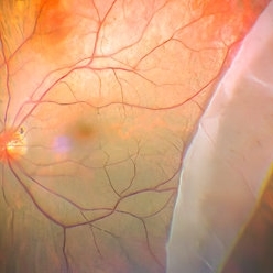
 Dislocation of the Crystalline Lens with a Retinal Detachment
Apr 21 2025 by Hrishikesh Naik, MS
An intraoperative screen grab shows a dislocation of the crystalline lens along with an associated rhegmatogenous retinal detachment in a case of Marfan’s syndrome. The case was managed by a combined PPV-SB procedure. A vitrectomy cutter is seen at the left.
Photographer: Hrishikesh Naik
Condition/keywords: intraoperative, lens dislocation, Marfan's syndrome, Retinal Detachment, vitrectomy
-
 Intraoperative Transillumination of Choroidal Melanoma
Apr 18 2025 by Virginia Gebhart
Intraoperative photo of transillumination of choroidal melanoma before plaque placement in 36 year old female.
Photographer: Chris Bergstrom, MD, OD
Imaging device: iphone
Condition/keywords: choroidal melanoma, intraoperative, transillumination
-
 Inadvert Globe Perfuration After Peribulbar Block
Mar 13 2025 by Bruno B Ribeiro
Fundus photograph of a 74-year-old woman who underwent pars plana vitrectomy OS due to rhegmatogenous retinal detachment. A horseshoe retinal tear can be seen at 5h. Intraoperative evaluation revealed a chorioretinal scar with the shape of the needle track at the same location. Despite rare, globe perfuration after peri or retrobulbar block may happen, even by the most experienced anesthesiologist.
Photographer: Bruno Barbosa Ribeiro, Angelina Meireles
Imaging device: Optos California
Condition/keywords: retinal detachment
-
 Inadvert Globe Perfuration After Peribulbar Block
Mar 13 2025 by Bruno B Ribeiro
Fundus photograph of a 74-year-old woman who underwent pars plana vitrectomy OS due to rhegmatogenous retinal detachment. A horseshoe retinal tear can be seen at 5h. Intraoperative evaluation revealed a chorioretinal scar with the shape of the needle track at the same location. Despite rare, globe perfuration after peri or retrobulbar block may happen, even by the most experienced anesthesiologist.
Photographer: Bruno Barbosa Ribeiro, Angelina Meireles
Imaging device: Optos California
Condition/keywords: hemmorhage
-
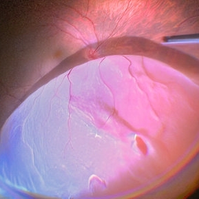
 Posterior Vitreous Detachment
Jan 31 2025 by Thirumalesh Mochi Basavaraj, MD
Intraoperative view of Triamcinolone-assisted posterior vitreous detachment.
Photographer: Thirumalesh Mochi Basavaraj
Condition/keywords: PVD induction, triamcinolone
-
 Intraoperative View of a Giant Retinal Tear
Dec 13 2024 by Thirumalesh Mochi Basavaraj, MD
Intraoperative view of 12 year old child with Giant retinal tear with Retinal detachment.
Photographer: Thirumalesh Mochi Basavaraj
Imaging device: Lumera Proveo 8
Condition/keywords: GIANT RETINAL TEAR, PVR, Retinal Detachment
-
 Dislocated Lens (IOL) Lying on the Retina
Sep 28 2024 by Anjana Mirajkar, MS Ophthalmology
An intraoperative image of right eye showing dislocated IOL with the capsular bag lying on the posterior pole with surrounding pigmentary changes.
Photographer: Dr. Anjana Mirajkar -Retina Foundation, Ahmedabad
Condition/keywords: dislocated IOL
-
 Dislocated Lens Lying on the Retina
Jul 3 2024 by Anjana Mirajkar, MS Ophthalmology
An intraoperative still showing us an dislocated crystalline lens lying on the retina.
Photographer: Dr. Anjana Mirajkar -Retina Foundation, Ahmedabad
Condition/keywords: dislocated crystalline lens
-
 Large Horseshoe Tear
Jun 14 2024 by Tejaswita Verma
Intraoperative still showing retinal detachment with a horse shoe tear.
Photographer: DR. TEJASWITA VERMA
-
 Starfolds
Jun 14 2024 by Tejaswita Verma
Intraoperative still depicting starfolds in a case of retinal detachment with PVR changes.
Photographer: DR. TEJASWITA VERMA
Condition/keywords: starfolds
-
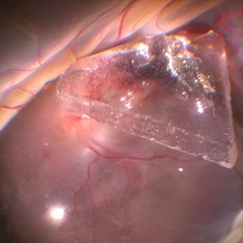 Glass Piece Inside Eye
Jun 14 2024 by Tejaswita Verma
Intraoperative still of a young male showing intraocular foreign body (glass piece) inside eye with retinal detachment.
Photographer: DR. TEJASWITA VERMA
Condition/keywords: intraocular foreign body
-
 IOFB Loosened Away From Imapct Site
Jun 14 2024 by Tejaswita Verma
Intraoperative still of a 21 year old male showing intraocular foreign body loosened away from the site of impact for easier removal.
Photographer: DR. TEJASWITA VERMA
Condition/keywords: intraocular foreign body, intraoperative
-
 Subretinal Silicon Oil With Inferior PVR
Jun 14 2024 by Tejaswita Verma
Intraoperative still of a 34 year old male with sub retinal silicon oil with proliferative vitreoretinopathy changes.
Photographer: DR. TEJASWITA VERMA
Condition/keywords: silicone oil
-
 Nucleus Drop
Jun 14 2024 by Tejaswita Verma
Intraoperative still of lens drop in a 63 year old female after an eventful cataract surgery.
Photographer: DR. TEJASWITA VERMA
Condition/keywords: intraoperative, lens matter, nucleus drop
-
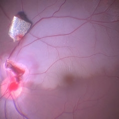
 ILM Peeling in Case of Optic Disc Pit Maculopathy
Jun 14 2024 by Tejaswita Verma
Intraoperative still of a 38 year old male post initiation of ILM peeling in a case of optic disc pit maculopathy.
Photographer: DR. TEJASWITA VERMA
Condition/keywords: intraoperative, optic pit
-
 Metallic Foreign Body Removed From Site of Impaction Intra Operatively
Jun 13 2024 by Anand Temkar
Intraoperative still of a 27 year old male showing metallic foreign body removed from site of impaction from retina.
Photographer: Dr.Anand Temkar- Retina Foundation, Ahmedabad
Condition/keywords: metallic foreign body
-
 IOL Drop
Jun 13 2024 by Anand Temkar
Intraoperative still of a 72 year old male showing dropped IOL.
Photographer: Dr.Anand Temkar- Retina Foundation, Ahmedabad
Condition/keywords: IOL drop
-
 Superior RD with HST Superiorly
Jun 13 2024 by Anand Temkar
Intraoperative still of a 48 year old male showing RD with superior HST and a break.
Photographer: Dr.Anand Temkar- Retina Foundation, Ahmedabad
Condition/keywords: RD
-
 Giant Retinal Tear With Retinal Fold
Jun 13 2024 by Anand Temkar
Intraoperative still of a 34 year old male showing giant retinal tear with retinal fold.
Photographer: Dr.Anand Temkar- Retina Foundation, Ahmedabad
Condition/keywords: giant retinal tear, GRT, retinal fold
-
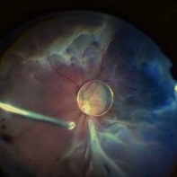
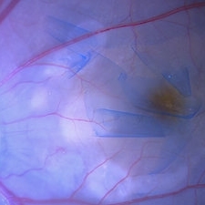
 ILM Peeling in a Case of Macular Hole
Jun 13 2024 by Anand Temkar
Intraoperative still of a 62 year old female with macular hole.
Photographer: Dr.Anand Temkar- Retina Foundation, Ahmedabad
Condition/keywords: ILM peeling, macular hole
-
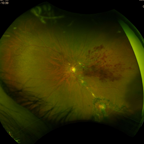
 Submacular Hemorrhage
Jun 12 2024 by Anand Temkar
Intraoperative still of a 58 year old male with submacular hemorrhage (LE).
Photographer: Dr.Anand Temkar- Retina Foundation, Ahmedabad
Condition/keywords: submacular hemorrhage, subretinal hemorrhage
-
 Nucleus Drop
Jun 12 2024 by Anand Temkar
Intraoperative still of a 58 year old female with nucleus drop (LE).
Photographer: Dr.Anand Temkar- Retina Foundation, Ahmedabad
Condition/keywords: nucleus drop
-
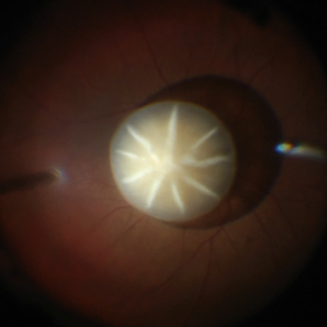
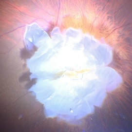
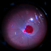 The Rose Of The Eye
Mar 14 2024 by SANDEEP KUMAR
Intraoperative capture of a failed full thickness macular hole being treated with human amniotic membrane graft and autologous blood under air.
Photographer: Sandeep Kumar , Shroff eye centre kailash colony New Delhi India
Imaging device: Callisto imaging system
Condition/keywords: autologous blood, full thickness macula hole, human amniotic graft
-
Time to Chill
Jan 23 2024 by SHISHIR VERGHESE, MS, FVRS, FAICO (Retina)
Intraoperative surgical video of a 65 year old female patient with advanced proliferative diabetic retinopathy showing neovascularization at the ora serrata for which a cryopexy is being done to cause regression. This video highlights a previously undocumented grape like Neovascularization at the ora serrata in this patient with advanced proliferative diabetic retinopathy.
Condition/keywords: Advanced Proliferative diabetic retinopathy, Cryopexy, neovascularization
 Loading…