Search results (124 results)
-
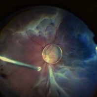
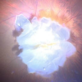
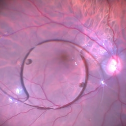
 Bacterial endophthalmitis
Jun 23 2020 by Thirumalesh Mochi Basavaraj, MD
Intraoperative pictures showing bacterial colonies, lab confirmed gram positive cocci(inset).
Imaging device: Leica microsurgery microscope
Condition/keywords: endophthalmitis, intraoperative
-
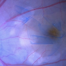
 Cutter Segmentation in a case of Diabetic Combined Retinal Detachment | Intra-Operative Still
Apr 25 2023 by Veer Singh, MS, FVRS, FMRF, FICO (Retina)
Cutter Segmentation in a case of Diabetic Combined Retinal Detachment | Intra-Operative Still Patient underwent Vitrectomy with Silicone Oil
Photographer: Dr. Veer Singh
Condition/keywords: combined retinal detachment, cutter, diabetic retinopathy, intraoperative, pars plana vitrectomy (PPV)
-
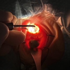
 Dislocation of the Crystalline Lens with a Retinal Detachment
Apr 21 2025 by Hrishikesh Naik, MS
An intraoperative screen grab shows a dislocation of the crystalline lens along with an associated rhegmatogenous retinal detachment in a case of Marfan’s syndrome. The case was managed by a combined PPV-SB procedure. A vitrectomy cutter is seen at the left.
Photographer: Hrishikesh Naik
Condition/keywords: intraoperative, lens dislocation, Marfan's syndrome, Retinal Detachment, vitrectomy
-
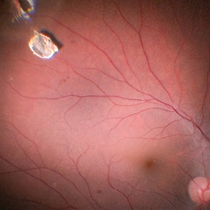
 Funnel Retinal Detachment
Jun 11 2023 by Ethan K Sobol, MD
Intraoperative view of a funnel retinal detachment with proliferative vitreoretinoapthy in an eye with previous open globe injury. PVR membranes were peeled, and the retina was flattened and re-attached with an inferior relaxing retinotomy and silicone oil tamponade
Condition/keywords: intraoperative, open funnel RD, open globe injury, proliferative vitreoretinopathy (PVR)
-
 ILM Peeling in Case of Optic Disc Pit Maculopathy
Jun 14 2024 by Tejaswita Verma
Intraoperative still of a 38 year old male post initiation of ILM peeling in a case of optic disc pit maculopathy.
Photographer: DR. TEJASWITA VERMA
Condition/keywords: intraoperative, optic pit
-
 Intra-op picture of SFIOL explantation with pars plana bleed
Aug 23 2023 by Harsh Vardhan Singh, MS
45-old-male with history of trauma & subluxated SFIOl - undergone IOL explantation & repeat SFIOL. Image showing pars plana bleed during IOL explantation
Photographer: Harsh Vardhan Singh
Condition/keywords: intraoperative, IOL explantation
-
 Intra-operative still of Rhegmatogenous Retinal Detachment
Apr 15 2023 by Veer Singh, MS, FVRS, FMRF, FICO (Retina)
Intra-operative still of Rhegmatogenous Retinal Detachment taken using an indigenous 1CMOS HD camera
Photographer: Dr. Veer Singh
Condition/keywords: intraoperative, vitrectomy
-
 Intra-operative still of PFCL bubble in RD Surgery
Apr 15 2023 by Veer Singh, MS, FVRS, FMRF, FICO (Retina)
Intra-operative still of PFCL bubble in RD Surgery. PFCL being used to flatten the detached retina.
Photographer: Dr. Veer Singh
Condition/keywords: intraoperative, pars plana vitrectomy (PPV), PFCL, Retinal Detachment
-
 Intraoperative Transillumination of Choroidal Melanoma
Apr 18 2025 by Virginia Gebhart
Intraoperative photo of transillumination of choroidal melanoma before plaque placement in 36 year old female.
Photographer: Chris Bergstrom, MD, OD
Imaging device: iphone
Condition/keywords: choroidal melanoma, intraoperative, transillumination
-
 IOFB Loosened Away From Imapct Site
Jun 14 2024 by Tejaswita Verma
Intraoperative still of a 21 year old male showing intraocular foreign body loosened away from the site of impact for easier removal.
Photographer: DR. TEJASWITA VERMA
Condition/keywords: intraocular foreign body, intraoperative
-
 Nucleus Drop
Jun 14 2024 by Tejaswita Verma
Intraoperative still of lens drop in a 63 year old female after an eventful cataract surgery.
Photographer: DR. TEJASWITA VERMA
Condition/keywords: intraoperative, lens matter, nucleus drop
-
 360 Degree Retinectomy
Feb 2 2022 by Manish Nagpal, MD, FRCS (UK), FASRS
Intraoperative photo of a case of retinal detachment with extensive PVR, which underwent 360 degree relaxing retinectomy followed by 360 laser barrage just prior to silicone oil injection.
Photographer: Manish Nagpal, Retina Foundation, Ahmedabad, India
Imaging device: Sony PMW -10 MD surgical camera
Condition/keywords: laser, laser photocoagulation, proliferative vitreoretinopathy (PVR), relaxing retinectomy, retinectomy, silicone oil
-
 360 Endolaser Barrage
Feb 2 2022 by Manish Nagpal, MD, FRCS (UK), FASRS
Intraoperative photo of a 360 laser barrage done for a case of retinal detachment with a large superior tear.
Photographer: Manish Nagpal, Retina Foundation, Ahmedabad, India
Imaging device: Sony PMW -10 MD surgical camera
Condition/keywords: laser, laser photocoagulation, tear
-
 Artisan Lens
Aug 23 2018 by Jessica G Lee, MD
Intraoperative photo of a 13 -year-old boy with Marfan's syndrome with dislocated crystalline lens, undergoing procedure for placement of Artisan lens.
Condition/keywords: intraocular lens (IOL), Marfan's syndrome
-
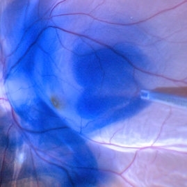
 Brilliant Blue Dye Injected in a Case of Macular Hole to Stain the ILM
Feb 4 2022 by Manish Nagpal, MD, FRCS (UK), FASRS
Intraoperative still of a brilliant blue dye being injected to stain the ILM.
Photographer: Manish Nagpal, Director, Retina Foundation, Ahmedabad
Imaging device: Sony PMW -10 MD surgical camera
Condition/keywords: brilliant blue, ILM flap, ILM staining, macular hole, retina, retina surgery
-
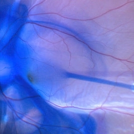
 Brilliant blue dye injection to stain ILM in a macular hole with retinal detachment
Feb 4 2022 by Manish Nagpal, MD, FRCS (UK), FASRS
Intraoperative still of brilliant blue dye injection in process to initiate ILM peel in a patient who has a retinal detachment with a macular hole
Photographer: Manish Nagpal, Director, Retina Foundation, Ahmedabad
Imaging device: Sony PMW -10 MD surgical camera
Condition/keywords: brilliant blue, ILM flap, macular hole
-
 Brilliant Blue Dye Injection to Stain ILM in a Macular Hole with Retinal Detachment
Feb 4 2022 by Manish Nagpal, MD, FRCS (UK), FASRS
Intraoperative still of a Brilliant blue dye injection being done to stain the ILM.
Photographer: Manish Nagpal, Director, Retina Foundation, Ahmedabad
Imaging device: Sony PMW -10 MD surgical camera
Condition/keywords: full thickness macular hole, macula, retina
-
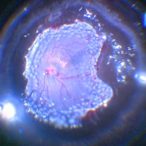
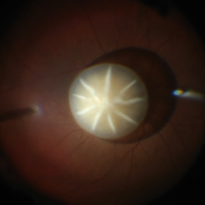
 Capillary Heamngioma of the Optic Disc
Jun 30 2022 by Thirumalesh Mochi Basavaraj, MD
fundus image showing a large Capillary Hemangioma at the Optic Disc
Photographer: Thirumalesh Mochi Basavaraj
Imaging device: intraoperative picture
Condition/keywords: Hemangioma, Von Hippel-Lindau
-
 Chronical Submacular Hemorrhage in the Setting of Neovascular AMD
Mar 23 2015 by Rita Couceiro, MD, MS
An 80-year-old male, with a history of hypertension and high cholesterol, complained of acute and painless vision loss in his left eye (OS) in the previous 5 months. On observation best corrected visual acuity in OS was hand motion. A dense vitreous opacity in OS precluded fundus examination. Ocular ultrasound revealed vitreous hemorrhage and thickening of the macular area. The patient was submitted to pars plana vitrectomy, which disclosed a large submacular hemorrhage with chronical features and disciform scarring in the setting of neovascular AMD.
Imaging device: Intraoperative fundus photograph
Condition/keywords: neovascular age-related macular degeneration (AMD), submacular hemorrhage, wet age-related macular degeneration (wet AMD)
-
 Dislocated Cataractous Lens
Jun 19 2025 by Mrinali Gupta, MD, FASRS
Intraoperative image of a chronically dislocated cataractous lens. The patient underwent pars plana vitrectomy, lensectomy, and placement of an anterior chamber intraocular lens, with improvement in vision from Count Fingers to 20/20 without correction.
Photographer: Mrinali Gupta, MD
Imaging device: Intraoperative surgical video (Zeiss Lumera scope, Resight lens)
Condition/keywords: dislocated crystalline lens
-
 Dislocated IOL and Lens Matter
Jan 11 2022 by Manish Nagpal, MD, FRCS (UK), FASRS
Intraoperative photo of dislocated IOL and lens matter in the vitreous.
Photographer: Manish Nagpal, Retina Foundation, Ahmedabad, india
Imaging device: Sony PMW -10 MD surgical camera
Condition/keywords: dislocated crystalline lens, dislocated intraocular lens (IOL), dislocated lens, dislocated posterior chamber intraocular lens (PCIOL)
-
 Dislocated IOL Over Macula
Jan 11 2022 by Manish Nagpal, MD, FRCS (UK), FASRS
Intraoperative photo of a dislocated IOL sitting over the macular area.
Photographer: Manish Nagpal, Director, Retina Foundation, Ahmedabad
Imaging device: Sony PMW -10 MD surgical camera
Condition/keywords: dislocated intraocular lens (IOL), dislocated lens, dislocated posterior chamber intraocular lens (PCIOL)
-
 Dropped Nucleus in Vitreous
Jan 12 2022 by Manish Nagpal, MD, FRCS (UK), FASRS
Intraoperative photo of a nucleus floating over the macula just prior to fragging it.
Photographer: Manish Nagpal, Retina Foundation, Ahmedabad, india
Imaging device: Sony PMW -10 MD surgical camera
Condition/keywords: dislocated crystalline lens, dropped nucleus
-
 Endogenous Candida Endophthalmitis
Apr 17 2014 by Shlomit Schaal, MD, PhD, MHCM
Intraoperative photo of vitrectomy for endogenous endopthalmitis.
Photographer: Shlomit Schaal MD, PhD and Nathan Podoll MD, University of Louisville, Louisville, KY
Condition/keywords: endogenous endophthalmitis
-
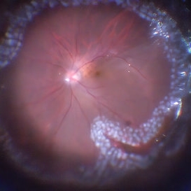
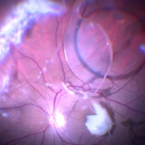
 Epiretinal Membrane
Feb 2 2022 by Manish Nagpal, MD, FRCS (UK), FASRS
Intraoperative photo of a epiretinal membrane, glistening reflex noted. Prior to this capture, PVD induction has been done, which has left a small splinter hemorrhage around the disc attachment of hyaloid.
Photographer: Manish Nagpal, Retina Foundation, Ahmedabad, India
Imaging device: Sony PMW -10 MD surgical camera
Condition/keywords: epiretinal membrane formation, ERM, ILM flap, PVD induction
 Loading…