Search results (124 results)
-
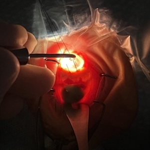
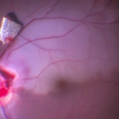
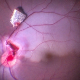 IOFB Over Disc BRAO Post Hyaloid Removal
Feb 25 2017 by Manish Nagpal, MD, FRCS (UK), FASRS
Intraoperative photo of a foreign body piercing the inferotemporal margin of disc revealing a infero temporal BRAO immediately after hyaloid removal. The foreign boy has just been removed from the disc and is freely lying on the retina.
Photographer: MANISH NAGPAL
Imaging device: STILL CAPTURED FROM A 3CHIP HD camera attached to microscope
Condition/keywords: intraocular foreign body
-
 Spontaneously Dropped Lens in a Congenital Rubella Syndrome
Apr 30 2022 by NEIFFER RABELO
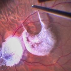
Intraoperative photograph of a 68-year-old patient with congenital rubella syndrome and light perception visual acuity since childhood. The image shows a pigmentary retinopathy and the lens spontaneously displaced into the vitreous cavity. The patient sought care complaining of a total and sporadic loss of vision that was hindering her in daily tasks. Surgery was indicated to remove the lens.
Photographer: Rodrigo Dos Anjos Versiani - Retina Institute - Belo Horizonte - Brazil
Imaging device: ZEISS OPMI LUMERA 700
Condition/keywords: dropped nucleus, retina surgery, rubella retinopathy
-
 Intraoperative Photo Taken During Vitrectomy
Jan 26 2017 by Manish Nagpal, MD, FRCS (UK), FASRS
Intraoperative photo while doing vitectomy near a horseshoe tear to clear the adherent vitreous enhanced by peripheral scleral indentation while using chandelier light.
Photographer: Manish Nagpal
Imaging device: Still captured from a 3 chip HD camera on microscope
Condition/keywords: cutter, scleral indentation, vitrectomy, vitreous
-
 Large Retinal Tear
Mar 24 2017 by Manish Nagpal, MD, FRCS (UK), FASRS
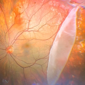
Intraoperative photo of a large retinal tear with everted edges.
Photographer: manish nagpal
Imaging device: Still captured from 3 Chip HD camera on microscope
Condition/keywords: retinal tear
-
 Large Retinal Tear
Mar 24 2017 by Manish Nagpal, MD, FRCS (UK), FASRS
Intraoperative photo of a large retinal tear with everted edges.
Photographer: Manish Nagpal
Imaging device: Still captured from a 3 chip HD camera on microscope
Condition/keywords: retinal tear
-
 Rescuing IOL CTR Bag Complex
Jun 14 2023 by Aditya S Kelkar, MS, FRCS, FASRS,FRCOphth
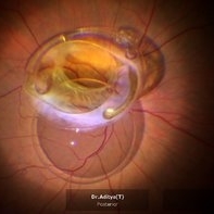
INTRAOPERATIVE SNAPSHOPT IN ZEISS ARTEVO 800 OF DROPPED IOL CTR BAG COMPLEX IN A 71 YEAR OLD MALE PATIENT
Photographer: SUBHASREE DUTTA, NATIONAL INSTITUTE OF OPHTHALMOLOGY, PUNE
Imaging device: ZEISS ARTEVO 800
Condition/keywords: dropped capsular IOL bag complex
-
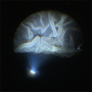
 Triamcinolone Stained Hyaloid
Feb 11 2017 by Manish Nagpal, MD, FRCS (UK), FASRS
Intraoperative photo of triamcinolone stained hyaloid being being lifted with vacuum generated by the cutter to detach it.
Photographer: Manish Nagpal
Imaging device: Still captured from a 3 chip HD camera on microscope
Condition/keywords: hyaloid, triamcinolone
-
 Chronical Submacular Hemorrhage in the Setting of Neovascular AMD
Mar 23 2015 by Rita Couceiro, MD, MS
An 80-year-old male, with a history of hypertension and high cholesterol, complained of acute and painless vision loss in his left eye (OS) in the previous 5 months. On observation best corrected visual acuity in OS was hand motion. A dense vitreous opacity in OS precluded fundus examination. Ocular ultrasound revealed vitreous hemorrhage and thickening of the macular area. The patient was submitted to pars plana vitrectomy, which disclosed a large submacular hemorrhage with chronical features and disciform scarring in the setting of neovascular AMD.
Imaging device: Intraoperative fundus photograph
Condition/keywords: neovascular age-related macular degeneration (AMD), submacular hemorrhage, wet age-related macular degeneration (wet AMD)
-
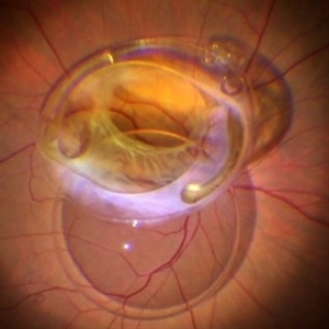
 Dislocation of the Crystalline Lens with a Retinal Detachment
Apr 21 2025 by Hrishikesh Naik, MS
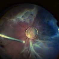
An intraoperative screen grab shows a dislocation of the crystalline lens along with an associated rhegmatogenous retinal detachment in a case of Marfan’s syndrome. The case was managed by a combined PPV-SB procedure. A vitrectomy cutter is seen at the left.
Photographer: Hrishikesh Naik
Condition/keywords: intraoperative, lens dislocation, Marfan's syndrome, Retinal Detachment, vitrectomy
-
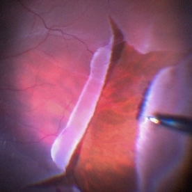
 Giant Retinal Tear
Jan 11 2022 by Manish Nagpal, MD, FRCS (UK), FASRS
Intraoperative photo a temporal giant retinal tear with everted flap and some laser marks noted on the bare choroid from previous barrage attempt elsewhere.
Photographer: Manish Nagpal, Retina Foundation, Ahmedabad, India
Imaging device: Sony PMW -10 MD surgical camera
Condition/keywords: giant retinal tear
-
Iatrogenic Macular Hole and Subretinal Migration of PFCL
Feb 7 2023 by Aditya S Kelkar, MS, FRCS, FASRS,FRCOphth
The video demonstrates a surgical scenario where the fovea gives away by the force imparted by the jet of an injecting PFCL (Perfluorocarbon heavy Liquid) and the PFCL migrates subfoveally. Intraoperative OCT confirms the presence of a macular hole. The situation is managed by ILM peeling and mobilizing subfoveal PFCL peripherally by injecting another bubble of PFCL over the posterior pole. A peripheral drainage retinotomy is then created to aspirate the subretinal PFCL followed by fluid-air exchange, PFCL-air exchange, and endolaser around the retinotomy. Post-operative OCT at 3 weeks’ follow-up shows a sealed macular hole.
Condition/keywords: Iatrogenic macular hole, Intraoperative complications, Subretinal PFCL
-
 ILM Peeling in Progress
Feb 4 2022 by Manish Nagpal, MD, FRCS (UK), FASRS
Intraoperative shot of ILM peeling in progress using forceps.
Photographer: Manish Nagpal, Director, Retina Foundation, Ahmedabad
Imaging device: Sony PMW -10 MD surgical camera
Condition/keywords: ILM flap, ILM staining, internal limiting membrane (ILM) peeling, macular hole, retina, retina surgery
-
 Intraoperative Photo During Vitrectomy
Jan 26 2017 by Manish Nagpal, MD, FRCS (UK), FASRS
Intraoperative photo while doing vitectomy for peripheral base shaving near the canula port enhanced by peripheral scleral indentation while using chandelier light.
Photographer: Manish Nagpal
Imaging device: Still captured from a 3 chip HD camera on microscope
Condition/keywords: canula, indentation, scleral depression
-
 Intraoperative Transillumination of Choroidal Melanoma
Apr 18 2025 by Virginia Gebhart
Intraoperative photo of transillumination of choroidal melanoma before plaque placement in 36 year old female.
Photographer: Chris Bergstrom, MD, OD
Imaging device: iphone
Condition/keywords: choroidal melanoma, intraoperative, transillumination
-
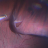
 Intraoperative View of a Giant Retinal Tear
Dec 13 2024 by Thirumalesh Mochi Basavaraj, MD
Intraoperative view of 12 year old child with Giant retinal tear with Retinal detachment.
Photographer: Thirumalesh Mochi Basavaraj
Imaging device: Lumera Proveo 8
Condition/keywords: GIANT RETINAL TEAR, PVR, Retinal Detachment
-
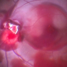 IOFB Over Disc With Blood Stained Hyaloid and BRAO
Feb 25 2017 by Manish Nagpal, MD, FRCS (UK), FASRS
Intraoperative photo of a foreign body piercing the inferotemporal margin of disc revealing a infero temporal BRAO and blood stained hyaloid just prior to removal of hyaloid and IOFB.
Photographer: manish nagpal
Imaging device: Still captured from 3 Chip HD camera on microscope
Condition/keywords: branch retinal artery occlusion (BRAO), intraocular foreign body
-
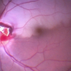 IOFB Over Disc With Blood Stained Hyaloid and BRAO
Feb 25 2017 by Manish Nagpal, MD, FRCS (UK), FASRS
Intraoperative photo of a foreign body piercing the inferotemporal margin of disc revealing a infero temporal BRAO immediately after hyaloid removal.
Photographer: MANISH NAGPAL
Imaging device: Still captured from a 3 chip HD camera attached to a microscope
Condition/keywords: branch retinal artery occlusion (BRAO), intraocular foreign body
-
 IOFB with BRAO
Feb 9 2017 by Manish Nagpal, MD, FRCS (UK), FASRS
Intraoperative photo of a IOFB impacting on the inferotemporal margin of disc leading to a BRAO. This picture is taken moments after dissecting the IOFB from the impact site and bringing it over the retinal surface for eventual removal.
Photographer: Manish Nagpal
Imaging device: Still captured from a 3 chip HD camera on microscope
Condition/keywords: branch retinal artery occlusion (BRAO), intraocular foreign body
-
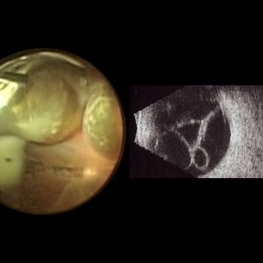 Macrocysts in Kickboxer
Nov 17 2023 by Bradley T. Smith, MD, FASRS
Intraoperative photo and preoperative b scan of chronic retinal detachment with macrocysts in a kickboxer
Condition/keywords: B scan ultrasound, chronic retinal detachment, ocular trauma, pars plana vitrectomy (PPV), retinal macrocyst
-
 RESCUING IOL CTR BAG COMPLEX
Jun 14 2023 by Aditya S Kelkar, MS, FRCS, FASRS,FRCOphth
INTRAOPERATIVE SNAPSHOPT IN ZEISS ARTEVO 800 OF DROPPED IOL CTR BAG COMPLEX IN A 71 YEAR OLD MALE PATIENT
Photographer: SUBHASREE DUTTA, NATIONAL INSTITUTE OF OPHTHALMOLOGY, PUNE
Imaging device: ZEISS ARTEVO 800
Condition/keywords: dropped capsular IOL bag complex
-
 Sub ILM Hemorrhage
Jan 12 2022 by Manish Nagpal, MD, FRCS (UK), FASRS
Intraoperative view of a non clearing sub ILM hemorrhage over the macula with partly de-hemoglobinized blood.
Photographer: Manish Nagpal, Retinal Foundation, Ahmedabad, India
Imaging device: Sony PMW -10 MD surgical camera
Condition/keywords: sub internal limiting membrane haemorrhage, subILM hemorrhage
-
Time to Chill
Jan 23 2024 by SHISHIR VERGHESE, MS, FVRS, FAICO (Retina)
Intraoperative surgical video of a 65 year old female patient with advanced proliferative diabetic retinopathy showing neovascularization at the ora serrata for which a cryopexy is being done to cause regression. This video highlights a previously undocumented grape like Neovascularization at the ora serrata in this patient with advanced proliferative diabetic retinopathy.
Condition/keywords: Advanced Proliferative diabetic retinopathy, Cryopexy, neovascularization
-
 Triamcinolone Stained Hyaloid
Feb 11 2017 by Manish Nagpal, MD, FRCS (UK), FASRS
Intraoperative photo of triamcinolone stained hyaloid showing the whitish haloes of its attachments on macula and disc.
Photographer: manish nagpal
Imaging device: Still captured from 3 Chip HD camera on microscope
Condition/keywords: hyaloid, triamcinolone
-
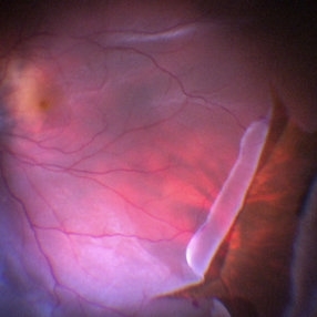
 Unfolding of GRT flap
May 31 2017 by Manish Nagpal, MD, FRCS (UK), FASRS
Intraoperative photo of unfolding of flap of giant retinal tear.
Photographer: MANISH NAGPAL
Imaging device: SONY 3 CHIP HD CAMERA
Condition/keywords: giant retinal tear, perfluorocarbon fluid
-
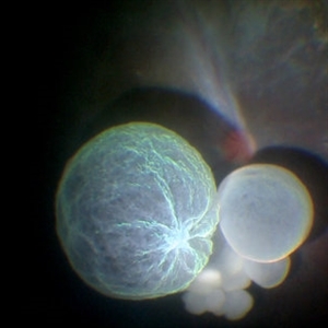 Intraocular Multiple Cysticercus
Oct 10 2018 by Vishal Agrawal, MD, FRCS,FACS,FASRS
Intraoperative fundus picture of right eye of a 18-year-old boy with complaints of DOV for the past 2 months. There were 12 intravitreal cysts in total with vitritis sclerosis retinal vessels and TRD. To note here, the largest cyst has a flimsy wall and no scolex (possibly ruptured) and the rest of the smaller cysts have a scolex and a taut wall.
Photographer: Vishal Agrawal MD,FRCS
Imaging device: SONY PMW-10 MD HD
Condition/keywords: cysticercosis, scolex
 Loading…