Search results (17 results)
-
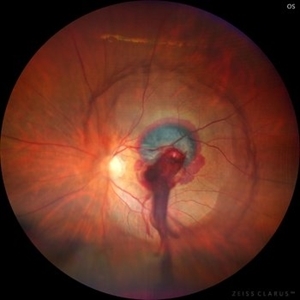
 Retinal Arteriovenous Malformation
Oct 7 2025 by Korey Starkey
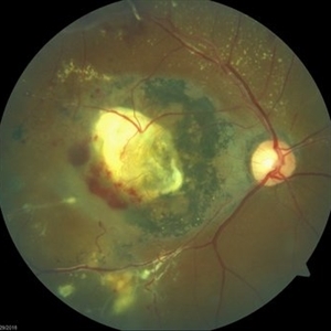
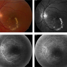
55 year-old patient presents with retinal arteriovenous malformation in the left eye and BRVO w/retinal neovascularization. Patient is asymptomatic. No edema or treatment necessary today, signs of old RVO with MAs along inferior arcade and dot heme.
Photographer: Korey Starkey
Imaging device: Topcon
Condition/keywords: branch retinal vein occlusion (BRVO), fundus photography, inferior arcade, microaneurysms, retinal arteriovenous malformations, retinal neovascularization, Topcon
-
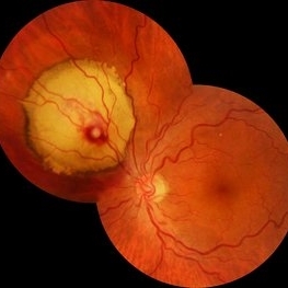
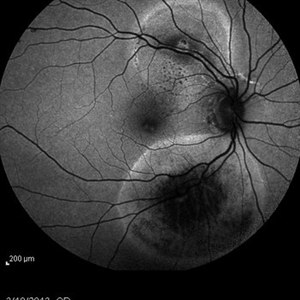
 Ruptured Retinal Artery Macroaneurysm
Jun 18 2024 by KANWALJEET HARJOT MADAN, M.S. (Ophthalmology); FAICO (Vitreous - Retina)
This is a fundus photo depicting ruptured Retinal Artery Macroaneurysm (RAM) in the left eye of a 63 years old female. RAM is an acquired saccular or fusiform dilatation of the retinal arterioles that usually occur within the first three orders of bifurcation. The Superotemporal artery is the most common location. RAM may be asymptomatic or cause a number of complications such as macular edema, serous macular detachment, and hemorrhages.
Photographer: Dr Kanwaljeet Harjot Madan
Condition/keywords: Haemorrhage, macroaneurysm, retinal arteriole
-
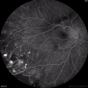 Coats Disease Fluorescein Angiography
Sep 2 2022 by FLOR ANGELICA JACOME GUTIERREZ
Fluorescein angiography of a patient with Coats disease where we found telangiectatic vessels, aneurysms, peripheral capillary nonperfusion and perivascular leak.
Photographer: Dr.Guillermo Salcedo Villanueva
Imaging device: Zeiss CLARUS 700 (FA)
Condition/keywords: Coats' disease, epiretinal membrane (ERM)
-
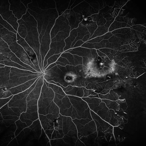
 Idiopathic retinal vasculitis, aneurysms and neuroretinitis
Apr 24 2022 by Aniruddha K Agarwal, MD
Ultra-wide field fundus fluorescein angiography (FFA) of the left eye from an asymptomatic, healthy 33-year-old woman who was referred to the retina clinic from a refractive surgery unit due to the presence of vascular anomalies and hard exudates in both eyes. FFA revealed the characteristic sacular aneurysms at the bifurcation of retinal arterioles in the posterior pole, together with microvascular anomalies and capillary closure peripherally.
Photographer: Julio J GONZALEZ-LOPEZ, MD, PhD, FEBO and Teresa GONZALEZ-LOMAS, RN
Imaging device: Optos California
Condition/keywords: IRVAN Syndrome, IUSG, neuroretinitis, retinal vasculitis, uveitis
-
 Coats Disease
Feb 18 2022 by Ahmad B. Tarabishy, MD
43 year old gentleman with poor vision in his left eye for many years. Examination shows multiple retinal telangiectasias and aneurysms. Ultrawide field fluorescein angiography shows light-bulb aneurysms, telangiectasias, and extensive vascular remodeling and non-perfusion.
Photographer: Dr. Angela Rico, Retina Specialists of Tampa
Condition/keywords: Coats' disease
-
 Coats' Disease
Feb 2 2021 by Niloofar Piri, MD
#2 Fluorescein angiography of the same patient in lamellar arteriovenous phase, demonstrating temporal peripheral telangiectatic vessels, as well as hyperfluorescent aneurysma lesions. Note the anterior capillary non perfusion. Posterior hypofluorescence is secondary to blocking effect from hard exudates.
Condition/keywords: Coats' disease, Leber's miliary aneurysm
-
 Ruptured Macroaneurysm
May 22 2019 by Nichole Lewis
FA of a 91-year-old woman with a ruptured macroaneurysm, intraretinal hemorrhage and subretinal hemorrhage. VA 20/400.
Photographer: Nichole Lewis
Condition/keywords: intraretinal hemorrhage, ruptured macroaneurysm, subretinal hemorrhage
-
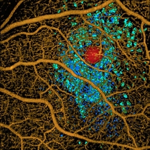 Volume Rendering Structural and Angiographic Optical Coherence Tomography Angiography Image of a Retinal Capillary Microaneurysm, A Newly Described Entity.
May 21 2019 by Richard F. Spaide, MD
This is a newly described entity in which patients develop solitary aneurysms that are much larger than typical microaneurysms and they are supplied by capillaries. The aneurysm is shown in red. The associated macular edema produced cystoid spaces in Henle’s fiber layer, rendered as teal and in the inner nuclear layer as blue.
Photographer: Richard F. Spaide, MD
Condition/keywords: aneurysm, optical coherence tomography (OCT), volume rendering
-
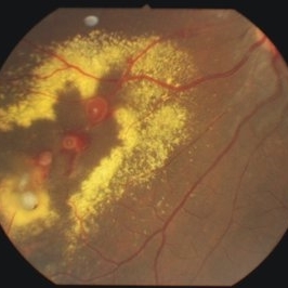
 Coats' Disease Color Fundus Photograph
Apr 19 2019 by Ahmad B. Tarabishy, MD
57-year-old man referred for macular hemorrhage. Examination reveals a large macular scar along with vascular sheathing, vascular irregularity, and extensive exudation. Fluorescein angiogram revealed findings of lightbulb vascular aneurysms and capillary non-perfusion.
Imaging device: Zeiss Cirrus Photo
Condition/keywords: Coats' disease
-
 Laser Induced BRAO in IRVAN Syndrome
May 3 2019 by Deependra Vikram Singh, MD FASRS
Fundus photograph of a 26-year-old man with IRVAN syndrome referred for vitreous surgery in OS for secondary rhegmatogenous retinal detachment. OD has received laser photocoagulation for capillary nonperfusion areas and retinal artery macroaneurysm associated with retinal vasculitis. Fundus photograph of OD shows laser induced nasal BRAO. Case re-emphasizes why laser for macroaneurysm should be avoided in cases with IRVAN.
Photographer: Deependra V Singh, Eye-Q Superspecialty Eye Hospitals. Gurugram, India
Imaging device: Zeiss Visucam 500
Condition/keywords: arteriolar macroaneurysm, branch retinal artery occlusion (BRAO), laser photocoagulation
-
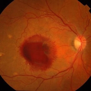
 Retinal Arterial Macroaneurysm
Sep 18 2018 by Somnath Chakraborty, MD
Left eye fundus photo of a 72-year-old hypertensive, female with a hemorrhagic retinal arterial macroaneurysm with sub-retinal blood.
Photographer: Pulok Chandra Roy, Retina Institute of Bengal
Condition/keywords: retinal arterial macroaneurysm
-
 Bilateral Lebers Miliary Aneurysm in a Female
Sep 5 2017 by Ogugua Ndubuisi Okonkwo, MD, FRCS (Edin), FASRS
Fundus photograph of the active left eye of a 26-year-old female with bilateral LMA. Shows severe exudation in the nasal retina by leaking aneurysms.
Condition/keywords: aneurysm
-
 Macroaneurysm
Apr 1 2017 by Manish Nagpal, MD, FRCS (UK), FASRS
Case of a ruptured macroaneurysm with subhyaloid and subretinal blood.
Photographer: Avijit Vishnoi
Condition/keywords: macroaneurysm, ruptured macroaneurysm
-
 RAMA
RAMA
Jun 20 2016 by John S. King, MD
RAMA with 2 w co decreased vision; htn, afib using anticoag; light laser applied; 20/400.
Condition/keywords: ruptured macroaneurysm
-
 Vascular Anormalities
Jan 6 2016 by Andrea Arriola-Lopez, MD MSc
77-year-old man. Decrease of visual acuity OS. VA 20/30 IOP 14mmHg. Fundus examination findings: Hard exudates, microaneurysms near to fovea. OCT shows IRF. Late leakage on FA.
Photographer: Andrea Elizabeth Arriola-Lopez, MSc MD
Condition/keywords: abnormal retinal vessel, aneurysm, hard exudates, vascular anomaly
-
 Retinal Arterial Macroaneurysms
Jan 22 2015 by Darrell E. Baskin, MD
Fundus autofluorescence image of a 60-year-old woman with two retinal arterial macroaneurysms--one recent and one not.
Photographer: Darrell Baskin, Wilford Hall, Lackland Air Force Base, Texas
Imaging device: Heidelberg Spectralis
Condition/keywords: retinal arterial macroaneurysm
-
 Proliferative Diabetic Retinopathy - Neovascularization on the Disc
Aug 23 2012 by Gerardo Garcia-Aguirre, MD
Fluorescein angiogram, early phase, showing microaneurysms, wide areas of capillary nonperfusion, and leakage secondary to neovascularization on the disc.
Photographer: Noemí Hernández, Asociación para Evitar la Ceguera en México
Condition/keywords: microaneurysms, neovascularization of the disc (NVD)
 Loading…