Search results (445 results)
-
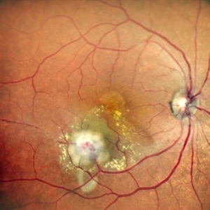
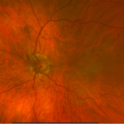
 Leber’s Miliary Aneurysm
Dec 12 2025 by KANWALJEET HARJOT MADAN, M.S. (Ophthalmology); FAICO (Vitreous - Retina)
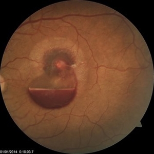
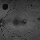
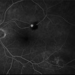
A 34 year-old male presented with decrease vision in right eye for 3 months. Anterior segment exam was normal. Fundus exam in RE revealed presence of macular edema which was evident on OCT. Multiple retinal vascular aneurysmal dilatations with telangiectasia of the retina blood vessels noted superiorly which was evident on FFA. These aneurysms were multiple, tiny and leaky on FFA. He was diagnosed to have Leber’s miliary aneurysms. It is a rare, typically unilateral eye condition, often seen in young males, characterized by multiple tiny, leaky aneurysms in the retinal blood vessels, leading to deposits of hard exudates and potential vision loss, especially if it affects the macula. It is considered a milder form of Coats' disease.
Photographer: Dr. Kanwaljeet Harjot Madan, Thind Eye Hospital, Jalandhar City (Punjab) INDIA.
Imaging device: Zeiss Fundus Camera
Condition/keywords: FFA, Leber's miliary aneurysm
-
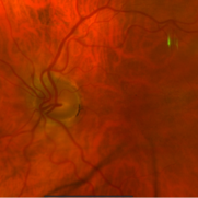
 Proliferative Diabetic Retinopathy
Nov 20 2025 by Oftalmontt Clínica Láser
Multimodal examination of a 39-year-old patient presenting with decreased visual acuity in both eyes due to clear proliferative diabetic retinopathy. Presence of neovascularization at the papillary level, microaneurysms in all four quadrants, a vascular loop in the inferior temporal arcade, and an altered ZAF due to low blood perfusion is observed.
Photographer: Ophthalmic Medical Technologist
Imaging device: Canon cx-1 and Avanti XR Optovue
Condition/keywords: proliferative diabetic retinopathy (PDR)
-
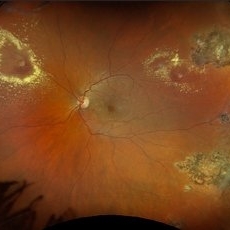
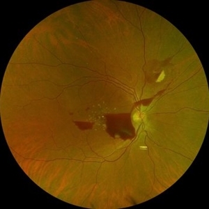
 All That Glows Yellow Isn’t Mellow: Coats' Disease Unveiled
Nov 4 2025 by SHRADDHA RAJ SHRIVASTAVA
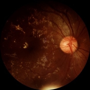
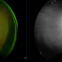
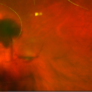
Montage fundus image of an 11 year old boy diagnosed with left eye Coats' disease (stage 3A1), reveals a hyperemic disc and surrounding intra-retinal exudates superior to the disc. There is a single fibroglial nodule at the macula causing submacular fibrosis with exudation. We can see areas of pigmentary changes and RPE atrophy in posterior pole and mid-peripheral retina supero-temporally. There is massive yellowish subretinal exudation in all the quadrants, which are associated with telangiectatic aneurysmal capillary dilation, more prominently seen in the nasal periphery. Supero-nasally we can also see an orange-red elevated vaso-proliferative mass with overlying dilated capillaries, which has likely developed secondary to untreated long standing disease. We can also see associated extrafoveal subtotal exudative retinal detachment in the inferior and nasal quadrants.
Photographer: Dr. Shraddha Raj Shrivastava
Imaging device: Nidek Mirante SLO/OCT (Confocal scanning/Spectral domain OCT)
Condition/keywords: COATS DISEASE, exudative detachment, leukocoria, subretinal exudates, Xanthocoria, yellow exudate
-
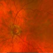
 Retinal Arteriovenous Malformation
Oct 7 2025 by Korey Starkey
55 year-old patient presents with retinal arteriovenous malformation in the left eye and BRVO w/retinal neovascularization. Patient is asymptomatic. No edema or treatment necessary today, signs of old RVO with MAs along inferior arcade and dot heme.
Photographer: Korey Starkey
Imaging device: Topcon
Condition/keywords: branch retinal vein occlusion (BRVO), fundus photography, inferior arcade, microaneurysms, retinal arteriovenous malformations, retinal neovascularization, Topcon
-
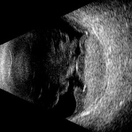 Type 1 Aneurysmal Neovascularization
Sep 22 2025 by Gustavo Uriel Fonseca Aguirre
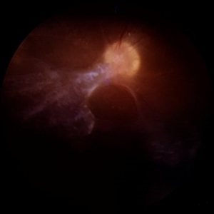
This transverse B-scan demonstrates vitreous hemorrhage, a bullous retinal detachment involving the macula, and dense subretinal hemorrhage, consistent with type 1 aneurysmal neovascularization. The scan reveals significant exudative activity with multi-level bleeding.
Photographer: Gustavo U. Fonseca Aguirre, Hospital Conde de Valenciana, Ciudad de México
Condition/keywords: polypoidal choroidal vasculopathy (PCV), Type 1 Aneurysmal Neovascularization
-
 Lasered Retinal Artery Macroaneurysm
Sep 22 2025 by Tejaswita Verma
Fundus image of a 73 year old hypertensive female status post focal laser for exudative RAM. There was associated macular edema on OCT. Vision was 6/18.Patient was also planned for intravitreal anti VEGF injection on the same day.
Photographer: DR. TEJASWITA VERMA
Imaging device: MIRANTE
Condition/keywords: focal laser, RAM, retinal artery macroaneurysm
-
 Retinal Artery Macroaneurysm With Macular Edema
Sep 12 2025 by Tejaswita Verma
Fundus photo of a 73 year old hypertensive female with 6/18 vision, presenting with RAM ,with surrounding hard exudates and macular edema. She was advised focal laser, anti VEGF injection.
Photographer: DR. TEJASWITA VERMA
Imaging device: MIRANTE
Condition/keywords: RAM, retinal arterial macroaneurysm
-
 Coats' Disease
Sep 2 2025 by Drew Mitchell
Optos color photograph of a young boy with Coats disease. Extensive subretinal exudation that is encroaching towards macula. There are peripheral berry aneurysms with localized area of subretinal fluid. Discussed treatment options including laser photocoagulation of aneurysms. Risks benefits and alternatives discussed including possible need for cryo.
Photographer: Drew Mitchell, OCT-C
Imaging device: Optos California
Condition/keywords: Coats' disease
-
 Retinal Macroaneurysm OS (RAM)
Aug 20 2025 by Drew Mitchell
Optos Color photograph of a 79 year old woman with non central macular edema and exudates around RAM inferotemporally.
Photographer: Drew Mitchell OCT-C
Imaging device: Optos Silverstone
Condition/keywords: color photo, exudates, OPTOS, RAM, retinal macroaneurysm
-
 Retinal Aneurysms
Aug 6 2025 by Korey Starkey
54 year-old patient presents with scattered peripheral aneurysms with exudates. FA was performed showing peripheral nonperfusion and aneurysms. Treated patient with PRP and focal laser to aneurysms and continued observation.
Photographer: Kore Starkey
Imaging device: Optos
Condition/keywords: aneurysm, branch retinal vein occlusion (BRVO), chorioretinal scar, circinate ring, exudates, fundus photography, lesion, Optos, retinal aneurysms
-
 Hemorrhagic Retinal Arterial Macroaneurysm
Jul 4 2025 by Julian Navarro Saucedo
Fundus photograph of a 52-year-old woman with sudden vision loss showing a hemorrhagic retinal arterial macroaneurysm.
Photographer: Julian Navarro, Tecnologico de Monterrey, Escuela de Medician y Ciencias de la Salud.
Imaging device: VISUCAM 524 ZEISS
Condition/keywords: macroaneurysm
-
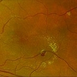
 Diabetic Retinopathy
Jun 4 2025 by Paulina Araujo
The 55-degree central fundus photograph of the right eye demonstrates numerous hard exudates, dot intraretinal hemorrhages, and microaneurysms.
Photographer: Paulina D.Araujo Martínez, Asociación para Evitar la Ceguera en México I.A.P., Hospital Dr Luis Sánchez Bulnes.
Condition/keywords: diabetic retinopathy
-
 Tractional Retinal Detachment
Jun 4 2025 by Paulina Araujo
The 55-degree central fundus photograph of the right eye reveals a thickened and opacified hyaloid exerting traction on the optic disc and posterior pole of the retina, along with hard exudates and microaneurysms consistent with advanced proliferative diabetic retinopathy.
Photographer: Paulina D.Araujo Martínez, Asociación para Evitar la Ceguera en México I.A.P., Hospital Dr Luis Sánchez Bulnes.
Condition/keywords: tractional retinal detachment
-
 Neovascularization of the Disc
Jun 3 2025 by Scott D Walter, MD, MSc, FASRS
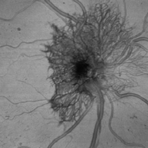
Near-infrared (NIR) en face OCT image showing neovascularization of the disc (NVD) in a patient with type II diabetes mellitus, complicated by proliferative diabetic retinopathy (PDR).
Imaging device: Heidelberg Spectralis
Condition/keywords: Diabetes, Heidelburg Spectralis, microaneurysms, Neovascularisation at the Disc (NVD), NEOVASCULARISATION OF DISC, OCT EN FACE, proliferative diabetic retinopathy (PDR)
-
 Retinal Microaneurysms & Dot/Blot Hemes Autofluorescence OS
May 12 2025 by Briana Hernandez
OS Autofluorescence Optos Image of Retinal Microaneurysms & Dot/Blot Hemes in 91-year-old female BRVO patient.
Photographer: Briana Hernandez, Hilton Head Retina Institute
Imaging device: Optos
Condition/keywords: Autoflourescence, branch retinal vein occlusion (BRVO)
-
 Retinal Microaneurysms & Dot/Blot Hemes Fundus Photo OS
May 12 2025 by Briana Hernandez
OS Optos Fundus Photo of Retinal Microaneurysms & Dot/Blot Hemes in 91-year-old female BRVO patient.
Photographer: Briana Hernandez, Hilton Head Retina Institute
Imaging device: Optos
Condition/keywords: macular
-
 Retinal Microaneurysm FA Image
May 12 2025 by Briana Hernandez
Fluorescein Angiogram OS Image of Retinal Microaneurysm in 81-year-old female BRVO patient.
Photographer: Briana Hernandez
Imaging device: Optos
Condition/keywords: FA
-
 Retinal Macroaneurysm (Left Eye)
Apr 29 2025 by Daniela Bogenschutz
72 year-old female has visual complaints of central vision changes ongoing for 4 days. Patient was acutely symptomatic with an intraretinal hemorrhage due to the retinal macroaneurysm. We had a fun little laugh as this retinal macroaneurysm form a shape of a tick in her left eye. This photo is a side-by-side of the color photos and the autofluorescence done. She is being treated by her general doctor for elevated blood pressure.
Photographer: Daniela Bogenschutz, OSC; Retina Consultants of Carolina, P.A.
Imaging device: Optos
Condition/keywords: retinal macroaneurysm
-
 MacroAneurysm - 1 Day After Rupture
Mar 31 2025 by Max Whitmeyer
Fundus photograph of a macroaneurysm progression prior to and following rupture.
Photographer: Natasa Stankovich, Edward Hines Jr. VA Hospital
Imaging device: Zeiss Clarus 500
Condition/keywords: color fundus photograph, macroaneurysm
-
 MacroAneurysm - 1 Week Before Rupture
Mar 31 2025 by Max Whitmeyer
Fundus photograph of a macroaneurysm progression prior to and following rupture.
Photographer: Natasa Stankovich, Edward Hines Jr. VA Hospital
Imaging device: Zeiss Clarus 500
Condition/keywords: color fundus photograph, macroaneurysm
-
 MacroAneurysm - 2 Months Before Rupture
Mar 31 2025 by Max Whitmeyer
Fundus photograph of a macroaneurysm progression prior to and following rupture.
Photographer: Natasa Stankovich, Edward Hines Jr. VA Hospital
Imaging device: Zeiss Clarus 500
Condition/keywords: color fundus photograph, macroaneurysm
-
 MacroAneurysm - 3 Months Before Rupture
Mar 31 2025 by Max Whitmeyer
Fundus photograph of a macroaneurysm progression prior to and following rupture.
Photographer: Natasa Stankovich, Edward Hines Jr. VA Hospital
Imaging device: Zeiss Clarus 500
Condition/keywords: color fundus photograph, macroaneurysm
-
 The Pouring RAM
Mar 25 2025 by Shrishti mishra
A 63 year old male with RAM lesion in the right eye associated with multilayered hemorrhage.
Imaging device: Optos nikon
Condition/keywords: FFA, retinal arterial macroaneurysm, subhyaloid hemorrhage
-
 Retinal Macroaneurysm (RAM)
Mar 19 2025 by Drew Mitchell
3x3 OCT-A of a Retinal Macroaneurysm in the left eye along the IT arcade that has surrounding edema and exudates
Photographer: Drew Mitchell, OCT-C
Imaging device: Zeiss Cirrus 5000
Condition/keywords: CIRRUS 5000 ANGIOPLEX, OCT Angiography, RAM, retinal macroaneurysm
-
 Retinal Macroaneurysm (RAM)
Mar 19 2025 by Drew Mitchell
3x3 OCT-A of a Retinal Macroaneurysm in the left eye along the IT arcade that has surrounding edema and exudates.
Photographer: Drew Mitchell OCT-C
Imaging device: Zeiss Cirrus 5000
Condition/keywords: OCT Angiography, RAM, retinal macroaneurysm
 Loading…