Search results (445 results)
-
 Macroaneurysm
Apr 1 2017 by Manish Nagpal, MD, FRCS (UK), FASRS
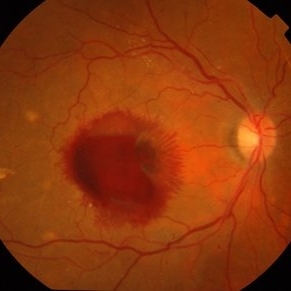
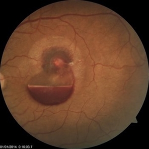
Case of a ruptured macroaneurysm with subhyaloid and subretinal blood.
Photographer: Avijit Vishnoi
Condition/keywords: macroaneurysm, ruptured macroaneurysm
-
 Coats' Disease
Feb 2 2021 by Niloofar Piri, MD
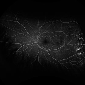
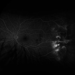
#2 Fluorescein angiography of the same patient in lamellar arteriovenous phase, demonstrating temporal peripheral telangiectatic vessels, as well as hyperfluorescent aneurysma lesions. Note the anterior capillary non perfusion. Posterior hypofluorescence is secondary to blocking effect from hard exudates.
Condition/keywords: Coats' disease, Leber's miliary aneurysm
-
 Bilateral Lebers Miliary Aneurysm in a Female
Sep 5 2017 by Ogugua Ndubuisi Okonkwo, MD, FRCS (Edin), FASRS
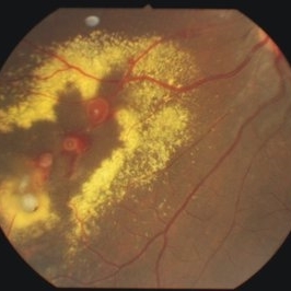
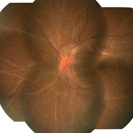
Fundus photograph of the active left eye of a 26-year-old female with bilateral LMA. Shows severe exudation in the nasal retina by leaking aneurysms.
Condition/keywords: aneurysm
-
 Arterial Macroaneurysm
Mar 29 2013 by Henry J. Kaplan, MD
Typical arterial macroaneurysm surrounded by lipid exudates and edema.
Condition/keywords: macroaneurysm, retinal arterial macroaneurysm
-
 Coats' Disease
Feb 2 2021 by Niloofar Piri, MD
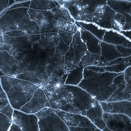
#3 Mid AV phase fluorescein angiography of the same patient demonstrating increasing hyper fluorescence of aneurysmal lesions.
Condition/keywords: Coats' disease, Leber's miliary aneurysm
-
 Coats' Disease
Feb 2 2021 by Niloofar Piri, MD
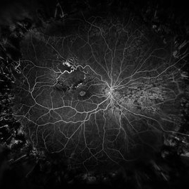
#4 Recirculation phase fluorescein angiography of the same patient demonstrating increased hyperfluorescence and leakage from abnormal vascular lesions in temporal periphery. Note the capillary non perfusion area anteriorly.
Condition/keywords: Coats' disease, Leber's miliary aneurysm
-
 Coats' Disease
Feb 25 2021 by Niloofar Piri, MD
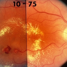
Collage color photo and FA image of the same patient with Coats' Disease demonstrating telangiectatic aneurysmal lesions in the temporal periphery, associated with hard exudate deposition posteriorly. FA (AV phase) demonstrating hyperfluorescent aneurysmal lesions as well as peripheral capillary non perfusion. Note the posterior hypofluorescence where the hard exudates are located.
Condition/keywords: Coats' disease, congenital retinal telangiectasis, retinal telangiectasia
-
 Coats' Disease
Feb 2 2021 by Niloofar Piri, MD
#1 16-year-old male with abnormal temporal peripheral telangiectatic and aneurysmal vascular lesions associated with hard exudate deposition posteriorly. Vision 20/20. Stage II Coats' disease.
Condition/keywords: Coats' disease, Leber's miliary aneurysm
-
 RAMA
RAMA
Jun 20 2016 by John S. King, MD
RAMA with 2 w co decreased vision; htn, afib using anticoag; light laser applied; 20/400.
Condition/keywords: ruptured macroaneurysm
-
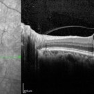
 Ruptured Macroaneurysm OCT
Mar 6 2024 by Mari Ann Z. Keithahn, MD, FASRS
OCT of 73 year-old female with ruptured macroaneurysm.
Photographer: JaTori Maxwell, Missouri Retina Consultants, PC
Imaging device: OPTOS Silverstone
Condition/keywords: Ruptured Macroaneurysm OCT
-
 Venous Beading
Apr 30 2021 by Shivani Reddy, MD
This is a fluorescein angiogram image capturing a beautiful example of different stages of venous beading in diabetic retinopathy all in one frame. This patient also has various microangiopathic findings including microaneurysms, venous loops and capillary dropout. This patient is a 41 y/o male with a history of type 1 diabetes, presenting for his first eye exam in years.
Imaging device: Optos FA
Condition/keywords: capillary dropouts, nonproliferative diabetic retinopathy, proliferative diabetic retinopathy (PDR), retinal ischemia, venous beading
-
 Advanced Proliferative Diabetic Retinopathy With Fibrovascular Proliferation
Jan 4 2019 by Isha Agarwalla
A 29-year-old female with a long-standing history of diabetes mellitus presented with a fibrovascular membrane (FVM) at the viteroretinal interface due to underlying inflammation and angiogenesis induced by ischemia. FVM involved the disc and extended towards the superior and inferior arcades along with extensive capillary drop out areas due to micro aneurysms.
Condition/keywords: fibrovascular proliferation, proliferative diabetic retinopathy (PDR)
-
 Arteriolar Macruaneurysm
Jul 11 2013 by Jerald A. Bovino, MD
No history, with hemorrhage.
Condition/keywords: arteriolar macroaneurysm
-
 Branch Retinal Vein Occlusion
Dec 9 2020 by Olivia Rainey
Ultra-widefield angiogram of a 78-year-old male with a branch retinal vein occlusion affecting his right eye. The patient was diagnosed on 12/17/12 at another practice. The physician noted that there wasn't NVE noted, however areas of micoaneurysmal dilation is present. She noted retinal ischemia secondary to BRVO. 12/8/20 leakage on FA noted to be worsening compared to his previous angiography. She has concern for progressing NVE and recommends sector PRP after injection of antiVEGF series of 3 for the health of the eye.
Photographer: Olivia Rainey, OCT-C, COA
Imaging device: Optos California
Condition/keywords: branch retinal vein occlusion (BRVO), macular branch retinal vein occlusion (BRVO), non-perfusion, scleral buckle, vitreoretinal surgery
-
 Coats Disease Fluorescein Angiography
Sep 2 2022 by FLOR ANGELICA JACOME GUTIERREZ
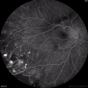
Fluorescein angiography of a patient with Coats disease where we found telangiectatic vessels, aneurysms, peripheral capillary nonperfusion and perivascular leak.
Photographer: Dr.Guillermo Salcedo Villanueva
Imaging device: Zeiss CLARUS 700 (FA)
Condition/keywords: Coats' disease, epiretinal membrane (ERM)
-
 Diabetic Macular Edema, Proliferative Diabetic Retinopathy, Neovascularization Elsewhere, DME, PDR, NVE
Apr 1 2013 by James B. Soque, CRA, OCT-C, COA, FOPS
39-year-old white female and long standing diabetis, c/o new peripheral symptoms of left eye. FA OS reveals diabetic macular edema, microaneurysms, and neovasculaization elsewhere. Fluorescein Angogram, Early Phase, 50 Deg, 2x Mag.
Photographer: James B Soque, CRA, COA
Imaging device: Topcon TRC 50DX with MERGE software, OIS 10.6.45
Condition/keywords: diabetic macular edema, neovascularization (NV), proliferative diabetic retinopathy (PDR)
-
 Diabetic Papillitis
Jun 28 2013 by Jason S. Calhoun
Patient was about to undergo surgery for CNS aneurysm. Patient woke up with little spots which never cleared up. Patients VA was 20/30-OD and 20/40-OS. Both eyes appeared to have disc edema with hemorrhages in the right eye. Ordered a CT of the brain to make sure the aneurysm didn't ruptured.
Photographer: Jason S. Calhoun, Mayo Clinic Jacksonville, Florida
Imaging device: TOPCON TRC 50-EX
Condition/keywords: diabetic mellitus
-
 Diabetic Retinopathy
Jun 4 2025 by Paulina Araujo
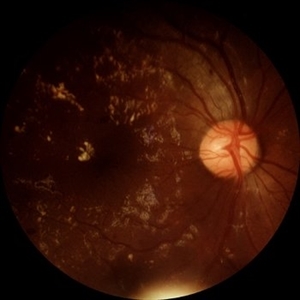
The 55-degree central fundus photograph of the right eye demonstrates numerous hard exudates, dot intraretinal hemorrhages, and microaneurysms.
Photographer: Paulina D.Araujo Martínez, Asociación para Evitar la Ceguera en México I.A.P., Hospital Dr Luis Sánchez Bulnes.
Condition/keywords: diabetic retinopathy
-
 Exudative Retinal Detachment and Branch Retinal Vein Occulsion
Oct 29 2020 by Olivia Rainey
Ultra-widefield fluorescein anigogram of a 51-year-old female with an exudative retinal detachment and branch retinal vein occlusion with retinal neovascularization affecting her right eye. The physician stated that the multiple aneurysmal dilations noted in the inferior periphery are responsible for the exudative RD seen on exam. He is considering Coat's vs FEVR given family history of aneurysms/congenital heart pathology per patient. He encouraged the patient to control their blood pressure, cholesterol, blood sugar, and co-morbidities which may have promoted the BRVO. He recommended antiVEGF injections to control the vascular leakage. Given the severe presentation and imminent threat to her vision, he recommended Eylea as first line therapy.
Photographer: Olivia Rainey, OCT-C, COA
Imaging device: Optos California
Condition/keywords: branch retinal vein occlusion (BRVO), chronic retinal detachment, fluorescein angiogram (FA), fluorescein leakage, inferior retina, inferior retinal detachment, Optos, ultra-wide field imaging
-
 Hemorrhagic Retinal Arterial Macroaneurysm
Jul 4 2025 by Julian Navarro Saucedo
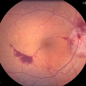
Fundus photograph of a 52-year-old woman with sudden vision loss showing a hemorrhagic retinal arterial macroaneurysm.
Photographer: Julian Navarro, Tecnologico de Monterrey, Escuela de Medician y Ciencias de la Salud.
Imaging device: VISUCAM 524 ZEISS
Condition/keywords: macroaneurysm
-
 Horizontal OCT Scan of Sub ILM Hemorrhage
Mar 8 2017 by Manish Nagpal, MD, FRCS (UK), FASRS
Patient with a macroaneurysm leading to a sub ILM hemorrhage near fovea showing an interesting horizontal scan passing through the central area.
Photographer: pranita chaudhary
Condition/keywords: hemorrhage, macroaneurysm
-
 Idiopathic retinal vasculitis, aneurysms and neuroretinitis
Apr 24 2022 by Aniruddha K Agarwal, MD
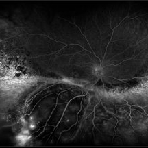
Ultra-wide field fundus fluorescein angiography (FFA) of the left eye from an asymptomatic, healthy 33-year-old woman who was referred to the retina clinic from a refractive surgery unit due to the presence of vascular anomalies and hard exudates in both eyes. FFA revealed the characteristic sacular aneurysms at the bifurcation of retinal arterioles in the posterior pole, together with microvascular anomalies and capillary closure peripherally.
Photographer: Julio J GONZALEZ-LOPEZ, MD, PhD, FEBO and Teresa GONZALEZ-LOMAS, RN
Imaging device: Optos California
Condition/keywords: IRVAN Syndrome, IUSG, neuroretinitis, retinal vasculitis, uveitis
-
 Idiopathic Retinal Vasculitis, Aneurysms, and Neuroretinitis (IRVAN)
Oct 16 2012 by S. Natarajan, MD, FASRS, FRCS (GLASGOW) , FICO, D.Sc, FELA
FFA photograph of a 28-year-old male with IRVAN Syndrome.
Photographer: Prof. Dr. S. Natarajan
Condition/keywords: aneurysm, neuroretinitis, retinal vasculitis
-
 Idiopathic Retinal Vasculitis, Aneurysms, and Neuroretinitis (IRVAN)
Oct 16 2012 by S. Natarajan, MD, FASRS, FRCS (GLASGOW) , FICO, D.Sc, FELA
Fundus photograph of a young male with IRVAN Syndrome
Photographer: Prof. Dr. S. Natarajan
Condition/keywords: aneurysm, neuroretinitis, retinal vasculitis
-
 Laser Induced BRAO in IRVAN Syndrome
May 3 2019 by Deependra Vikram Singh, MD FASRS
Fundus photograph of a 26-year-old man with IRVAN syndrome referred for vitreous surgery in OS for secondary rhegmatogenous retinal detachment. OD has received laser photocoagulation for capillary nonperfusion areas and retinal artery macroaneurysm associated with retinal vasculitis. Fundus photograph of OD shows laser induced nasal BRAO. Case re-emphasizes why laser for macroaneurysm should be avoided in cases with IRVAN.
Photographer: Deependra V Singh, Eye-Q Superspecialty Eye Hospitals. Gurugram, India
Imaging device: Zeiss Visucam 500
Condition/keywords: arteriolar macroaneurysm, branch retinal artery occlusion (BRAO), laser photocoagulation
 Loading…