Search results (47 results)
-
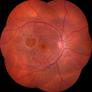
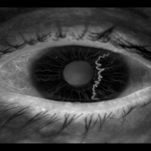
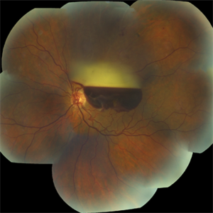
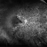
 MIDD (Maternally Inherited Diabetes and Deafness) - Right AF
Nov 30 2024 by John S. King, MD
Both right and left eyes have symmetrical ring of mottled hypo/hyper AF around the fovea and disc. The HyperAF areas correspond to RPE deposits on OCT as well as areas of blockage on FA, and drusenoid deposits seen on fundus photos. Disc drusen in right eye present as HyperAF spot 57 yo WF referred for AMD vs Pattern Dystrophy that was diagnosed 10 years ago. Reported some slow progressive vision loss in both eyes for distance and near. Denies nyctalopia or hemeralopia. Background medical history includes HTN, CVD, and DM. No family history of eye problems. Denied pentosan use. Anterior segment showed moderate cataracts (OD>OS). Posterior segment exam showed macular changes and mild NPDR. The macular appearance showed a symmetrical, paramacular ring of fleck-like drusenoid material with some faint focal areas of RPE hyperplasia. Fundus Photos, AF, OCT were performed as well as a gene test. Further questioning showed revealed that her mother and maternal grandmother had both diabetes mellitus and sensorineural hearing loss. The patient developed diabetes in her teens, and some high frequency hearing loss in her early twenties. She had not had a previous genetic test or diagnosis of MIDD. Gene testing is pending for the mitochondrial component. Invitae's retinal panel, which does not include mitochondrial disorders, only showed a variant of uncertain significance, HMCN1. I discussed this case with Dr. Freund, and it is similar to a the case report : Inoue M, Kiss S, Freund KB. MACULAR PIGMENT RINGS AS THE PRESENTING FINDING OF MITOCHONDRIAL MYOPATHY, ENCEPHALOPATHY, LACTIC ACIDOSIS, AND STROKELIKE EPISODES. Retin Cases Brief Rep. 2015 Fall;9(4):260-4. doi: 10.1097/ICB.0000000000000182. PMID: 26200388.
Photographer: Grace Melton and Carley Gunn
Imaging device: Clarus
Condition/keywords: Macular Dystrophy, Maternally Inherited Diabetes and Deafness, MIDD, Mitochondrial Disorder
-
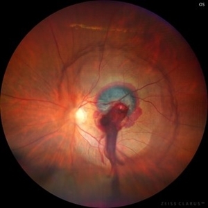
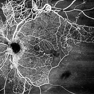
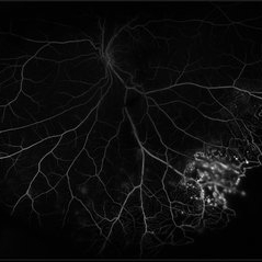
 Pericentral Retinitis Pigmentosa
Sep 6 2024 by Mauricio Bayram-Suverza, MD
A 65-year-old male patient reports experiencing bilateral blind spots that have gradually intensified over time. Genetic testing was unrevealing. The fundus autofluorescence image shows a hypoautofluorescent ring in the posterior pole, especially nasal to the nerve and along arcades.
Photographer: Mauricio Bayram-Suverza, Casey Eye Institute, OHSU.
Imaging device: Optos California
Condition/keywords: fundus autofluorescence (FAF), inherited retinal disease, nyctalopia, retinal dystrophy, retinitis pigmentosa
-
 Failure of Macular Hole Surgery
Jul 2 2024 by Abel Ramírez-Estudillo, MD
Fundus photograph of a 67-year-old woman with failed macular hole surgery, now referred to our clinic with 8 holes.
Photographer: Berenice Palafox, Centro Oftalmológico Mira, Mexico City
Imaging device: Zeiss
Condition/keywords: iatrogenic retinal tear, internal limiting membrane (ILM) peeling, macular hole, vitrectomy
-
 Ruptured Retinal Artery Macroaneurysm
Jun 18 2024 by KANWALJEET HARJOT MADAN, M.S. (Ophthalmology); FAICO (Vitreous - Retina)
This is a fundus photo depicting ruptured Retinal Artery Macroaneurysm (RAM) in the left eye of a 63 years old female. RAM is an acquired saccular or fusiform dilatation of the retinal arterioles that usually occur within the first three orders of bifurcation. The Superotemporal artery is the most common location. RAM may be asymptomatic or cause a number of complications such as macular edema, serous macular detachment, and hemorrhages.
Photographer: Dr Kanwaljeet Harjot Madan
Condition/keywords: Haemorrhage, macroaneurysm, retinal arteriole
-
 Venous Loop
Feb 20 2024 by Soobien Lee
A 77-year-old male with a history of bilateral optic neuropathy from bilateral optic nerve sheath meningiomas S/P radiation/proton-beam therapies. Presented with radiation retinopathy OS and a known venous loop OS.
Photographer: Gavin Bragdon, Elman Retina Group
Imaging device: Optos Ultra-Widefield Fluorescein Angiography
Condition/keywords: fluorescein angiogram (FA), Optos, radiation retinopathy, retinal vascular disease, venous loop
-
 Retinal Fold
Sep 26 2023 by Mauricio Bayram-Suverza, MD
A 38-year-old man underwent vitrectomy in the left eye due to a giant tear in the upper retina. SF6 gas was used as endotamponade. During the post-surgical check-up, it was identified that the patient developed a full-thickness retinal fold due to retinal slippage during fluid-air exchange. As the fold was away from the macular area, it was decided to observe the patient. Three weeks after the surgery, his best-corrected visual acuity was 20/30.
Photographer: Mauricio Bayram-Suverza, Fundación Hospital Nuestra Señora de la Luz
Imaging device: TRC-50DX
Condition/keywords: giant retinal tear, retina surgery complications, Retinal slippage, vitreoretinal surgery
-
 Proliferative Retinopathy
Mar 28 2023 by Harold Rodriguez
Fluorescein Angiogram on a 43 Year Old female with Proliferative Retinopathy.
Photographer: Harold Rodriguez
Condition/keywords: proliferative retinopathy
-
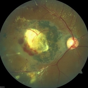
 Angioid Streaks
Dec 14 2022 by Pramod Kumar Suman, MBBS, MD
Fundus autofluorescence photograph of a 65-year-old male with numerous narrow, irregular streaks radiating in a circumferential pattern within the posterior pole with macular atrophy.
Photographer: Pramod Kumar Suman, Retina Foundation, Ahmedabad
Imaging device: Mirante
Condition/keywords: angioid streaks
-
 Iris Racemose Hemangioma
Jan 1 2023 by Maxwell J Wingelaar, MD
Fluorescein Angiogram of 66 year old female presented with an iris racemose hemangioma
Photographer: Ken Huff
Condition/keywords: Racemose hemangioma
-
 Central Retinal Vein Occlusion by OCT Angiography
Jun 13 2022 by JORGE SOBERANES
A 63 year old man with a central retinal vein oclussion. In the OCT angiogram we could observe retinal isquemia, neovascularization and arteriovenous shunts.
Photographer: Jorge I. Soberanes MD
Imaging device: PLEX Elite 9000, Zeiss
Condition/keywords: Central vein oclussion, neovascularization, OCT angiography, retina, Shunts
-
 Proliferative Diabetic Retinopathy
Oct 16 2021 by Timur Shaimov
32 y.o. female with Type 1 Diabetes with no glucose compensation for several years. A manual montage of several 8x8 mm OCT angiograms were obtained for this Widefield OCTA image.
Photographer: Timur Shaimov
Imaging device: RTVue xR Avanti
Condition/keywords: OCT Angiography, proliferative diabetic retinopathy (PDR)
-
 Diabetic Retinopathy Treated with PRP Laser
Jun 8 2021 by Ronald Coriasso
Diabetic retina treated with complete 360 PRP laser, taken during fluorescein angiogram.
Photographer: Ronald Coriasso
Imaging device: OPTOS
Condition/keywords: pan-retinal photocoagulation (PRP)
-
 Retinal Autograft Postoperative Day 1
Feb 29 2020 by Raja Rami P Reddy, MD FRCS FASRS
20-year-old boy presented with recurrent retinal detachment post silicone oil removal with a macular hole. During surgery a retinal graft was created from the detached retina and positioned on the macular hole. silicone oil was injected at the end of surgery. One can see the diathermy marks on all sides of the edges of the graft.
Condition/keywords: autograft, macular hole
-
 Branch Retinal Vein Occlusion with Acute on Chronic Subhyaloid Hemorrhage
Oct 24 2019 by Nichole Lewis
60-year-old male with a branch retinal vein occlusion and subhyaloid hemorrhage and retinal neovascularization. VA HM.
Photographer: Nichole Lewis
Condition/keywords: branch retinal vein occlusion (BRVO), retinal neovascularization, subhyaloid hemorrhage
-
 Coats' Disease
Jul 16 2019 by Kim Barrett
Ultra-wide field fluorescein angiogram of a 23-year-old male with Coats' disease, presented with distorted vision affecting his left eye. He reported seeing flashes and floaters since January of 2019, but the flashes had resolved. He was treated with Intravitreal Preservative Free Triamcinolone in the office and scheduled for PRP laser in the near future.
Photographer: Kim Barrett
Imaging device: Optos
Condition/keywords: Coats' disease, fluorescein angiogram (FA), fluorescein leakage, inferior retina, ischemia, left eye, Optos, ultra-wide field imaging
-
 Ruptured Macroaneurysm
May 22 2019 by Nichole Lewis
FA of a 91-year-old woman with a ruptured macroaneurysm, intraretinal hemorrhage and subretinal hemorrhage. VA 20/400.
Photographer: Nichole Lewis
Condition/keywords: intraretinal hemorrhage, ruptured macroaneurysm, subretinal hemorrhage
-
 Coats' Disease - Widefield Fundus Image
May 18 2019 by Tony Tsai, MD, FASRS
Widefield fundus image of 11-year-old male with peripheral lipid exudative retinopathy secondary to Coats' Syndrome.
Photographer: Chue Vang, Retinal Consultants, Sacramento, CA
Imaging device: Optos
Condition/keywords: Coats' disease
-
 PDR; High Myopia; PRP
May 2 2019 by Carissa Hurdstrom
PDR; high myopia; PRP
Imaging device: Optos
Condition/keywords: fluorescein angiogram (FA), high myopia, pan-retinal photocoagulation (PRP), proliferative diabetic retinopathy (PDR)
-
 Coats' Disease Color Fundus Photograph
Apr 19 2019 by Ahmad B. Tarabishy, MD
57-year-old man referred for macular hemorrhage. Examination reveals a large macular scar along with vascular sheathing, vascular irregularity, and extensive exudation. Fluorescein angiogram revealed findings of lightbulb vascular aneurysms and capillary non-perfusion.
Imaging device: Zeiss Cirrus Photo
Condition/keywords: Coats' disease
-
 Amelanotic Choroidal Melanoma
Apr 12 2019 by David L Kilpatrick, MD
Fundus photograph of a 69-year-old male with an amelanotic choroidal melanoma and corresponding exudative retinal detachment. Transvitreal biopsy was performed at the time of radioactive I-125 plaque placement. The genetic expression profile revealed a Class 1A, PRAME negative tumor.
Photographer: Retina Consultants of Alabama, P. C.
Imaging device: Optos
Condition/keywords: amelanotic melanoma
-
 Laser Induced BRAO in IRVAN Syndrome
May 3 2019 by Deependra Vikram Singh, MD FASRS
Fundus photograph of a 26-year-old man with IRVAN syndrome referred for vitreous surgery in OS for secondary rhegmatogenous retinal detachment. OD has received laser photocoagulation for capillary nonperfusion areas and retinal artery macroaneurysm associated with retinal vasculitis. Fundus photograph of OD shows laser induced nasal BRAO. Case re-emphasizes why laser for macroaneurysm should be avoided in cases with IRVAN.
Photographer: Deependra V Singh, Eye-Q Superspecialty Eye Hospitals. Gurugram, India
Imaging device: Zeiss Visucam 500
Condition/keywords: arteriolar macroaneurysm, branch retinal artery occlusion (BRAO), laser photocoagulation
-
 Ischemic Central Retinal Vein Occlusion
Jan 24 2019 by Nichole Lewis
76-year-old woman with an ischemic central retinal vein occlusion, severely attenuated and sclerotic vessels and scattered retinal hemorrhages. Vision decrease over 1 year. VA 20/CF. Patient is returning for pan retinal photocoagulation.
Photographer: Nichole Lewis
Imaging device: Optos
Condition/keywords: attenuated vessels, central retinal vein occlusion (CRVO), hemorrhage, ischemic CRVO, sclerotic vessels
-
 Albinotic Fundus
Oct 1 2018 by Rameez N Hussain, MD
Albinotic fundus
Photographer: THAMBI DURAI (Edited by Lafas DE)
Imaging device: TOPCON
Condition/keywords: ocular albinism, oculocutaneous albinism
-
 Choroidal Melanoma With a Serous Retinal Detachment
Aug 23 2018 by Nichole Lewis
63-year-old male with a large choroidal melanoma and a serous macula off retinal detachment. Vision is count fingers.
Photographer: Nichole Lewis
Condition/keywords: serous retinal detachment
-
 Branch Retinal Artery Occlusion With Calcium Embolus at the Disc - Fundus Photo
Apr 7 2018 by Rameez N Hussain, MD
Acute retinal artery occlusion with a calcium embolus at the disc and retinal whitening.
Photographer: DR RAMEEZ N HUSSAIN
Imaging device: zeiss
Condition/keywords: branch retinal artery occlusion (BRAO), embolus, fundus photograph, retinal edema
 Loading…