Search results (1522 results)
-
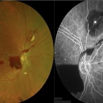
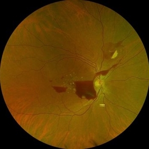
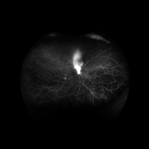
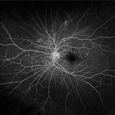
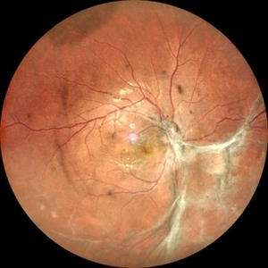
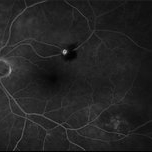 Retinal Microaneurysm FA Image
May 12 2025 by Briana Hernandez
Fluorescein Angiogram OS Image of Retinal Microaneurysm in 81-year-old female BRVO patient.
Photographer: Briana Hernandez
Imaging device: Optos
Condition/keywords: FA
-
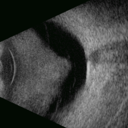 Synchysis Scintillans
Apr 21 2025 by Gustavo Uriel Fonseca Aguirre
This B-mode axial ultrasound scan demonstrates synchysis scintillans, characterized by multiple hyperechoic mobile opacities within the vitreous cavity. The particles exhibit a distinctive 'snowglobe' motion pattern during dynamic assessment, with gravitational settling in dependent areas. The vitreous framework appears liquefied without associated tractional changes.
Photographer: Gustavo U. Fonseca Aguirre, Hospital Conde de Valenciana, Ciudad de México
Condition/keywords: synchysis scintillans
-
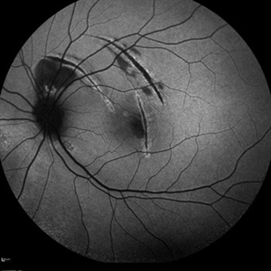 Choroidal Rupture
Apr 7 2025 by Ramses Rosales-Diaz
Autofluorescence image of a 39-year-old female patient who sustained blunt ocular trauma resulting in three choroidal ruptures.
Photographer: Ramses Rosales-Diaz, Asociación Para Evitar la Ceguera en México I.A.P., Mexico City
Imaging device: Heidelberg Spectralis
Condition/keywords: blunt trauma, Choroidal Rupture
-
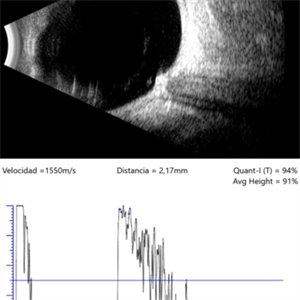 Melanocytoma
Mar 25 2025 by Gustavo Uriel Fonseca Aguirre
Longitudinal B-scan echogram shows mildly elevated lesion overlying surface of optic nerve. A-scan shows regular internal structure and high reflectivity of lesion.
Photographer: Gustavo U. Fonseca Aguirre, Hospital Conde de Valenciana, Ciudad de México
Condition/keywords: Melanocytoma
-
 The Pouring RAM
Mar 25 2025 by Shrishti mishra
A 63 old male with RAM lesion associated with multilayered hemmoraghe.
Photographer: Mrs Vinutha, Narayana Nethralaya
Imaging device: Nikon optos
Condition/keywords: FFA, RAM, SHH
-
 The Pouring RAM
Mar 25 2025 by Shrishti mishra
A 63 year old male with RAM lesion in the right eye associated with multilayered hemorrhage.
Imaging device: Optos nikon
Condition/keywords: FFA, retinal arterial macroaneurysm, subhyaloid hemorrhage
-
 Retinal Macroaneurysm (RAM)
Mar 19 2025 by Drew Mitchell
3x3 OCT-A of a Retinal Macroaneurysm in the left eye along the IT arcade that has surrounding edema and exudates
Photographer: Drew Mitchell, OCT-C
Imaging device: Zeiss Cirrus 5000
Condition/keywords: CIRRUS 5000 ANGIOPLEX, OCT Angiography, RAM, retinal macroaneurysm
-
 Retinal Macroaneurysm (RAM)
Mar 19 2025 by Drew Mitchell
3x3 OCT-A of a Retinal Macroaneurysm in the left eye along the IT arcade that has surrounding edema and exudates.
Photographer: Drew Mitchell OCT-C
Imaging device: Zeiss Cirrus 5000
Condition/keywords: OCT Angiography, RAM, retinal macroaneurysm
-
 Diabetic Tractional Retinal Detachment Involving the Macula OD
Feb 21 2025 by Kaitlyn Anderson
57-year-old female. Diabetic Tractional Retinal Detachment involving the Macula OD. Active Proliferative Diabetic Retinopathy
Photographer: Kaitlyn Anderson TN Retina Nashville TN
Imaging device: Optos Fluorescein Angiogram
Condition/keywords: Active PDR Tractional retinal Detachment
-
 Indocyanine Green (ICG) of Circumscribed Choroidal Hemangioma (CCH)
Feb 6 2025 by Jack B Margines, MD, MHCI
Peripheral patchy hyperfluorescence is seen on this early image of ICG-A on a 53-year-old asymptomatic with an extramacular circumscribed choroidal hemangioma.
Photographer: W Ryan Miliam, CRA, OCT-C, University of California, Irvine Gavin Herbert Eye Institute
Imaging device: Optos
Condition/keywords: choroidal hemangioma, indocyanine green (ICG) angiography
-
 CCH
CCH
Feb 6 2025 by Jack B Margines, MD, MHCI
SD-OCT of an extramacular Circumscribed Choroidal Hemangioma in an asymptomatic 53 year-old female
Photographer: Ryan Milam, University of California, Irvine Gavin Herbert Eye Institute
Imaging device: Zeiss Cirrus
Condition/keywords: Circumscribed Choroidal Hemangioma, OCT
-
 Sickle-Cell Retinopathy
Jan 22 2025 by Virginia Gebhart
Fluorescein angiogram of 54 year old female with non-diabetic proliferative retinopathy. Recent labs confirm sickle-cell disease. FA shows temporal peripheral non perfusion with NV. S/p PRP with retrobulbar block
Photographer: Virginia Gebhart, Retina Consultants of Carolina
Imaging device: Optos California
Condition/keywords: FA, Fluorescein angiography, Neovascularisation elsewhere (NVE), non-perfusion, Nose, pan-retinal photocoagulation (PRP), PRP, sickle cell retinopathy
-
 Stargardt Disease (FA)
Jan 22 2025 by Virginia Gebhart
Fluorescein angiogram of 19 year old female with confirmed Stargardt Disease. Hyperfluorescence in the macula with staining defect and silent choroid.
Photographer: Virginia Gebhart, Retina Consultants of Carolina
Imaging device: Optos California
Condition/keywords: fluorescein angiogram (FA), Silent Choroid, Stargardt disease
-
 Choroidal Melanoma 3 Ways
Jan 16 2025 by Virginia Gebhart
RGB/FA/ICG of 76 year old female with a new choroidal melanoma. Pt scheduled for plaque radiation. BCVA 20/400
Photographer: Virginia Gebhart, Retina Consultants of Carolina
Imaging device: Optos California
Condition/keywords: fluorescein angiogram (FA), indocyanine green (ICG) angiography, OPTOS CALIFORNIA RGB
-
 MEWDS
MEWDS
Dec 11 2024 by Virginia Gebhart
28 year old female with new Multiple Evanescent White Dot Syndrome. Patient reports gray spot in vision, OCT shows RPE disruption centrally but no edema. FA shows early hyperfluorescent punctate spots throughout the posterior pole, but no leakage. Normal findings OD. Will observe for now
Photographer: Virginia Gebhart, Retina Consultants of Carolina
Imaging device: Optos California
Condition/keywords: FA, fluorescein angiogram (FA), multiple evanescent white dot syndrome (MEWDS)
-
 Asteroid Hyalosis in Retinitis Pigmentosa
Dec 9 2024 by Mauricio Bayram-Suverza, MD
A 54 year-old male patient presented with asteroid hyalosis. Retinal examination revealed the presence of bone spicules, primarily located in the mid-periphery. Genetic testing identified a pathogenic variant in the RHO gene.
Photographer: Mauricio Bayram-Suverza, Casey Eye Institute, OHSU.
Imaging device: Optos California
Condition/keywords: Asteroid hyalosis, retinal dystrophy, Retinitis Pigmentosa, vitreous
-
 Extramacular TRD in Idiopathic Occlusive Vasculitis
Dec 5 2024 by Tejaswita Verma
Fundus photo showing extramacular TRD in a 16 year old boy with idiopathic occlusive vasculitis secondary to presumed IOTB. History of taking ATT for 6 months , Mantoux positive previously. Vision was 6/6P,other eye had funnel RD .
Photographer: DR. TEJASWITA VERMA
Imaging device: MIRANTE
Condition/keywords: tractional retinal detachment, vasculitis
-
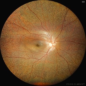 Benign Familial Fleck Retina
Dec 2 2024 by KANWALJEET HARJOT MADAN, M.S. (Ophthalmology); FAICO (Vitreous - Retina)
This is fundus picture of a 21 year old female patient who had come for refractive surgery consultation. Her best corrected vision in both eyes was 20/20. She had myopic astigmatism in both eyes. Fundus exam revealed presence of multiple yellowish white flecks spread throughout retina sparing macular area in both eyes. Her color vision was normal. Electroretinogram and electrooculogram were normal. She gave no history of night blindness. A diagnosis of Benign Familial Fleck Retina was made. She was also advised ocular exam of her parents and elder brother which was normal.
Photographer: Dr. Kanwaljeet Harjot Madan, M.S. (Ophthalmologist) Fellow in Vitrous & Retina. Thind Eye Hospital, Jalandhar City. Punjab. India
Imaging device: Zeiss Clarus
Condition/keywords: Benign familial fleck retina, Night Blindness
-
 MIDD (Maternally Inherited Diabetes and Deafness) - Left AF
Nov 30 2024 by John S. King, MD
Both right and left eyes have symmetrical ring of mottled hypo/hyper AF around the fovea and disc. The HyperAF areas correspond to RPE deposits on OCT as well as areas of blockage on FA, and drusenoid deposits seen on fundus photos 57 yo WF referred for AMD vs Pattern Dystrophy that was diagnosed 10 years ago. Reported some slow progressive vision loss in both eyes for distance and near. Denies nyctalopia or hemeralopia. Background medical history includes HTN, CVD, and DM. No family history of eye problems. Denied pentosan use. Anterior segment showed moderate cataracts (OD>OS). Posterior segment exam showed macular changes and mild NPDR. The macular appearance showed a symmetrical, paramacular ring of fleck-like drusenoid material with some faint focal areas of RPE hyperplasia. Fundus Photos, AF, OCT were performed as well as a gene test. Further questioning showed revealed that her mother and maternal grandmother had both diabetes mellitus and sensorineural hearing loss. The patient developed diabetes in her teens, and some high frequency hearing loss in her early twenties. She had not had a previous genetic test or diagnosis of MIDD. Gene testing is pending for the mitochondrial component. Invitae's retinal panel, which does not include mitochondrial disorders, only showed a variant of uncertain significance, HMCN1. I discussed this case with Dr. Freund, and it is similar to a the case report : Inoue M, Kiss S, Freund KB. MACULAR PIGMENT RINGS AS THE PRESENTING FINDING OF MITOCHONDRIAL MYOPATHY, ENCEPHALOPATHY, LACTIC ACIDOSIS, AND STROKELIKE EPISODES. Retin Cases Brief Rep. 2015 Fall;9(4):260-4. doi: 10.1097/ICB.0000000000000182. PMID: 26200388.
Photographer: Grace Melton and Carley Gunn
Imaging device: Clarus
Condition/keywords: Macular Dystrophy, Maternally Inherited Diabetes and Deafness, MIDD, Mitochondrial Disorder
-
 MIDD (Maternally Inherited Diabetes and Deafness) - Right AF
Nov 30 2024 by John S. King, MD
Both right and left eyes have symmetrical ring of mottled hypo/hyper AF around the fovea and disc. The HyperAF areas correspond to RPE deposits on OCT as well as areas of blockage on FA, and drusenoid deposits seen on fundus photos. Disc drusen in right eye present as HyperAF spot 57 yo WF referred for AMD vs Pattern Dystrophy that was diagnosed 10 years ago. Reported some slow progressive vision loss in both eyes for distance and near. Denies nyctalopia or hemeralopia. Background medical history includes HTN, CVD, and DM. No family history of eye problems. Denied pentosan use. Anterior segment showed moderate cataracts (OD>OS). Posterior segment exam showed macular changes and mild NPDR. The macular appearance showed a symmetrical, paramacular ring of fleck-like drusenoid material with some faint focal areas of RPE hyperplasia. Fundus Photos, AF, OCT were performed as well as a gene test. Further questioning showed revealed that her mother and maternal grandmother had both diabetes mellitus and sensorineural hearing loss. The patient developed diabetes in her teens, and some high frequency hearing loss in her early twenties. She had not had a previous genetic test or diagnosis of MIDD. Gene testing is pending for the mitochondrial component. Invitae's retinal panel, which does not include mitochondrial disorders, only showed a variant of uncertain significance, HMCN1. I discussed this case with Dr. Freund, and it is similar to a the case report : Inoue M, Kiss S, Freund KB. MACULAR PIGMENT RINGS AS THE PRESENTING FINDING OF MITOCHONDRIAL MYOPATHY, ENCEPHALOPATHY, LACTIC ACIDOSIS, AND STROKELIKE EPISODES. Retin Cases Brief Rep. 2015 Fall;9(4):260-4. doi: 10.1097/ICB.0000000000000182. PMID: 26200388.
Photographer: Grace Melton and Carley Gunn
Imaging device: Clarus
Condition/keywords: Macular Dystrophy, Maternally Inherited Diabetes and Deafness, MIDD, Mitochondrial Disorder
-
 MIDD (Maternally Inherited Diabetes and Deafness) - Left FP
Nov 30 2024 by John S. King, MD
Both the right and left Eye have fairly symmetrical, extrafoveal drusenoid-like flecks and focal and faint areas of RPE hyperplasia (in addition to mild NPDR and PPA) 57 yo WF referred for AMD vs Pattern Dystrophy that was diagnosed 10 years ago. Reported some slow progressive vision loss in both eyes for distance and near. Denies nyctalopia or hemeralopia. Background medical history includes HTN, CVD, and DM. No family history of eye problems. Denied pentosan use. Anterior segment showed moderate cataracts (OD>OS). Posterior segment exam showed macular changes and mild NPDR. The macular appearance showed a symmetrical, paramacular ring of fleck-like drusenoid material with some faint focal areas of RPE hyperplasia. Fundus Photos, AF, OCT were performed as well as a gene test. Further questioning showed revealed that her mother and maternal grandmother had both diabetes mellitus and sensorineural hearing loss. The patient developed diabetes in her teens, and some high frequency hearing loss in her early twenties. She had not had a previous genetic test or diagnosis of MIDD. Gene testing is pending for the mitochondrial component. Invitae's retinal panel, which does not include mitochondrial disorders, only showed a variant of uncertain significance, HMCN1. I discussed this case with Dr. Freund, and it is similar to a the case report : Inoue M, Kiss S, Freund KB. MACULAR PIGMENT RINGS AS THE PRESENTING FINDING OF MITOCHONDRIAL MYOPATHY, ENCEPHALOPATHY, LACTIC ACIDOSIS, AND STROKELIKE EPISODES. Retin Cases Brief Rep. 2015 Fall;9(4):260-4. doi: 10.1097/ICB.0000000000000182. PMID: 26200388.
Photographer: Grace Melton and Carley Gunn
Imaging device: Clarus
Condition/keywords: Macular Dystrophy, Maternally Inherited Diabetes and Deafness, MIDD, Mitochondrial Disorder
-
 MIDD (Maternally Inherited Diabetes and Deafness) - Right FP
Nov 30 2024 by John S. King, MD
Both the right and left Eye have fairly symmetrical, extrafoveal drusenoid-like flecks and focal and faint areas of RPE hyperplasia (in addition to mild NPDR and PPA) 57 yo WF referred for AMD vs Pattern Dystrophy that was diagnosed 10 years ago. Reported some slow progressive vision loss in both eyes for distance and near. Denies nyctalopia or hemeralopia. Background medical history includes HTN, CVD, and DM. No family history of eye problems. Denied pentosan use. Anterior segment showed moderate cataracts (OD>OS). Posterior segment exam showed macular changes and mild NPDR. The macular appearance showed a symmetrical, paramacular ring of fleck-like drusenoid material with some faint focal areas of RPE hyperplasia. Fundus Photos, AF, OCT were performed as well as a gene test. Further questioning showed revealed that her mother and maternal grandmother had both diabetes mellitus and sensorineural hearing loss. The patient developed diabetes in her teens, and some high frequency hearing loss in her early twenties. She had not had a previous genetic test or diagnosis of MIDD. Gene testing is pending for the mitochondrial component. Invitae's retinal panel, which does not include mitochondrial disorders, only showed a variant of uncertain significance, HMCN1. I discussed this case with Dr. Freund, and it is similar to a the case report : Inoue M, Kiss S, Freund KB. MACULAR PIGMENT RINGS AS THE PRESENTING FINDING OF MITOCHONDRIAL MYOPATHY, ENCEPHALOPATHY, LACTIC ACIDOSIS, AND STROKELIKE EPISODES. Retin Cases Brief Rep. 2015 Fall;9(4):260-4. doi: 10.1097/ICB.0000000000000182. PMID: 26200388.
Photographer: Grace Melton and Carley Gunn
Imaging device: Clarus
Condition/keywords: Macular Dystrophy, Maternally Inherited Diabetes and Deafness, MIDD, Mitochondrial Disorder
-
 MIDD (Maternally Inherited Diabetes and Deafness) - OCT OD
Nov 30 2024 by John S. King, MD
OCT shows mild RPE deposit inferiorly (corresponds to area of FA blockage and HyperAF) and a focal area of iRORA with loss of EZ more superiorly (possibly due to regression of RPE deposit). No choroidal thickening (like in pachychoroid pigment epitheliopathy or cscr) 57 yo WF referred for AMD vs Pattern Dystrophy that was diagnosed 10 years ago. Reported some slow progressive vision loss in both eyes for distance and near. Denies nyctalopia or hemeralopia. Background medical history includes HTN, CVD, and DM. No family history of eye problems. Denied pentosan use. Anterior segment showed moderate cataracts (OD>OS). Posterior segment exam showed macular changes and mild NPDR. The macular appearance showed a symmetrical, paramacular ring of fleck-like drusenoid material with some faint focal areas of RPE hyperplasia. Fundus Photos, AF, OCT were performed as well as a gene test. Further questioning showed revealed that her mother and maternal grandmother had both diabetes mellitus and sensorineural hearing loss. The patient developed diabetes in her teens, and some high frequency hearing loss in her early twenties. She had not had a previous genetic test or diagnosis of MIDD. Gene testing is pending for the mitochondrial component. Invitae's retinal panel, which does not include mitochondrial disorders, only showed a variant of uncertain significance, HMCN1. I discussed this case with Dr. Freund, and it is similar to a the case report : Inoue M, Kiss S, Freund KB. MACULAR PIGMENT RINGS AS THE PRESENTING FINDING OF MITOCHONDRIAL MYOPATHY, ENCEPHALOPATHY, LACTIC ACIDOSIS, AND STROKELIKE EPISODES. Retin Cases Brief Rep. 2015 Fall;9(4):260-4. doi: 10.1097/ICB.0000000000000182. PMID: 26200388.
Photographer: Grace Melton and Carley Gunn
Imaging device: Zeiss Cirrus
Condition/keywords: Macular Dystrophy, Maternally Inherited Diabetes and Deafness, MIDD, Mitochondrial Disorder
-
 MIDD (Maternally Inherited Diabetes and Deafness) - OCT OS
Nov 30 2024 by John S. King, MD
Magnified section of radial scan through the left eye showing a focal nodular RPE deposit that corresponds to a focal drusenoid deposit in temporal macula, that HypoFLs and HyperAFs. Choroid not significantly thickened or thinned, and the nodular thickening may be just above a large outer choroid vessel?) 57 yo WF referred for AMD vs Pattern Dystrophy that was diagnosed 10 years ago. Reported some slow progressive vision loss in both eyes for distance and near. Denies nyctalopia or hemeralopia. Background medical history includes HTN, CVD, and DM. No family history of eye problems. Denied pentosan use. Anterior segment showed moderate cataracts (OD>OS). Posterior segment exam showed macular changes and mild NPDR. The macular appearance showed a symmetrical, paramacular ring of fleck-like drusenoid material with some faint focal areas of RPE hyperplasia. Fundus Photos, AF, OCT were performed as well as a gene test. Further questioning showed revealed that her mother and maternal grandmother had both diabetes mellitus and sensorineural hearing loss. The patient developed diabetes in her teens, and some high frequency hearing loss in her early twenties. She had not had a previous genetic test or diagnosis of MIDD. Gene testing is pending for the mitochondrial component. Invitae's retinal panel, which does not include mitochondrial disorders, only showed a variant of uncertain significance, HMCN1. I discussed this case with Dr. Freund, and it is similar to a the case report : Inoue M, Kiss S, Freund KB. MACULAR PIGMENT RINGS AS THE PRESENTING FINDING OF MITOCHONDRIAL MYOPATHY, ENCEPHALOPATHY, LACTIC ACIDOSIS, AND STROKELIKE EPISODES. Retin Cases Brief Rep. 2015 Fall;9(4):260-4. doi: 10.1097/ICB.0000000000000182. PMID: 26200388.
Photographer: Grace Melton and Carley Gunn
Imaging device: Zeiss Cirrus
Condition/keywords: Macular Dystrophy, Maternally Inherited Diabetes and Deafness, MIDD, Mitochondrial Disorder
-
 MIDD (Maternally Inherited Diabetes and Deafness) - Right FA (4 min)
Nov 30 2024 by John S. King, MD
Both eyes had similar FA findings. There was no dark choroid or signs of leakage. Granular staining around the fovea and disc were present, and the HypoAF areas corresponded to the drusenoid deposits that showed HyperAF. Mild MAs present due to NPDR 57 yo WF referred for AMD vs Pattern Dystrophy that was diagnosed 10 years ago. Reported some slow progressive vision loss in both eyes for distance and near. Denies nyctalopia or hemeralopia. Background medical history includes HTN, CVD, and DM. No family history of eye problems. Denied pentosan use. Anterior segment showed moderate cataracts (OD>OS). Posterior segment exam showed macular changes and mild NPDR. The macular appearance showed a symmetrical, paramacular ring of fleck-like drusenoid material with some faint focal areas of RPE hyperplasia. Fundus Photos, AF, OCT were performed as well as a gene test. Further questioning showed revealed that her mother and maternal grandmother had boith diabetes mellitus and sensorineural hearing loss. The patient developed diabetes in her teens, and some high frequency hearing loss in her early twenties. She had not had a previous genetic test or diagnosis of MIDD. Gene testing is pending for the mitochondrial component. Invitae's retinal panel, which does not include mitochondrial disorders, only showed a variant of uncertain significance, HMCN1. I discussed this case with Dr. Freund, and it is similar to a the case report : Inoue M, Kiss S, Freund KB. MACULAR PIGMENT RINGS AS THE PRESENTING FINDING OF MITOCHONDRIAL MYOPATHY, ENCEPHALOPATHY, LACTIC ACIDOSIS, AND STROKELIKE EPISODES. Retin Cases Brief Rep. 2015 Fall;9(4):260-4. doi: 10.1097/ICB.0000000000000182. PMID: 26200388.
Photographer: Grace Melton and Carley Gunn
Imaging device: Clarus
Condition/keywords: Macular Dystrophy, Maternally Inherited Diabetes and Deafness, MIDD, Mitochondrial Disorder
 Loading…