Search results (1522 results)
-
 Ozurdex implant
Aug 23 2012 by Daniel A. Adelberg, MD, FASRS
Anterior Segment photograph of a 50 year old with Uveitis and Cystoid Macular Edema status post Intravitreal injection of an Ozurdex dexamethasone implant
Photographer: Robert Ramsey, Southwestern Eye Center, Mesa Arizona
Condition/keywords: Ozurdex implant
-
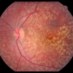
 Sickle Cell Retinopathy
Sep 14 2012 by Michael P. Kelly, FOPS
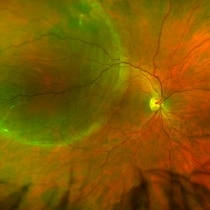
Fluorescein angiogram image of an individual with sickle cell retinopathy using an Optos P200MA ultra-wide field imaging device.
Photographer: Michael P. Kelly, FOPS Director, Duke Eye Center Labs, Duke University Hospital
Imaging device: Optos P200MA
Condition/keywords: Optos, sea fan, sickle cell retinopathy, ultra-wide field imaging
-
 Retinal Detachment Right Eye Optomap
Mar 31 2014 by James B. Soque, CRA, OCT-C, COA, FOPS
36-year-old white male presented with non traumatic retinal detachment OD, with six very distinct demarcation lines and isolated tear, and detachment parameters. Patient underwent PPV OD on 12/3/13 with 20% SF6 gas placement and face down in his first 1 month post op period.
Photographer: James Soque, CRA, COA
Imaging device: Optos Daytona
Condition/keywords: Cryopexy, demarcation line, gas pneumatic displacement, Optomap, Optos, pars plana vitrectomy (PPV), retinal tear, scanning laser ophthalmoscope
-
 Diabetic Macular Edema, Proliferative Diabetic Retinopathy, Neovascularization Elsewhere, DME, PDR, NVE
Apr 1 2013 by James B. Soque, CRA, OCT-C, COA, FOPS
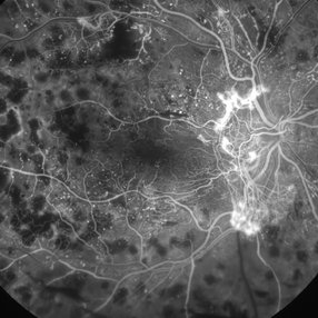
39-year-old white female and long standing diabetis, c/o new peripheral symptoms of left eye. FA OS reveals diabetic macular edema, microaneurysms, and neovasculaization elsewhere. Fluorescein Angogram, Early Phase, 50 Deg, 2x Mag.
Photographer: James B Soque, CRA, COA
Imaging device: Topcon TRC 50DX with MERGE software, OIS 10.6.45
Condition/keywords: diabetic macular edema, neovascularization (NV), proliferative diabetic retinopathy (PDR)
-
 Retinitis Pigmentosa - Fundus Autofluorescence
Sep 20 2014 by Rameez N Hussain, MD
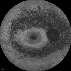
Fundus autofluorescence of retinitis pigmentosa showing hyperautofluorescent rings or foveal hyperautofluorescence.
Photographer: Dr.Rameez N Hussain, MD, Central Imaging Center, Vitreo Retinal Services, Giridhar Eye Institute, Cochin, India
Imaging device: Heidelberg Blue Peak Autofluorescence imaging.
Condition/keywords: bone spicule, cystoid macular edema (CME), fundus autofluorescence (FAF), retinitis pigmentosa
-
 Vitelliform Macular Dystrophy or Best Disease
Dec 16 2016 by Young Hee Yoon, MD, PhD
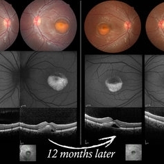
Bilateral fundus photographs and autofluorescence images of 15-year-old girl who was diagnosed as vitelliform macular dystrophy or Best disease. Vitelliform macular lesion showed morphologic change during one year.
Photographer: Hyejin Jo, Sunghyun Kim, Heoni Hong, Minjung Chae, Mihwa Shin, Asan medical center, Seoul
Imaging device: Topcon TRC-500X fundus camera, Heidelberg HRA 2 autofluorescence, Heldelberg Spectralis OCT
Condition/keywords: Best disease, pseudohypopyon, scrambled-egg, vitelliform macular dystrophy
-
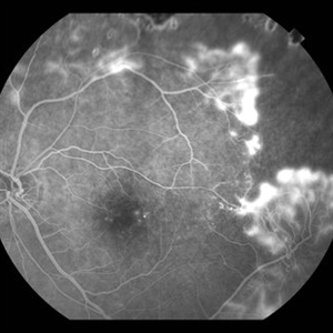
 Ischemic BRVO with neovascularization
Aug 23 2012 by Gerardo Garcia-Aguirre, MD
Fluorescein angiogram of the temporal periphery showing wide areas of capillary nonperfusion and leakage secondary to neovascularization.
Photographer: Noemí Hernández, Asociación para Evitar la Ceguera en México
Condition/keywords: branch retinal vein occlusion (BRVO), capillary nonperfusion, neovascularization (NV)
-
 Fluorescein Angiogram of the Case with Proliferative Diabetic Retinopathy
Mar 21 2013 by Yusuke Oshima, MD, PhD
Fluorescein angiography demonstrates a prominent neovascular network at the disc with an enlarged avascular zone at the macula.
Photographer: Yusuke Takada, Osaka University Graduate School of Medicine
-
---thumb.JPG/image-square;max$300,300.ImageHandler) Retinal Pigment Epithelial Detachment With No Subretinal Fluid
Retinal Pigment Epithelial Detachment With No Subretinal Fluid
Jun 29 2013 by Jason S. Calhoun
A 38-year-old male who comes in with blurred vision in the left eye. VA is 20/30. Noticed a defect inferior of his central vision. Did an fluorescein angiogram to determine an RPE with no sub retinal fluid. Also OCT confirms. Patient was injected with Avastin.
Photographer: Jason S. Calhoun, Mayo Clinic Jacksonville, Florida
Imaging device: TOPCON TRC 50-EX
Condition/keywords: central serous retinopathy (CSR), retinal pigment epithelium (RPE) detachment
-
 Central Retinal Artery Occlusion
Aug 23 2012 by Gerardo Garcia-Aguirre, MD
Fluorescein angiogram, late phase, of a central retinal artery occlusion, showing very delayed filling and wide areas of capillary nonperfusion.
Photographer: Noemí Hernández, Asociación para Evitar la Ceguera en México
Condition/keywords: capillary nonperfusion, central retinal artery occlusion (CRAO), vessel sheathing
-
 Retinoblastoma Ultrasound
Oct 2 2015 by Aparna Ramasubramanian
Ultrasonography of a retinoblastoma tumor shows hyperreflective echoes suggestive of calcification. It is seen in 90% of retinoblastoma patients and is an important diagnostic sign.
Photographer: Aparna Ramasubramanian
Condition/keywords: A-scan ultrasound, B scan ultrasound, calcification, retinoblastoma
-
 Ocular Toxoplasmosis Scar, Fluorescein Angiogram
Aug 23 2012 by Gerardo Garcia-Aguirre, MD
Fluorescein angiogram showing a large hypofluorescent round lesion with well-defined borders, where the fluorescence of the choroidal vessels is observed.
Photographer: Noemí Hernández, Asociación para Evitar la Ceguera en México
Imaging device: Zeiss FF4
Condition/keywords: toxoplasmosis
-
 Proliferative Diabetic Retinopathy - Neovascularization on the Disc
Aug 23 2012 by Gerardo Garcia-Aguirre, MD
Fluorescein angiogram, early phase, showing microaneurysms, wide areas of capillary nonperfusion, and leakage secondary to neovascularization on the disc.
Photographer: Noemí Hernández, Asociación para Evitar la Ceguera en México
Condition/keywords: microaneurysms, neovascularization of the disc (NVD)
-
 Ischemic BRVO with Neovascularization
Aug 23 2012 by Gerardo Garcia-Aguirre, MD
Fluorescein angiogram of the macula showing wide areas of capillary nonperfusion and leakage in the superotemporal quadrant.
Photographer: Noemí Hernández, Asociación para Evitar la Ceguera en México
Condition/keywords: branch retinal vein occlusion (BRVO), capillary nonperfusion, neovascularization (NV)
-
 Dry Age-Related Macular Degeneration, Fluorescein Angiogram
Aug 23 2012 by Gerardo Garcia-Aguirre, MD
Fluroescein angiogram of a 66 year-old patient with several hyperfluorescent spots corresponding to drusen.
Photographer: Noemí Hernández, Asociación para Evitar la Ceguera en México
Imaging device: FF4
Condition/keywords: age-related macular degeneration (AMD), dry age-related macular degeneration (dry AMD)
-
---thumb.jpg/image-square;max$300,300.ImageHandler) Sturge-Weber Diffuse Hemangioma and Retinal Detachment on B-scan
Sturge-Weber Diffuse Hemangioma and Retinal Detachment on B-scan
Apr 18 2014 by Susanna S. Park, MD, PhD
B-scan ultrasonogram of the right eye of an 8 year old Hispanic boy with Sturge -Weber Syndrome showing diffuse choroidal thickening from diffuse choroidal hemangioma and associated total exudative retinal detachment.
Photographer: Ellen Redenbo, University of California Davis Eye Center
Condition/keywords: B scan ultrasound, diffuse choroidal hemangioma, Sturge-Weber syndrome
-
 Central Serous Chorioretinopathy, Fluorescein Angiogram
Aug 23 2012 by Gerardo Garcia-Aguirre, MD
Fluorescein angiogram, late phase, showing hyperfluorescent spot, larger than earlier phases.
Photographer: Noemí Hernández, Asociación para Evitar la Ceguera en México
Imaging device: Zeiss FF4
Condition/keywords: central serous chorioretinopathy (CSCR)
-
 Familial drusen LE
Dec 29 2012 by Barbara Parolini, MD
Fundus photograph of a 25-year-old woman with familial drusen observed over time for growth. BCVA is 20\20. Electrooculogram is abnormal. Electroretinogram is normal.
Photographer: Fausto Lorenzi, MD
Condition/keywords: familial drusen
-
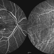 Multifocal CSCR 2
Sep 2 2012 by Hamid Ahmadieh, MD
Early-phase FA and ICG angiograms of a 36-year-old man with an active multifocal CSCR.
Photographer: Hamid Ahmadieh, Ophthalmic Research Center, Labbafinejad Medical Center
Imaging device: Heidelberg Spectralis
Condition/keywords: central serous chorioretinopathy (CSCR), indocyanine green (ICG) angiography
-
 Presumed Ocular Histoplasmosis Syndrome
May 3 2018 by Nichole Lewis
39-year-old female with presumed ocular histoplasmosis syndrome. Patient presented with vision of 20/200 in 10/2014. Vision CF @ 1 ft on 7/2015. Right eye vision is 20/20.
Photographer: Nichole Lewis
Condition/keywords: ocular histoplasmosis syndrome (OHS), presumed ocular histoplasmosis syndrome (POHS)
-
 Chronic Central Serous Chorioretinopathy re af
Dec 29 2012 by Barbara Parolini, MD
Panoramic autofluorescence fundus photograph of a 56 year old man with chronic central serous chorioretinopathy. BCVA is 20\200.
Photographer: Barbara Parolini, MD
Condition/keywords: bilateral chronic central serous retinopathy
-
 Sickle Cell Retinopathy with Sea Fans (angiogram)
Aug 24 2012 by Geoffrey G. Emerson, MD, PhD, FASRS
Fluorescein angiography (mid phase) of a 40-year-old man with African heritage and sickle SC disease. Sea fans are present temporal to the macula.
Photographer: Geoffrey Emerson, MD, PhD, Retina Center, Minneapolis
Condition/keywords: sea fan, sickle cell retinopathy
-
 Reticular Drusen, Doyne's Honeycomb Retinal Dystrophy, Malattia Leventinese, Familial Dominant Drusen
Feb 22 2018 by Nichole Lewis
Reticular Drusen, Doyne's Honeycomb Retinal Dystrophy, Malattia Leventinese, Familial Dominant Drusen
Photographer: Nichole Lewis
Condition/keywords: Doyne's Honeycomb, Familial Dominant Drusen, Malattia Leventinese, reticular drusen
-
 Choroidal Neovascularization, Idiopathic
Aug 23 2012 by Gerardo Garcia-Aguirre, MD
Fluoresein Angiogram of a 40 year-old patient showing a hyperfluorescent lesion with irregular margins corresponding to a choroidal neovascularization, surrounded by hypofluorescence corresponding to subretinal hemorrhage.
Photographer: Noemí Hernández, Asociación para Evitar la Ceguera en México
Imaging device: Zeiss FF4
Condition/keywords: choroidal neovascularization (CNV)
-
 Scleral Buckle and Cryo Color
Dec 29 2012 by Barbara Parolini, MD
Panoramic fundus photograph of a 55-year-old man after episcleral sugary for retinal detachment. An encircling scleral buckle and a superotemporal cryotherapy scar are visible.
Photographer: Barbara Parolini, MD
Imaging device: Daytona
Condition/keywords: scleral buckle
 Loading…