Search results (18 results)
-
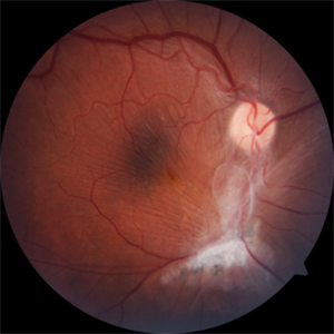
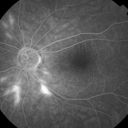
 Choroidal Metastasis With Orange Pigment in a Patient With Endometrial Carcinoma
Aug 8 2024 by Guilherme Sturzeneker, MD, MSc
Ultra-widefield fundus photograph and autofluorescence of a 62-year-old woman with endometrial cancer, denoting choroidal metastasis with unusual orange pigment. This presentation is a reminder that the development of orange pigment is not pathognomonic for choroidal melanoma, as it may be seen in other lesions such as carcinoma metastasis.
Photographer: Andrea Almeida
Imaging device: Optos Silverstone
Condition/keywords: choroidal metastasis, metastatic cancer, orange pigment
-
 B-FAF in Stargardt's Disease
Jul 4 2024 by Tejaswita Verma
Blue fundus autofluorescence showing hypoautofluorescence picture of a 28 year old male with 6/60 vision in BE in a case of Stargardt's disease.
Photographer: DR. TEJASWITA VERMA
Imaging device: MIRANTE
Condition/keywords: fundus autofluorescence (FAF), hereditary macular dystrophy, Stargardt disease
-
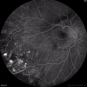 Coats Disease Fluorescein Angiography
Sep 2 2022 by FLOR ANGELICA JACOME GUTIERREZ
Fluorescein angiography of a patient with Coats disease where we found telangiectatic vessels, aneurysms, peripheral capillary nonperfusion and perivascular leak.
Photographer: Dr.Guillermo Salcedo Villanueva
Imaging device: Zeiss CLARUS 700 (FA)
Condition/keywords: Coats' disease, epiretinal membrane (ERM)
-
 Thioridazine-toxicity
Apr 30 2022 by Niloofar Piri, MD
61 yo male with PMH of longstanding schizophrenia since 20s with secondary intellectual disability presented with decreased vision following a recent stroke. He was found to have bilateral chorio-retinal atrophy involving posterior pole with scalloped edges and coin shaped atrophic area at margins extending into mid-periphery, diagnosis most concerning for intermediate stage thioridazine toxicity given the history. Mother could find handwritten prescriptions from 1990s when he was on Thioridazine 800 mg daily for unknown period of time. Patient had better vision in the left eye which was affected by recent stroke and prompted him to seek medical care. Fundus photograph of the right eye is demonstrated here.
Photographer: Jacob Grodsky, MD
Condition/keywords: drug toxicity, thioridazine toxicity, toxic retinopathy
-
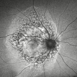
 Epiretinal Membrane
Sep 6 2021 by Ricardo Leitão Guerra
65-year-old woman with an asymptomatic ERM (BCVA=20/20).
Imaging device: Zeiss Clarus 700
Condition/keywords: epiretinal membrane (ERM)
-
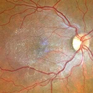
 Macular Traction Related to Toxoplasma Chorioretinitis
Jan 7 2021 by Lucas Zago Ribeiro, MD
Fundus image of a 50-year-old woman with macular traction and epiretinal membrane after toxoplasma chorioretinitis.
Photographer: Lucas Zago Ribeiro, UNIFESP / EPM, Brazil
Condition/keywords: epiretinal membrane (ERM), toxoplasmosis
-
 Retinal Autograft Postoperative Day 1
Feb 29 2020 by Raja Rami P Reddy, MD FRCS FASRS
20-year-old boy presented with recurrent retinal detachment post silicone oil removal with a macular hole. During surgery a retinal graft was created from the detached retina and positioned on the macular hole. silicone oil was injected at the end of surgery. One can see the diathermy marks on all sides of the edges of the graft.
Condition/keywords: autograft, macular hole
-
 Large, Dome-Shaped Peripheral Choroidal Melanoma - Widefield Color
Feb 13 2020 by Michael Seider, MD
Large, dome-shaped peripheral choroidal melanoma of the left eye with inferior exudative retinal detachment. Note the lack of obvious orange pigment over the tumor and apparent drusen anteriorly. A lack of ophthalmoscopically obvious lipofuscin is not uncommon among larger choroidal melanomas. B-Scan ultrasonography (transverse, 10 o’clock) confirms a low-moderate internally reflective dome-shaped choroidal lesion with a small adjacent retinal detachment. Ultrasound biomicroscopy (radial, 10 o’clock) confirms no ciliary body involvement of the tumor.
-
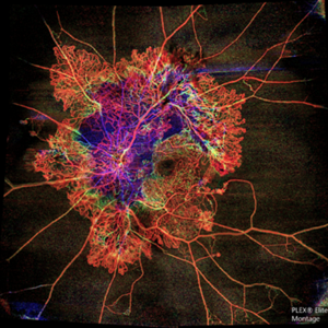 Flame of the Forest
Apr 9 2020 by Daraius N Shroff, MS FMRF FRCS
A 54-year-old man with DM for 15 years. The left eye had a visual acuity of 20/40. Wide field swept source OCTA revealed branching out central neovascular trunk vessels from the disc with terminal loops, along with exuberant proliferation of irregular small-calibre fine new vessels. The patient underwent OCTA guided pan retinal photocoagulation.
Photographer: Anuj Choudhary, Shroff Eye Centre, New Delhi
Imaging device: Zeiss Plex Elite 9000
Condition/keywords: proliferative diabetic retinopathy (PDR)
-
 Weiss Ring
Jan 9 2019 by John S. King, MD
77-year-old white male with ERM and PVD OD; sheet of vitreous with weiss ring in the nasal mid-vitreous cavity.
Photographer: Macey Highfill, RN
Imaging device: Topcon 50
Condition/keywords: posterior vitreous detachment, Weiss ring
-
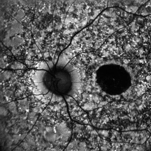
 Epiretinal Membrane
Jun 1 2018 by vitor borges porfirio pereira pereira
Epiretinal membrane by multicolor
Photographer: Marcos Avila, CBV Hospital de Olhos, Brasilia
Condition/keywords: epiretinal membrane (ERM)
-
 Submacular Hemorrhage
Apr 24 2018 by Pauline T Merrill, MD, FASRS
Fundus photo of left eye of a 65-year-old AMD patient who presented with sudden drop of vision from 20/30 to CF due to a large submacular hemorrhage, 7 months following her last Eylea injection. She underwent immediate injection of C3F8 in the office, with little effect. 10 days later vitrectomy with subretinal tPA and air-fluid exchange was performed, with successful displacement of the hemorrhage.
Photographer: Ermelinda Diaz, Illinois Retina Associates, Chicago, Illinois
Imaging device: Topcon 50DX
Condition/keywords: neovascular age-related macular degeneration (AMD), submacular hemorrhage
-
 Susac's Syndrome
Feb 13 2018 by John S. King, MD
Background: 46-year-old WF with CML (stable on Sprycel) saw her PCP for headaches without known cause; Headaches worsened and became confused, disoriented, off balance, and impaired short term memory. Heme-oncology ordered MRI that showed abnormal signal in the cerebellum and other parts of the brain, and LP has elevated protein. LP did show positive tau test, but fortunately, was a false positive for CJD. IV and PO steroids started and symptoms improved. MRI showed much improvement one month since starting steroids. 3 weeks later had a scotoma in right eye and eye doctor did not find anything at that time to cause it. Tinnitus developed (and some intermittent vertigo before that) and ENT referred back to eye doctor, who then referred the patient to Dr. Zocchi. He found a CWS and BRAO OD, and bilateral arteritis. She had some additional work-up for vasculitis. Given the retinal arteritis, cochlear issues, and MRI findings, Dr.Zocchi suspected Susac's Syndrome. She was started on multiple regimens including prednisone, IVIG, azathiprine, and MTX, and has had the best reponse to IVIG (FA shows a recurrence/worsening while adjusting IMT). She is stable and doing well with 20/20 vision in both eyes.
Photographer: Kay Dalby
Imaging device: Topcon
Condition/keywords: retinal vasculitis, Susac's syndrome
-
 Toxoplasmosis Associated Epiretinal Membrane
Oct 27 2016 by Gabriel Costa Andrade, PhD
Fundus photograph of a 26-year-old woman with a chorioretinal scar due to toxoplasmosis and secondary epiretinal membrane.
Photographer: Gabriel Andrade, Federal University of São Paulo, São Paulo, Brazil
Condition/keywords: epiretinal membrane (ERM), toxoplasmosis
-
 Epiretinal Membrane/Macular Pucker With Combined Hamartoma of Retina and RPE
Jul 8 2015 by Emmanuel Chang, MD PhD FACS FASRS
10-year-old with history of progressive severe distortion in the left eye over the past year.
Photographer: Retina and Vitreous of Texas
Imaging device: Heidelberg Autofluorescence
Condition/keywords: combined hamartoma, epiretinal membrane (ERM), retinal pigment epithelium (RPE) hamartoma
-
 PDR
PDR
Mar 17 2015 by Jason Griffith
Photograph of a 43-year-old female with QPDR and an early/mild ERM.
Photographer: Jason Griffith, Tennessee Retina, Nashville, TN
Imaging device: Topcon TRC-50EX
Condition/keywords: proliferative diabetic retinopathy (PDR)
-
 Fungal Endophthalmitis Associated With Intravenous Drug Abuse
Apr 16 2014 by Scott D. Schoenberger, MD
Fundus photograph of a 20-year-old male with pain and decreased vision OS for 3 days. His visual acuity was counting fingers and he had conjunctival injection, anterior chamber cells and vitreous cells. He admitted to intermittent use of intravenous heroin. A vitrectomy was performed and cultures were positive for candida albicans.
Condition/keywords: endogenous endophthalmitis, fungal endophthalmitis
-
 OCT Myopic Staphyloma With Schisis and ERM
Apr 24 2014 by Scott E. Pautler, MD
OCT of high myope with asymptomatic macular schisis.
Imaging device: Heidelberg Spectralis
Condition/keywords: foveal schisis, maculopathy, maculoschisis, optical coherence tomography (OCT), pathologic myopia, staphyloma
 Loading…