Search results (645 results)
-
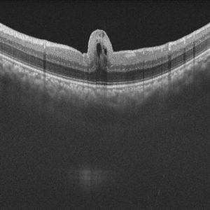
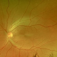
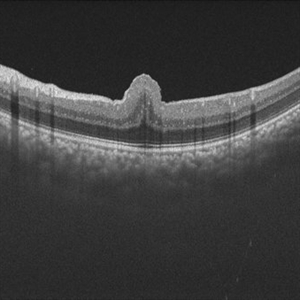
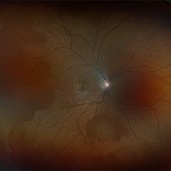
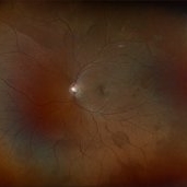
 Epiretinal Membrane
Jun 25 2025 by Kimberly Wakester
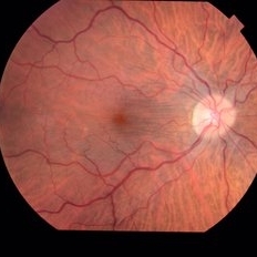
Fundus photograph of a 32-year-old woman with a stable epiretinal membrane in the right eye. Patients vision remains stable. No intervention is required at this time.
Photographer: Kimberly Wakester, COA, OCT-C
Imaging device: Topcon TRC 50DX
Condition/keywords: ERM
-
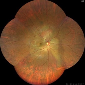
 Fractal Pattern of Chronic Serpiginous Choroiditis
Jun 17 2025 by Guilherme Sturzeneker, MD, MSc
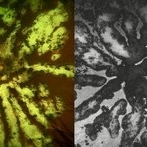
Ultra-widefield fundus photograph and autofluorescence of a 33-year-old woman with longstanding serpiginous choroiditis in the right eye. The image reveals centrifugal chorioretinal atrophy forming a dramatic fractal-like pattern, sparing the fovea. The patient is several years post-onset, with repeated negative workups, including for tuberculosis. Despite extensive lesions, the patient retains 20/20 vision in both eyes. Management included azathioprine monotherapy, as systemic steroids were contraindicated due to bipolar disorder.
Photographer: Andrea Almeida, IPEPO - Instituto da Visão
Imaging device: Optos Silverstone
Condition/keywords: autoimmune uveitis, azathioprine, chorioretinal atrophy, serpiginous choroiditis, ultra-wide field imaging
-
 Vitreous Cavity Inhabitant
Jun 2 2025 by Poornachandra B, MS, FVRS
A 36-year-old male presented with a 6-week history of intermittent ocular redness, now accompanied by the recent onset of floaters for the past 2 days. Fundus examination revealed the presence of a nematode in the vitreous cavity.
Photographer: Mr Dhikshith
Condition/keywords: parasite
-
 VKH Pseudotumor – Fluorescein Angiography
May 11 2025 by Felipe Murati
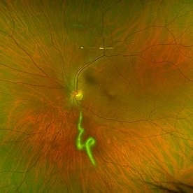
Fluorescein angiography image from a 36-year-old woman with chronic Vogt-Koyanagi-Harada (VKH) syndrome showing a pseudotumor-like lesion with late-phase staining and no active leakage. The image highlights subretinal fibrosis in the right eye, stable under long-term immunosuppressive therapy with mycophenolate mofetil and adalimumab. No signs of active choroiditis are present, confirming a quiescent phase.
Photographer: Felipe A. Murati, MD, University of Arizona
Imaging device: Optos California, fluorescein angiography modality
Condition/keywords: choroiditis, Fluorescein angiography, granulomatous uveitis, Optos FA, pseudotumor, subretinal fibrosis, VKH, Vogt-Koyanagi-Harada
-
 A Vessel That Would Not Let Go
May 5 2025 by Malvika Singh
Fundus photograph of a retinal detachment showing a horse shoe shaped tear and a bridging vessel.
Photographer: Dr Tejaswita Verma, Retina Foundation, Ahmedabad, India
Imaging device: Mirante SLO/OCT
Condition/keywords: bridging vessel, horseshoe tear
-
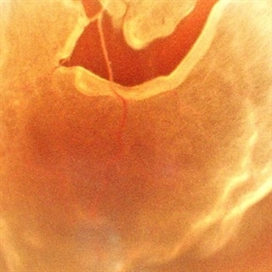
 Cilioretinal Artery Sparing CRAO
May 1 2025 by Tejaswita Verma
Fundus photo of a middle aged male with CRAO partially sparing cilioretinal artery and papillomacular bundle. Vision 6/60.
Photographer: Dr. Tejaswita Verma
Imaging device: MIRANTE
Condition/keywords: CRAO with cilioretinal sparing
-
 CRAO Sparing Cilioretinal Artery
May 1 2025 by Tejaswita Verma
Fundus photo of a middle aged male with 6/60 vision in left eye showing CRAO partially sparing cilioretinal artery.
Photographer: Dr. Tejaswita Verma
Imaging device: MIRANTE
Condition/keywords: cilioretinal sparing, CRAO
-
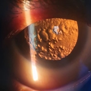 Posterior Subcapsular Cataract
Apr 18 2025 by DR Rohit Gupta
Slit lamp image on Retroillumination showing posterior subcapsular cataract of a female patient who was on long term corticosteroids inhaler medications.
Photographer: Dr Rohit gupta
Imaging device: Samsung S21
Condition/keywords: posterior capsule opacification, posterior subcapsular cataract, posterior subcapsular changes, posterior subcapsular polar senile cataract, steroids
-
 Toxic Maculopathy (Elmiron)
Apr 9 2025 by Virginia Gebhart
79 year old male with toxic maculopathy from long term use of Elmiron (15+ yrs.) On exam there is stippled RPE changes, pigment clumping, and subretinal deposits. BCVA 20/100 | 20/40.
Photographer: Virginia Gebhart, Retina Consultants of Carolina
Imaging device: Optos California
Condition/keywords: autofluorescence imaging, cystoid macular degeneration, Elmiron Toxicity, Toxic Maculopathy
-
 CRVO with RD
Apr 4 2025 by Tejaswita Verma
Fundus photo of a 58 year-old hypertensive male who presented with RE 6/60 vision with CRVO, rhegmatogenous RD.H/O DOV in RE since 3 days, H/O receiving anti VEGF X 2 injections 2 months ago
Photographer: DR. TEJASWITA VERMA
Imaging device: MIRANTE
Condition/keywords: central retinal vein occlusion (CRVO), Retinal Detachment
-
 Neovascularisation Disc
Apr 4 2025 by Tejaswita Verma
Fundus photo of a 38 year-old Type 1 diabetic male with 6/18P vision in RE, presenting with TRD, NVD.
Photographer: DR. TEJASWITA VERMA
Imaging device: MIRANTE
Condition/keywords: neovascularization of the disc (NVD), TRD
-
 Hereditary Macular Dystrophy with CNVM
Apr 4 2025 by Tejaswita Verma
Fundus photo of a 46 y/o male with 6/36 vision, Stargardt's disease with CNVM
Photographer: DR. TEJASWITA VERMA
Imaging device: MIRANTE
Condition/keywords: CNVM, hereditary macular dystrophy, HMD
-
 Large Horseshoe Tear
Apr 4 2025 by Tejaswita Verma
Fundus photo of a 29 year old myopic male with RE 6/12P vision with large ragged tear on the posterior edge of lattice degeneration
Photographer: DR. TEJASWITA VERMA
Imaging device: MIRANTE
Condition/keywords: horseshoe tear
-
 Macular Schisis with RD
Apr 4 2025 by Tejaswita Verma
Fundus photo of a 29 year-old male with RRD, breaks in periphery, macular schisis . Vision CF2.5 mts
Photographer: DR. TEJASWITA VERMA
Imaging device: MIRANTE
Condition/keywords: macular schisis, retinal detachment
-
 Intravitreal Ozurdex Implant
Apr 3 2025 by Tejaswita Verma
Fundus photo of a middle-aged diabetic male showing Ozurdex implant in situ with laser marks.
Photographer: Tejaswita Verma
Imaging device: MIRANTE
Condition/keywords: dexamethasone implant, ozurdex
-
 Solar Retinopathy
Apr 1 2025 by Isaac Agranoff
OCT scan of 18-year-old male presenting with 20/40 BCVA OU and bilateral focal outer retinal subfoveal defects. Patient reported long-term history of frequent sungazing, has stopped within past 6-9 months.
Photographer: Isaac Agranoff
Imaging device: Heidelberg Spectralis
Condition/keywords: solar retinopathy
-
 Choroidal Melanoma Masquerading as PEHCR
Mar 3 2025 by Tejaswita Verma
A 65 year old diabetic male presented with large nasal retinal mass giving the appearance of organized dehaemoglobinized subretinal hemorrhage with breakthrough vitreous hemorrhage , with 6/6P vision. Enucleation specimen showed histopathology confirmed choroidal melanoma.
Photographer: DR. TEJASWITA VERMA
Imaging device: MIRANTE
Condition/keywords: vitreous hemorrhage
-
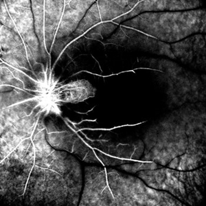
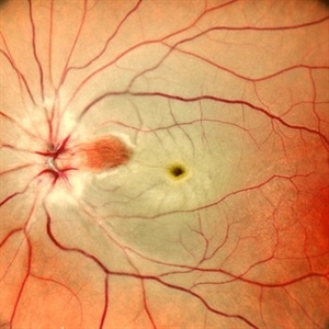
 Retinal Fold in Posterior Microphthalmos
Mar 1 2025 by Hemanth Murthy, MBBS, MD, FASRS
Swept source OCT image of left eye of 34 year male patient with high hypermetropia(+14). BCVA 20/20 in right eye and 20/60 in left eye. Anterior segment was normal. There is loss of foveal pit with omega shaped elevation of inner retinal layers.
Photographer: Mr Veda Vyas
Condition/keywords: posterior microphthalmos
-
 Retinal Fold in Posterior Microphthalmos
Mar 1 2025 by Hemanth Murthy, MBBS, MD, FASRS
Fundus photo of left eye of 34 year male patient with high hypermetropia(+14). BCVA 20/20 in right eye and 20/60 in left eye. Anterior segment was normal. There is loss of foveal pit with omega shaped elevation of inner retinal layers.
Photographer: Mr Veda Vyas
Condition/keywords: posterior microphthalmos
-
 Retinal Fold in Posterior Microphthalmos
Mar 1 2025 by Hemanth Murthy, MBBS, MD, FASRS
Fundus photo of Right eye of 34 year male patient with high hypermetropia(+14). BCVA 20/20 in right eye and 20/60 in left eye. Anterior segment was normal. There is loss of foveal pit with omega shaped elevation of inner retinal layers.
Photographer: Mr Veda Vyas
Condition/keywords: posterior microphthalmos
-
 Retinal Fold in Posterior Microphthalmos
Mar 1 2025 by Hemanth Murthy, MBBS, MD, FASRS
Swept source OCT image of Right eye of 34 year male patient with high hypermetropia(+14). BCVA 20/20 in right eye and 20/60 in left eye. Anterior segment was normal. There is loss of foveal pit with omega shaped elevation of inner retinal layers.
Photographer: Mr Veda Vyas
Condition/keywords: posterior microphthalmos
-
 Sickle Cell Retinopathy
Feb 24 2025 by Kimberly Wakester
Optomap RGB image of an 24-year-old woman with sickle cell retinopathy in both eyes. There is overall progression of the ischemic vessels and vascular drops out compared to previous images completed in 2021. Oral FA was completed and shows possible progression of peripheral non-perfusion but difficult to determine due to drinking FA dye and images not being as bright. On Clinical exam there is no evidence of NV, RD, or RT in either eye. Patient understands the need for continued follow up care and the likely need for PRP laser in both eyes.
Photographer: Kimberly Wakester, COA
Imaging device: Optos California
Condition/keywords: sickle cell retinopathy
-
 Vortex-pattern Exudative Retinal Detachment
Feb 22 2025 by CUI YUELING
Patient: Male, 40 years old. Chief Complaint: Blurred vision and metamorphopsia in the left eye for more than 10 days. Past Medical History Hypertension for 4 years, with a highest recorded blood pressure of 160/80 mmHg. Currently controlled with oral "Nifedipine Sustained-Release Tablets, 2 tablets daily." Long-term history of heavy alcohol consumption and smoking. Ophthalmic Examination: Visual Acuity: Right eye (OD): 0.4 (uncorrected, no improvement with correction). Left eye (OS): 0.5 (-1.5DS = 1.0). Intraocular Pressure (IOP): OD: 15 mmHg. OS: 17 mmHg. Anterior Segment:Unremarkable. Fundus Examination: Right eye: Optic disc margins are clear, with a slightly reddish hue. Cup-to-disc ratio (C/D) = 0.2. A scalloped, orange-red elevated lesion is observed superior to the optic disc, with anterior displacement of the focal point. This is accompanied by a secondary, turbine-like exudative retinal detachment centered around the optic disc, involving the macula. The macular region shows scattered punctate yellow-white exudates. Diagnosis: Choroidal hemangioma with secondary exudative retinal detachment(OD).
Photographer: Cui yueling The First People's Hospital of Zunyi, Guizhou, Zunyi, China
Imaging device: Zeiss Clarus 500
Condition/keywords: choroidal hemangioma, exudative retinal detachment
-
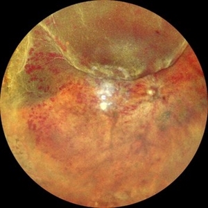
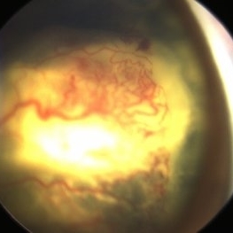 Advance Coats' Disease
Feb 15 2025 by Theinchai Pasurakul, MD
From the fundus image, the peripheral retina exhibits telangiectatic vessels accompanied by light bulb aneurysms at their terminal ends.
Photographer: Michael J. Shapiro MD, Advocate Lutheran General Hospital, Des Plaines
Imaging device: Retcam
Condition/keywords: Coats' disease, light-bulb aneurysms
 Loading…