Search results (645 results)
-
 BBG Dye injection to Stain ILM during Vitrectomy Surgery | Intra-Operative Still
Apr 28 2023 by Veer Singh, MS, FVRS, FMRF, FICO (Retina)
BBG Dye injection to Stain ILM during Vitrectomy Surgery | Intra-Operative Still
Photographer: Dr. Veer Singh
Condition/keywords: brilliant blue staining, ERM, ILM staining
-
 Choroidal rupture
Jun 7 2023 by Prerana Shah
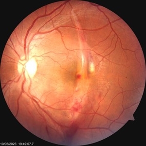
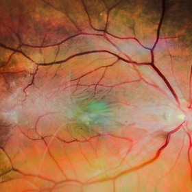
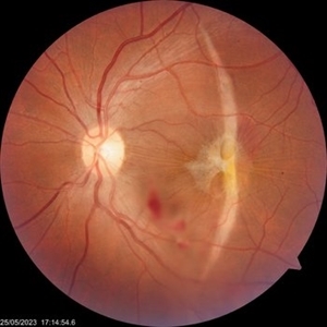
Fundus photograph of a 35 year old gentleman on the day of blunt trauma.
Photographer: Swati Kadam and Disha Malusare, Shreeramkrishna Netralaya, Thane , Maharashtra ,India
Imaging device: Zeiss Visucam
Condition/keywords: choroidal rupture, ERM
-
 EPIRETINAL MEMBRANE
Jun 6 2023 by Akansha Sharma
COLOUR FUNDUS PHOTOGRAPH OF A 40 YAER OLD MALE WITH EPIRETINAL MEMBRANE FORMATION
Photographer: Dr. Akansha Sharma, Dr. Denish Patel, Dr. Urmil Shah,
Condition/keywords: epiretinal membrane (ERM), ERM
-
 Epiretinal Membrane
Jan 30 2024 by Akansha Sharma
Color fundus photograph of a 65 year old hypertensive male with an epiretinal membrane in a case of old branch retinal vein occlusion with a subhyaloid hemorrhage seen inferiorly.
Photographer: Dr. Akansha Sharma, Bharati Eye Hospital
Condition/keywords: epiretinal membrane (ERM), ERM
-
 Epiretinal membrane
Nov 28 2021 by Jorge Berganza
22 yo female with metamorphopsia in the left eye.
Photographer: Jorge Berganza MD
Imaging device: Iphone XR
Condition/keywords: ERM, macular traction
-
 Epiretinal Membrane
Aug 22 2023 by Vaidehi Sathaye
Fundus photograph of RE of a 57 year old female with Epiretinal membrane
Photographer: Dr. Vaidehi Sathaye
Imaging device: Mirante
Condition/keywords: ERM
-
 Epiretinal Membrane
Mar 14 2024 by Mario R. Ventresca, MD, FRCS(C), FASRS
Fundus photo and OCT of an 80-year-old man with Stage 2 ERM with mild SUKIMA.
Photographer: Mario R Ventresca MD, Ontario, Canada
Imaging device: eidon
Condition/keywords: ERM
-
 Epiretinal Membrane
Feb 2 2022 by Manish Nagpal, MD, FRCS (UK), FASRS
Intraoperative photo of a epiretinal membrane, glistening reflex noted. Prior to this capture, PVD induction has been done, which has left a small splinter hemorrhage around the disc attachment of hyaloid.
Photographer: Manish Nagpal, Retina Foundation, Ahmedabad, India
Imaging device: Sony PMW -10 MD surgical camera
Condition/keywords: epiretinal membrane formation, ERM, ILM flap, PVD induction
-
 Epiretinal Membrane
Jun 25 2025 by Kimberly Wakester
Fundus photograph of a 32-year-old woman with a stable epiretinal membrane in the right eye. Patients vision remains stable. No intervention is required at this time.
Photographer: Kimberly Wakester, COA, OCT-C
Imaging device: Topcon TRC 50DX
Condition/keywords: ERM
-
 Epiretinal Membrane Peeling during Vitrectomy Surgery | Intra-Operative Still
Apr 28 2023 by Veer Singh, MS, FVRS, FMRF, FICO (Retina)
Epiretinal Membrane Peeling during Vitrectomy Surgery | Intra-Operative Still
Photographer: Dr. Veer Singh
Condition/keywords: ERM, peeling, vitreomacular surgery
-
Epiretinal membrane removal
Oct 24 2022 by Manish Nagpal, MD, FRCS (UK), FASRS
This video highlights the surgical technique of tangentially removing the epiretinal membrane using a forceps
Photographer: Manish Nagpal
Condition/keywords: epiretinal membrane, ERM, macular pucker, staining, video, vitrectomy
-
---thumb.jpg/image-square;max$300,300.ImageHandler) ERM
ERM
Oct 8 2012 by David R. Chow, MD, FRCS(C)
Condition/keywords: epiretinal membrane (ERM)
-
---thumb.JPG/image-square;max$300,300.ImageHandler) ERM
ERM
Oct 8 2012 by David R. Chow, MD, FRCS(C)
Condition/keywords: epiretinal membrane (ERM)
-
 ERM
ERM
Oct 8 2012 by David R. Chow, MD, FRCS(C)
Condition/keywords: epiretinal membrane (ERM)
-
 ERM
ERM
Nov 26 2020 by Priya Rasipuram Chandrasekaran, MBBS, DO, DNB, FRCS
A 58-year-old female presented with distortion of images 1 month following cataract surgery in the right eye and fundus examination showed epiretinal membrane extending from the disc to the macula and OCT macula showing epiretinal membrane with disorganization of the foveal architecture.
Condition/keywords: epiretinal membrane (ERM)
-
 ERM
ERM
Aug 30 2018 by Dhaivat Shah
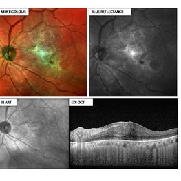
55-year-old female presented with left eye BCVA 6/24 N18, early cataract changes on slit lamp, fundus showing ERM with retinal thickening. Multi-color image (MCI) depicting an ERM (green hue) with retinal thickening. Note how beautifully the extent of ERM is captured, which can help the surgeon to decide the area of surgical peeling. The BR particularly provides details of the inner retina and the vitreoretinal interface, hence showing the ERM. This platform utilizes confocal technology and thus has unique advantages over CFP. MCI provides good image quality in hazy media and in small pupil. It does not use bright white light and thus is not discomforting to the patient. Images obtained with MCI have better contrast and sharper borders as compared to CFP. Definitely the new tech in for the next generation!
Photographer: Miss Moupiya Das
Imaging device: SPECTRALIS
Condition/keywords: blue reflectance, epiretinal membrane (ERM)
-
 ERM
ERM
Jan 9 2025 by Richa Chaudhary, Mbbs,ms
52 year old male presented with idipathic ERM, with pucker showing, retinal folds. Planned for surgical removal of the same.
Condition/keywords: ERM
-
 ERM under Silicon oil
Nov 9 2022 by Vaidehi Sathaye
Fundus photograph of LE of a 45 year male patient with ERM under Silicon Oil. Patient had a history of undergoing Vitrectomy with Silicon Oil Infusion for a redetachment after failure of Vitrectomy with C3F8 tamponade for a macula-off Rhegmatogenous Retinal Detachment.
Photographer: Dr. Vaidehi Sathaye
Imaging device: Mirante
Condition/keywords: ERM, silicone oil
-
 Familial Exudative Vitreoretinopathy
Nov 25 2022 by Aditya S Kelkar, MS, FRCS, FASRS,FRCOphth
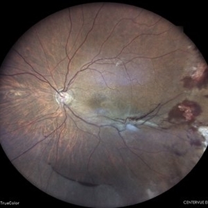
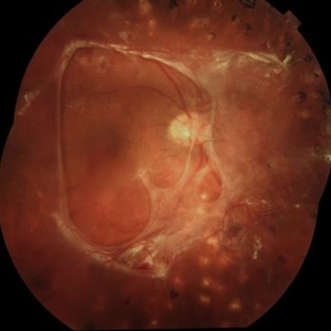
Colour fundus photograph of the right eye of a 56-year-old lady showing lasered FEVR with epiretinal membrane and vitreous band.
Photographer: Dr. Pranali Surawase. National Institute of Ophthalmology, Pune, Maharashtra, India
Imaging device: Zeiss Clarus 500
Condition/keywords: ERM, familial exudative vitreoretinopathy (FEVR), laser photocoagulation
-
Macular Hematoma Secondary Valsalva Maneuver
Oct 14 2021 by Islam bechakh
A 32-year-old man, who has presented for 02 months, a macular hematoma secondary to a Valsalva maneuver. He benefited from an attempt to open the hematoma with a Yag laser, but to no avail. We operated on and performed a 23G vitrectomy with posterior vitreous detachment, and discovered an epiretinal membrane which separated the hematoma from the posterior hyaloid. After removal of this membrane and aspiration of red blood cells and fibrin, the macula regained a normal appearance with good functional recovery.
Photographer: Islam Bechakh
Condition/keywords: epiretinal membrane (ERM), ERM, Macular hematoma, Valsalva maneuver
-
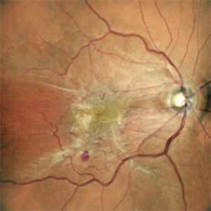
 Preretinal Fibrosis
Jan 12 2024 by Virginia Gebhart
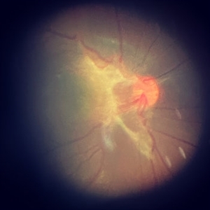
53 year old diabetic male with significant persistent ERM due to fibrotic NV superiorly. Possibly developing a tractional MH. Vitreous Hemorrhage secondary to traction on the fibrosis
Photographer: Virginia Gebhart
Imaging device: Topcon 50DX
Condition/keywords: epiretinal membrane, ERM, fibrosis, macular pseudohole, neovascularization (NV)
-
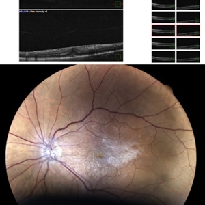
 Retinal Detachment with Multiple OCT Overlays
Jan 7 2025 by Drew Mitchell
Optos 360* Color photo montage with multiple Zeiss Cirrus OCT scan overlays. Retinal Detachment with multiple breaks and a Epiretinal Membrane.
Photographer: Drew Mitchel, OCT-C
Imaging device: Optos California
Condition/keywords: ERM, macular pucker, montage, Optos, OPTOS CALIFORNIA, RD, Retinal Detachment
-
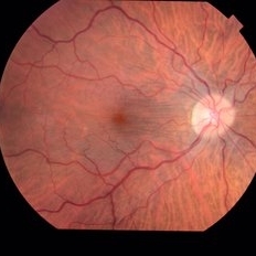
 The sun, the star and the crescent moon
Jun 7 2023 by Prerana Shah
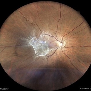
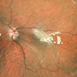
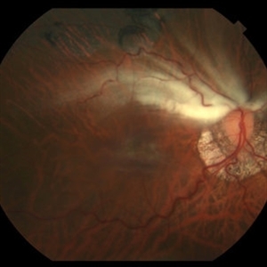
Fundus photograph of a 35 year old gentleman one week post blunt trauma.
Photographer: Swati Kadam and Disha Malusare, Shreeramkrishna Netralaya, Thane , Maharashtra ,India
Imaging device: Zeiss Visucam
Condition/keywords: choroidal rupture, ERM
-
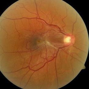
 ERM / Myelinated NFL
Jun 10 2016 by John S. King, MD
High myope who dev ERM post-RD repair.
Condition/keywords: epiretinal membrane (ERM), myelinated nerve fibers, myopic degeneration
-
---thumb.jpg/image-square;max$300,300.ImageHandler) ERM and Dry AMD OCT
ERM and Dry AMD OCT
Apr 18 2014 by Susanna S. Park, MD, PhD
OCT image of the macula of this 78-year-old man with progressive loss of vision in this left eye from vitreo-macular traction, epiretinal membrane and dry AMD. BCVA 20/100
Photographer: Ellen Redenbo, UC Davis Eye Center
Condition/keywords: dry age-related macular degeneration (dry AMD), epiretinal membrane (ERM), optical coherence tomography (OCT), vitreomacular traction (VMT)
 Loading…