Search results (645 results)
-
 Macular Pseudohole - OCT
Jan 11 2013 by Gerardo Garcia-Aguirre, MD
OCT scan showing a hyperreflective line that is partially separated from the retina in the fovea and temporal macula, corresponding to an epiretinal membrane. Note the discontinuity of the line just above the fovea, which clinically corresponds to the pseudohole.
Photographer: Gerardo Garcia-Aguirre, MD
Imaging device: Topcon 3DOCT 1000
Condition/keywords: epiretinal membrane (ERM), macular pseudohole
-
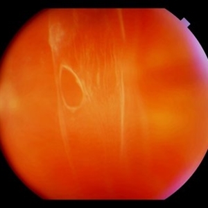 Weiss Ring
Jan 9 2019 by John S. King, MD
77-year-old white male with ERM and PVD OD; sheet of vitreous with weiss ring in the nasal mid-vitreous cavity.
Photographer: Macey Highfill, RN
Imaging device: Topcon 50
Condition/keywords: posterior vitreous detachment, Weiss ring
-
 Epiretinal Membrane
Oct 26 2012 by Sharon Fekrat, MD FACS FASRS
39-year-old female with long standing epiretinal membrane in the left eye and good vision
Photographer: Jim Crowell, Ophthalmic Photographer, Duke Eye Imaging, Durham, NC
Condition/keywords: epiretinal membrane (ERM), macular pucker
-
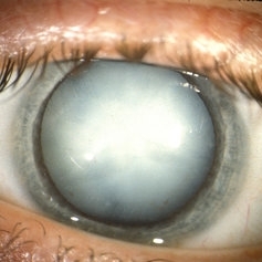 Hypermature Cataract
Oct 8 2012 by Jeffrey G. Gross, MD, FASRS
Hypermature cataract HM.
Condition/keywords: hypermature cataract
-
 ERM that Spontaneously Peeled
Oct 8 2012 by David R. Chow, MD, FRCS(C)
An ERM that through follow-up sponateously separated with the development of PVD.
Condition/keywords: epiretinal membrane (ERM), posterior vitreous detachment
-
 Epiretinal Membrane
Oct 11 2012 by Michael P. Kelly, FOPS
This is a patient with idiopathic panuveitis who developed a visually significant epiretinal membrane. Pars plana vitrectomy with membrane peeling was performed to remove the epiretinal proliferation. I recommend magnifying the image to see the exquisite detail centrally.
Photographer: Michael P. Kelly, FOPS Director, Duke Eye Center Labs, Duke Universtiy Hospital
Imaging device: Zeiss 450Plus
Condition/keywords: epiretinal membrane (ERM), panuveitis
-
---thumb.JPG/image-square;max$300,300.ImageHandler) Retinal Pigment Epithelial Detachment With No Subretinal Fluid
Retinal Pigment Epithelial Detachment With No Subretinal Fluid
Jun 29 2013 by Jason S. Calhoun
A 38-year-old male who comes in with blurred vision in the left eye. VA is 20/30. Noticed a defect inferior of his central vision. Did an fluorescein angiogram to determine an RPE with no sub retinal fluid. Also OCT confirms. Patient was injected with Avastin.
Photographer: Jason S. Calhoun, Mayo Clinic Jacksonville, Florida
Imaging device: TOPCON TRC 50-EX
Condition/keywords: central serous retinopathy (CSR), retinal pigment epithelium (RPE) detachment
-
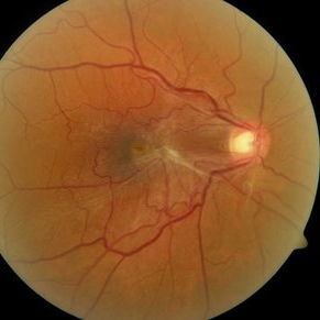
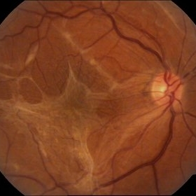 Epiretinal membrane - Fundus photograph
Feb 5 2014 by Gerardo Garcia-Aguirre, MD
Fundus photograph of a 62 year old female with metamorphopsia and decreased visual acuity. A stage 2 epiretinal membrane is observed, causing distortion of the retinal vasculature.
Photographer: Gerardo Garcia-Aguirre, MD
Condition/keywords: epiretinal membrane (ERM)
-
 OCT Myopic Staphyloma With Schisis and ERM
Apr 24 2014 by Scott E. Pautler, MD
OCT of high myope with asymptomatic macular schisis.
Imaging device: Heidelberg Spectralis
Condition/keywords: foveal schisis, maculopathy, maculoschisis, optical coherence tomography (OCT), pathologic myopia, staphyloma
-
---thumb.JPG/image-square;max$300,300.ImageHandler) TID (Trans Illumination Defect)
TID (Trans Illumination Defect)
Jul 8 2013 by Jason S. Calhoun
74-year-old patient who VA 20/70 OD, 20/50 OS. Complaints of blurred vision. Iris atrophy, both eyes. ERM, right eye, Patient to have cataract surgery to improve distance vision.
Photographer: Jason S. Calhoun, Department of Ophthalmology, Mayo Clinic Jacksonville, Florida
Condition/keywords: translucency of iris
-
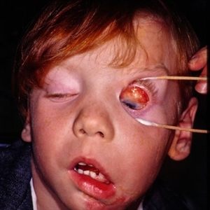
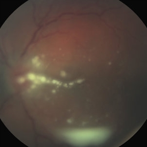 Fungal Endophthalmitis Associated With Intravenous Drug Abuse
Apr 16 2014 by Scott D. Schoenberger, MD
Fundus photograph of a 20-year-old male with pain and decreased vision OS for 3 days. His visual acuity was counting fingers and he had conjunctival injection, anterior chamber cells and vitreous cells. He admitted to intermittent use of intravenous heroin. A vitrectomy was performed and cultures were positive for candida albicans.
Condition/keywords: endogenous endophthalmitis, fungal endophthalmitis
-
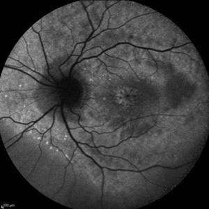
 Epiretinal Membrane/Macular Pucker With Combined Hamartoma of Retina and RPE
Jul 8 2015 by Emmanuel Chang, MD PhD FACS FASRS
10-year-old with history of progressive severe distortion in the left eye over the past year.
Photographer: Retina and Vitreous of Texas
Imaging device: Heidelberg Autofluorescence
Condition/keywords: combined hamartoma, epiretinal membrane (ERM), retinal pigment epithelium (RPE) hamartoma
-
 Superior Peripapillary Hemorrhage
Jul 13 2013 by Jason S. Calhoun
Patient was seen for acute vision loss in the right eye. Patient has glaucoma. VA was 20/70 in the right eye. Had vitrectomy back in May 2012 for ERM stripping. Also had trabectome with cataract surgery in December of 2012. Fundus photos presents a superior peripapillary Hemorrhage of the optic nerve. Patient will be followed up in one month.
Photographer: Jason S. Calhoun, Department of Ophthalmology, Mayo Clinic Jacksonville, Florida
Imaging device: TOPCON TRC 50-EX
Condition/keywords: peripapillary hemorrhage
-
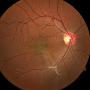
 Epiretinal Membrane
Oct 15 2012 by Sharon Fekrat, MD FACS FASRS
Fundus photograph of an epiretinal membrane
Photographer: Michael P. Kelly, FOPS, Director, Duke Eye Labs, Duke University Eye Center, Durham, NC
Condition/keywords: epiretinal membrane (ERM)
-
 Epiretinal Membrane
Sep 14 2012 by Michael P. Kelly, FOPS
Epiretinal membrane imaged using a high magnification retinal fundus camera and red free illumination.
Photographer: Michael P. Kelly, FOPS, Director, Duke Eye Center Labs, Duke Universtiy Hospital
Condition/keywords: epiretinal membrane (ERM), high magnification, monochromatism, red-free
-
---thumb.JPG/image-square;max$300,300.ImageHandler) ERM
ERM
Oct 8 2012 by David R. Chow, MD, FRCS(C)
Condition/keywords: epiretinal membrane (ERM)
-
 ERM
ERM
Oct 8 2012 by David R. Chow, MD, FRCS(C)
Condition/keywords: epiretinal membrane (ERM)
-
 Cystoid Macular Edema (CME)
Sep 11 2012 by Hamid Ahmadieh, MD
Autofluorescence imaging of the left eye of a 17-year-old boy with chronic intermediate uveitis showing CME.
Photographer: Hamid Ahmadieh, MD, Ophthalmic Research Center, Labbafinejad Medical Center, Shahid Beheshti University of Medical Sciences
Imaging device: Heidelberg Spectralis
Condition/keywords: autofluorescence imaging, cystoid macular edema (CME), intermediate uveitis
-
 Pucker
Oct 8 2012 by David R. Chow, MD, FRCS(C)
Condition/keywords: epiretinal membrane (ERM)
-
 EDI OCT Detachment With No Subretinal Fluid
Jun 29 2013 by Jason S. Calhoun
A 38-year-old male came in with blurred vision in the left eye. VA is 20/30. Notice a defect inferior of his central vision. Did an fluorescien angiogram to determine an RPE with no subretinal fluid. Also OCT confirms. Patient was injected with Avastin.
Photographer: Jason S. Calhoun, Mayo Clinic Jacksonville, Florida
Imaging device: TOPCON TRC 50-EX/CIRRUS HD OCT
Condition/keywords: central serous retinopathy (CSR), retinal pigment epithelium (RPE) detachment
-
---thumb.jpg/image-square;max$300,300.ImageHandler) ERM
ERM
Oct 8 2012 by David R. Chow, MD, FRCS(C)
Condition/keywords: epiretinal membrane (ERM)
-
 Retinal Schisis Detachment
Nov 9 2012 by Norman Byer
This 57-year-old man has a combined retinal schisis detachment caused by an outer layer hole in the upper right. On the right half of this photograph, the outer layer is detached and represented by the prominent yellow line which is lying against the inner layer. On the left half the inner layer appears very thin and the schisis cavity lies just behind it as it was originally. This, therefore, represents a localized detachment of the outer layer and thus a true secondary retinal detachment. The reason these cases remain localized and nonprogressive is that the only fluid available to the subretinal space is that which is contained within the schisis cavity. Furthermore, this fluid tends to be quite viscous and is not readily passed through the retinal breaks. A clinical symptomatic progressive retinal detachment cannot occur unless the retinal schisis cavity is very large or a break occurs in the inner layer also.
Condition/keywords: intact inner layer, localized detachment of outer layer, outer layer hole, retinal schisis detachment, retinoschisis, secondary retinal detachment
-
---thumb.jpg/image-square;max$300,300.ImageHandler) Retinoblastoma To Chemothermotherapy
Retinoblastoma To Chemothermotherapy
Oct 4 2013 by Maurice F. Rabb
A 7 week old girl with a family history of retinoblastoma was found to have a small retinoblastoma in each eye. In the right eye the tumor was adjacent to the optic disc in the papillomacular bundle and measured 2 X 2 X 2 mm. Its temporal margin was 1.0 mm from the foveola and it overhung 20% of the optic disc surface. There was not clinical or ultrasonographic evidence of vitreous seeking or optic nerve invation. In the left eye there was a solitary tumor 1mm superonasal to the optic disc. The tumor measured 1 X 1 X 1 mm. The foveal reflex was normal in both eyes. Both tumors showed a fluorescein angiographic pattern compatible with retinoblastoma with rapid filling and late hyperfluorescence.
Condition/keywords: retina
-
 Purtscher retinopathy 2 right eye
Jan 11 2013 by Alex P. Hunyor, MD
Purtscher - like retinopathy in a patient with dermatomyositis, right eye.
Condition/keywords: Purtscher's retinopathy
-
 Linear Nevus Sebaceous Syndrome
Feb 20 2015 by H. Michael Lambert, MD
Color photo of conjuctival lipodermoid in linear sebaceous nevus syndrome .
Condition/keywords: conjunctiva, linear nevus sebaceous syndrome, lipodermoid
 Loading…