Search results (645 results)
-
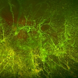
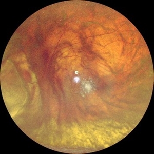
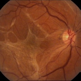
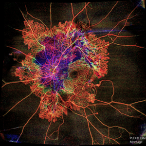 Flame of the Forest
Apr 9 2020 by Daraius N Shroff, MS FMRF FRCS
A 54-year-old man with DM for 15 years. The left eye had a visual acuity of 20/40. Wide field swept source OCTA revealed branching out central neovascular trunk vessels from the disc with terminal loops, along with exuberant proliferation of irregular small-calibre fine new vessels. The patient underwent OCTA guided pan retinal photocoagulation.
Photographer: Anuj Choudhary, Shroff Eye Centre, New Delhi
Imaging device: Zeiss Plex Elite 9000
Condition/keywords: proliferative diabetic retinopathy (PDR)
-
 Epiretinal Membrane/Macular Pucker With Combined Hamartoma of Retina and RPE
Jul 8 2015 by Emmanuel Chang, MD PhD FACS FASRS
10-year-old with history of progressive severe distortion in the left eye over the past year.
Photographer: Retina and Vitreous of Texas
Imaging device: Heidelberg Autofluorescence
Condition/keywords: combined hamartoma, epiretinal membrane (ERM), retinal pigment epithelium (RPE) hamartoma
-
 Pucker
Oct 8 2012 by David R. Chow, MD, FRCS(C)
Condition/keywords: epiretinal membrane (ERM)
-
 A Vessel That Would Not Let Go
May 5 2025 by Malvika Singh
Fundus photograph of a retinal detachment showing a horse shoe shaped tear and a bridging vessel.
Photographer: Dr Tejaswita Verma, Retina Foundation, Ahmedabad, India
Imaging device: Mirante SLO/OCT
Condition/keywords: bridging vessel, horseshoe tear
-
 Epiretinal Membrane
Oct 15 2012 by Sharon Fekrat, MD FACS FASRS
Fundus photograph of an epiretinal membrane
Photographer: Michael P. Kelly, FOPS, Director, Duke Eye Labs, Duke University Eye Center, Durham, NC
Condition/keywords: epiretinal membrane (ERM)
-
 Epiretinal Membrane
Sep 14 2012 by Michael P. Kelly, FOPS
Epiretinal membrane imaged using a high magnification retinal fundus camera and red free illumination.
Photographer: Michael P. Kelly, FOPS, Director, Duke Eye Center Labs, Duke Universtiy Hospital
Condition/keywords: epiretinal membrane (ERM), high magnification, monochromatism, red-free
-
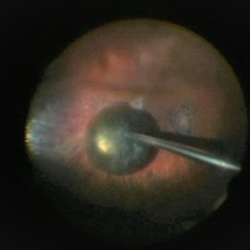
 ERM With Retinal Detachment
May 25 2017 by Manish Nagpal, MD, FRCS (UK), FASRS
Per operative photo prior to ERM removal in a case of retinal detachment with ERM.
Photographer: MANISH NAGPAL
Imaging device: SONY 3 CHIP HD CAMERA
Condition/keywords: epiretinal membrane (ERM), internal limiting membrane (ILM) peeling
-
 Extreme Asteroid Hyalosis
Apr 27 2016 by Matt Poe, COA
This patient was sent for a possible retinal detachment. Extreme difficult view of posterior pole due to asteroid hyalosis. After B-Scan was performed it was determined patient did not have a retinal detachment, only posterior vitreous detachment.
Photographer: Matt Poe, COA. Northwest Arkansas Retina Associates, Springdale, AR.
Condition/keywords: asteroid hyalosis, posterior vitreous detachment
-
 OCT Image of Epiretinal Membrane
Aug 29 2017 by Carolyn Daley
OCT photograph of a 64-year-old women with an epiretinal membrane in the right eye. Patient has not noticed any decline in vision so surgery was not recommended at this time.
Photographer: Carolyn Daley
Imaging device: Heidelberg Spectralis
Condition/keywords: epiretinal membrane (ERM), optical coherence tomography (OCT)
-
 PPV retained cataract
Apr 19 2023 by Denica Rodriguez
A 46-year-old male with hypermature dense cataract. Patient got a piece of metal in his eye when he was 5 years old and was not able to see since. Patient was having cataract surgery and phacodonesis was present. The lens dropped to the back of the eye. Patient had to have another surgery to do vitrectomy. The lens removal was done with a fragmatome handpiece.
Photographer: Denica Rodriguez COA, ST
Imaging device: Zeiss Microscope with resight
Condition/keywords: cataract, dropped nucleus, fragmatome, lens capsule, ocular trauma, pars plana vitrectomy (PPV), retained lens fragments, Retina, retina surgery, traumatic cataract
-
 Thioridazine-toxicity
Apr 30 2022 by Niloofar Piri, MD
61 yo male with PMH of longstanding schizophrenia since 20s with secondary intellectual disability presented with decreased vision following a recent stroke. He was found to have bilateral chorio-retinal atrophy involving posterior pole with scalloped edges and coin shaped atrophic area at margins extending into mid-periphery, diagnosis most concerning for intermediate stage thioridazine toxicity given the history. Mother could find handwritten prescriptions from 1990s when he was on Thioridazine 800 mg daily for unknown period of time. Patient had better vision in the left eye which was affected by recent stroke and prompted him to seek medical care. Fundus photograph of the right eye is demonstrated here.
Photographer: Jacob Grodsky, MD
Condition/keywords: drug toxicity, thioridazine toxicity, toxic retinopathy
-
 B-FAF in Stargardt's Disease
Jul 4 2024 by Tejaswita Verma
Blue fundus autofluorescence showing hypoautofluorescence picture of a 28 year old male with 6/60 vision in BE in a case of Stargardt's disease.
Photographer: DR. TEJASWITA VERMA
Imaging device: MIRANTE
Condition/keywords: fundus autofluorescence (FAF), hereditary macular dystrophy, Stargardt disease
-
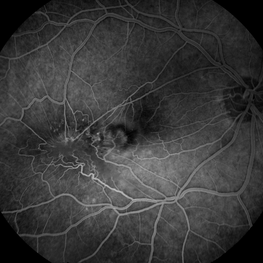
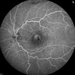 Branch Retinal Vein Occlusion with Multifactorial Macular Edema and Epiretinal Membrane
Oct 3 2024 by Logan ryzenga
Fluorescein angiogram of a 62 year old woman with cystoid macular edema from concurrent Epiretinal Membrane and Branch Retinal Vein occlusion. She has an extensive history of anti-VEGF injections with stable but unresolved macular edema. Following angiography, it was determined that an epiretinal membrane peel would be indicated in an attempt to achieve resolution of macular edema.
Photographer: Logan Ryzenga
Imaging device: Heidelberg Spectralis
Condition/keywords: 55-degrees, branch retinal vein occlusion (BRVO), cystoid macular edema (CME), epiretinal membrane (ERM), Fluorescein angiography, heidelberg spectralis, hyperfluorescence, leakage, left eye, OS, wide angle imaging
-
 Central Retinal Artery Occlusion
Apr 10 2024 by Tejaswita Verma
Left eye fundus photo of a 75 year old male with pale edematous retina with cherry red spot in a case of central retinal artery occlusion.
Photographer: DR. TEJASWITA VERMA
Imaging device: MIRANTE
Condition/keywords: central retinal artery occlusion (CRAO), cherry red spot
-
 Choroidal Melanoma Masquerading as Subretinal Hemorrhage With Breakthrough VH
Jan 23 2025 by Tejaswita Verma
A 65 year old diabetic male presented with large nasal retinal mass giving the appearance of organized dehaemoglobinized subretinal hemorrhage with breakthrough vitreous hemorrhage , with 6/6P vision. Enucleation specimen showed histopathology confirmed choroidal melanoma.
Photographer: DR. TEJASWITA VERMA
Imaging device: MIRANTE
-
 Cilioretinal Artery Sparing CRAO
May 1 2025 by Tejaswita Verma
Fundus photo of a middle aged male with CRAO partially sparing cilioretinal artery and papillomacular bundle. Vision 6/60.
Photographer: Dr. Tejaswita Verma
Imaging device: MIRANTE
Condition/keywords: CRAO with cilioretinal sparing
-
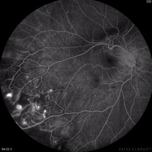 Coats Disease Fluorescein Angiography
Sep 2 2022 by FLOR ANGELICA JACOME GUTIERREZ
Fluorescein angiography of a patient with Coats disease where we found telangiectatic vessels, aneurysms, peripheral capillary nonperfusion and perivascular leak.
Photographer: Dr.Guillermo Salcedo Villanueva
Imaging device: Zeiss CLARUS 700 (FA)
Condition/keywords: Coats' disease, epiretinal membrane (ERM)
-
 CRAO Sparing Cilioretinal Artery
May 1 2025 by Tejaswita Verma
Fundus photo of a middle aged male with 6/60 vision in left eye showing CRAO partially sparing cilioretinal artery.
Photographer: Dr. Tejaswita Verma
Imaging device: MIRANTE
Condition/keywords: cilioretinal sparing, CRAO
-
 Deferoxamine Retinopathy slide 1
Oct 22 2012 by Ronald C. Gentile, MD
44-year-old man complained of decreased vision in both eyes with chromotopsia. He has a history of thalassemia intermedia with iron overload treated with deferoxamine.
Photographer: The New York Eye & Ear Infirmary Department of Medical Imaging
Condition/keywords: angioid streaks, Deferoxamine retinopathy, Thalassemia intermedia
-
 Dislocated Intraocular Lens
Nov 15 2024 by Tejaswita Verma
Fundus image of a spontaneously posteriorly dislocated IOL 10 years following surgery. Other eye had a subluxated opacified IOL.
Photographer: DR. TEJASWITA VERMA
Imaging device: MIRANTE
Condition/keywords: dislocated intraocular lens (IOL)
-
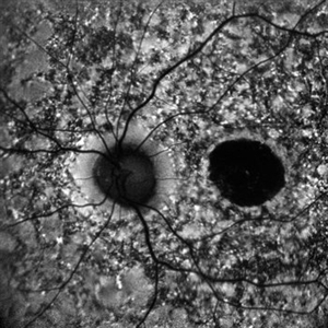
 Disseminated Chorioretinitis With Unknown Etiology
Apr 5 2018 by Kim Barrett
Ultra-wide field fluorescein angiogram of a 31-year-old female with intermittent pain in her left eye. Her condition has been managed in Liberia until recently when she moved to the United States. She suffers from multiple modalities including central retinal artery occlusion, posterior synechiae of the iris, interstitial keratitis, disseminated chorioretinitis, as well as HIV. An infectious cause is high on the differential in light of her HIV status. DDx: hypertensive crisis, an embolism (? IV drug use), coagulopathy, trauma, infectious. Blood work was normal. Her current vision is 20/30 right eye and 20/400 left eye.
Photographer: Kim Barrett, COA
Imaging device: Optos
Condition/keywords: central retinal artery occlusion (CRAO), chorioretinal scar, ciliary artery sparring, disseminated chorioretinitis, HIV, left eye, optic atrophy, staining
-
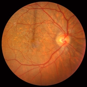
 Epiretinal Membrane
Sep 6 2021 by Ricardo Leitão Guerra
65-year-old woman with an asymptomatic ERM (BCVA=20/20).
Imaging device: Zeiss Clarus 700
Condition/keywords: epiretinal membrane (ERM)
-
 Epiretinal Membrane
May 10 2017 by Nichole Lewis
Epiretinal membrane.
Photographer: Nichole Lewis
Condition/keywords: epiretinal membrane (ERM)
-
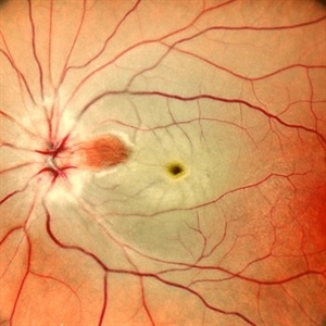
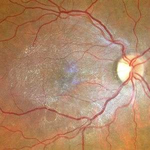
 Epiretinal Membrane
Oct 11 2012 by Michael P. Kelly, FOPS
This is a patient with idiopathic panuveitis who developed a visually significant epiretinal membrane. Pars plana vitrectomy with membrane peeling was performed to remove the epiretinal proliferation. I recommend magnifying the image to see the exquisite detail centrally.
Photographer: Michael P. Kelly, FOPS Director, Duke Eye Center Labs, Duke Universtiy Hospital
Imaging device: Zeiss 450Plus
Condition/keywords: epiretinal membrane (ERM), panuveitis
-
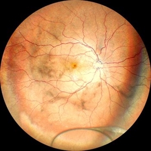
 Epiretinal membrane - Fundus photograph
Feb 5 2014 by Gerardo Garcia-Aguirre, MD
Fundus photograph of a 62 year old female with metamorphopsia and decreased visual acuity. A stage 2 epiretinal membrane is observed, causing distortion of the retinal vasculature.
Photographer: Gerardo Garcia-Aguirre, MD
Condition/keywords: epiretinal membrane (ERM)
 Loading…