 Initializing download.
-
 By John S. King, MD
Retina Associates, PA
Co-author(s): Jennifer Doyle, MD, Neuro-ophthalmologist at the Little Rock Eye Clinic - Uploaded on Jun 1, 2019.
- Last modified by Caroline Bozell on Jun 4, 2019.
- Rating
- Appears in
- Miscellaneous
- Condition/keywords
- optic neuropathy, bear tracks
- Photographer
- Karin Aletter
- Imaging device
-
Fundus camera
Topcon 50 - Description
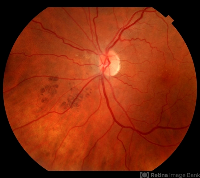
- 84-year-old white female with acute loss of vision in the left eye one day ago was sent here after going to the ED per primary eye provider. She described vision loss as a grey curtain that became total darkness. She had left sided temporal tenderness and some left sided neck pain. In the ED the cardiac work-up was u/r, the ESR and CRP were normal, and the CTH showed some non-specific opacification in the L ethmoid sinus. Acuity was HM OS with RAPD, normal EOMs, no proptosis or ptosis, posteriorly no SVPs were noted; the optic discs were pink and flat; no emboli or retinal whitening present; some bear tracks located nasally (see photo). She was referred to Dr. Doyle, who ordered an MRI, which showed a large mucocele with bony erosion into the left orbit, along with some ON enhancement possibly from compression (see images). She was operated that night and later recovered to 20/40 in that eye with a residual, inferior arcuate scotoma.


---thumb.JPG/image-square;max$79,0.ImageHandler "Radiation maculopathy")


---thumb.JPG/image-square;max$79,0.ImageHandler "Bear Tracks")

