Search results (281 results)
-
 Asymptomatic Eye in FEVR
Jul 7 2015 by Hamid Ahmadieh, MD
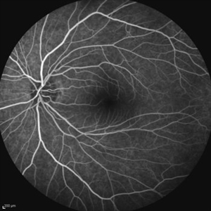
FA image of the asymptomatic left eye of a 28-year-old man with total RD secondary to advanced FEVR in his right eye. Notice straightening of the retinal vessels.
Photographer: Soulmaz Shahmohammad, Negah Eye Center, Tehran, Iran
Imaging device: Specteralis
Condition/keywords: asymptomatic, familial exudative vitreoretinopathy (FEVR)
-
 Asymptomatic Lesion
Nov 9 2012 by Norman Byer
This asymptomatic lesion in a 27-year-old woman is a very interesting example of a cystic retinal tuft. Note the discrete white nubbin, which is the chief characteristic of this lesion. In this case, it is surrounded by a small area of subretinal fluid. The next slide pair will reveal the reason for this.
Condition/keywords: asymptomatic, cystic retinal tuft, subretinal fluid
-
 Asymptomatic Lesion
Nov 9 2012 by Norman Byer
This is the same lesion as seen in the previous slide pair. Here the scleral indentation is carried more posterior revealing a tiny, round, full thickness retinal hole. This is not a tear produced by traction even though vitreous is always attached to these flaps. You will note that the hole is round and is separated by a slight distance from the flap itself. It is probably the result of long continued atrophy and devitalization of the retina. A posterior vitreous was not detached. This lesion has not changed its appearance for more than a year of observation, but the age of the hole is actually unknown.
Condition/keywords: asymptomatic, atrophy, full thickness retinal hole, posterior scleral indentation, retinal hole, round hole
-
 Asymptomatic Rhegmatogenous Retinal Detachment
Sep 14 2012 by Sharon Fekrat, MD FACS FASRS
Fundus photograph of a 25-year-old emmetropic male graduate student with an inferotemporal phakic chronic asymptomatic rhegmatogenous retinal detachment with a demarcation line in the right eye. His sister who is an ophthalmology resident discovered this incidental finding. Vision 20/20.
Photographer: Brian Lutman CRA, Duke University Eye Center, Durham, NC
Condition/keywords: asymptomatic, demarcation line
-
 Asymptomatic Superior Retinal Detachment
May 5 2016 by Steven J Ryder, MD
38-year-old African American female with moderate myopia (-4.50 Sph OU) and asymptomatic superior retinal detachment in the right eye. Montage fundus photography showing localized retinal detachment superiorly with single full-thickness retinal break at 12:00.
Photographer: Luis Bernhard, Miami VA Healthcare System
Imaging device: Topcon
Condition/keywords: asymptomatic, full thickness retinal hole, myopia, retinal break, retinal detachment with retinal defect
-
 Asymptomatic Superior Retinal Detachment
May 5 2016 by Steven J Ryder, MD
38-year-old African American female with moderate myopia (-4.50 Sph OU) and asymptomatic superior retinal detachment in the right eye. Zeiss Cirrus OCT capturing full-thickness retinal break at 12:00 and temporal vitreoretinal traction.
Photographer: Luis Bernhard, Miami VA Healthcare System
Imaging device: Zeiss Cirrus
Condition/keywords: asymptomatic, full thickness retinal hole, retinal break, retinal detachment with retinal defect
-
 Asymptomatic Superior Retinal Detachment
May 5 2016 by Steven J Ryder, MD
38-year-old African American female with moderate myopia (-4.50 Sph OU) and asymptomatic superior retinal detachment in the right eye. Zeiss OCT capturing vertical raster scans through border of retinal detachment.
Photographer: Luis Bernhard, Miami VA Healthcare System
Imaging device: Cirrus
Condition/keywords: asymptomatic, full thickness retinal hole, retinal detachment with retinal defect
-
 Asymptomatic Tractional Tear
Nov 9 2012 by Norman Byer
This 38-year-old man was found to have this asymptomatic tractional tear in which the vitreoretinal traction had completely avulsed this tiny fragment of retina as a free operculum. Note how the examination and also the photography of this tiny lesion is made easier by scleral indentation.
Condition/keywords: asymptomatic, free operculum, scleral indentation, vitreoretinal traction
-
 Retinal Tear in Aphakic Fellow Eye
Nov 9 2012 by Norman Byer
This 59-year-old man presented with sudden symptoms of retinal detachment in his opposite aphakic eye secondary to a tiny retinal tear about 1/8th disc diameter in size. During the examination, the fellow eye shown here was found to have this much larger tractional tear approximately 2 disc diameters in total length. If you look carefully, you will see that this is really a series of three separate tears with a common flap. The tears are separated by tiny bridges of remaining tissue which cause the edges of the apparent large tear to be serrated. This was also an aphakic eye with a posterior vitreous detachment but the lesion had produced no symptoms.
Condition/keywords: asymptomatic, bridge of tissue between tears, posterior vitreous detachment, tractional retinal tear
-
---thumb.jpg/image-square;max$300,300.ImageHandler) Retinoschisis
Retinoschisis
Dec 25 2012 by Philip J. Polkinghorne, MD
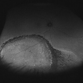
Asymptomatic inferotemporal retinoschisis with large outer layer break
Condition/keywords: asymptomatic
-
 White Retinal Tuft
Nov 9 2012 by Norman Byer
This white retinal tuft was seen in a 20-year-old man. It is associated with an asymptomatic retinal tear posterior to the tuft and with a tiny adjacent amount of subretinal fluid. It remained just like this for six years and then underwent the change shown in the next slide pair.
Condition/keywords: asymptomatic, subretinal fluid, white retinal tuft
-
 Aquired Vitelliform Maculopathy
Jun 29 2014 by John S. King, MD
Asymptomatic MA healthy female consulted for wet AMD. Plan: observe. Photo and AF initially and year afterwards. Health MA female; no vitritis or other lesions; similar findings in both eyes.
Photographer: Wayne A Ladlee Jr
Condition/keywords: aquired vitelliform maculopathy
-
 Asteroid Hyalosis
Aug 22 2023 by Angela Rico
Asymptomatic 83 y/o F
Photographer: Angela Rico M.D.
Imaging device: optos
Condition/keywords: asteroid hyalosis
-
 Asymptomatic Chronic Retinal Detachment With Demarcation Line
Jun 11 2016 by Philip J. Polkinghorne, MD
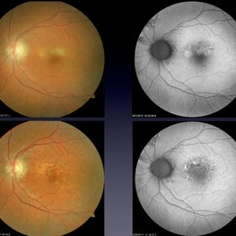
A 65-year-old emmetrope with asymptomatic chronic retinal detachment with demarcation line.
Photographer: Alex Fraser, Greenlane Clinical Center, Auckland, New Zealand
Condition/keywords: chronic retinal detachment, fundus autofluorescence (FAF)
-
 Asymptomatic Eye in FEVR
Jul 7 2015 by Hamid Ahmadieh, MD
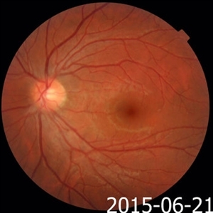
Color fundus photograph of the asymptomatic eye of a patient with FEVR. Notice straightening of the retinal vessels.
Photographer: Soulmaz Shahmohammad, Negah Eye Center, Tehran, Iran
Condition/keywords: color fundus photograph, familial exudative vitreoretinopathy (FEVR)
-
 Bilateral Central Serous Retinopathy
Mar 26 2019 by Gary R. Cook, MD, FACS
Asymptomatic left eye of a 37-year-old white male with a history of previous CSR OS showing some focal RPE depigmentation perifoveally and subretinic deposits temporally; no NSRD is present; VA = 20/15+3.
Imaging device: Topcon VT-50
Condition/keywords: central serous retinopathy (CSR), resolved subretinal fluid, retinal pigment epithelium (RPE) changes
-
 Bilateral Metastatic Lesions Secondary to Breast Cancer
Feb 18 2014 by Gabriela Lopezcarasa Hernandez, MD
Asymptomatic 44-year-old woman who went to a general exam to the ophthalmologist.
Photographer: Araceli Rojas Arriaga, Hospital Angeles Lomas, Mexico
Imaging device: ZEISS FF4
Condition/keywords: metastatic lesion
-
 Horse Shoe Tear
Sep 16 2017 by Purva Patwari
Asymptomatic horse shoe tear found on preoperative cataract assessment of a 54-year-old male patient. Laser barrage was done and he underwent Phacoemulsification surgery a month later.
Photographer: Dr Purva Patwari,Patwari Retina Center,Ahmedabad,India
Imaging device: Zeiss Visucam 500
-
 Human Shoe Tear
Jan 2 2018 by Purva Patwari
Asymptomatic horse shoe tear found on preoperative cataract assessment of a 54-year-old male patient. Laser barrage was done and he underwent phacoemulsification surgery a month later.
Photographer: Dr Purva Patwari, Patwari Retina Center, Ahmedabad, Gujarat , India
Imaging device: ZEISS VISU 500
-
 Inferior Rhegmatogenous Retinal Detachment with Subretinal Fibrosis
Aug 23 2012 by Gabriela Lopezcarasa Hernandez, MD
Asymptomatic 25-year-old woman with high myopia.
Photographer: Gabriela Lopezcarasa Hernandez, Hospital Angeles Lomas
Imaging device: FF4
Condition/keywords: high myopia, subretinal fibrosis
 Loading…