Search results (281 results)
-
 Pseudoduplication of the Optic Disc
Jul 9 2025 by Hrishikesh Naik, MS
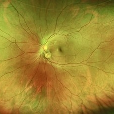
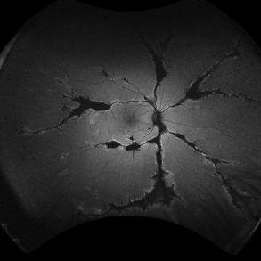
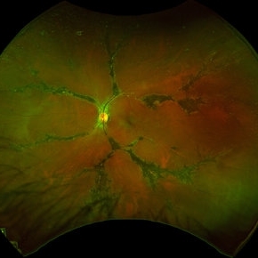
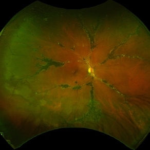
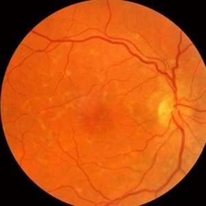
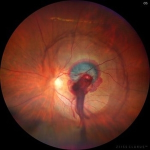
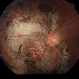
A peripapillary colobomatous pseudo-duplication of the optic disc as seen in an asymptomatic 23 year old female with myopia referred for routine retinal periphery screening. Rest retinal exam was normal. Duplication of the optic disc can be classified as either true duplication or pseudoduplication, both of which are rare clinical conditions. Pseudodoubling of the optic disc is commonly caused by optic disc or peripapillary colobomas, characterized by a circumscribed, disc-like lesion with radiating vessels but only one normal optic nerve. A few cases have involved pathological myopia, moderate myopia, proliferative diabetic retinopathy and CHARGE syndrome. The lesion is often found inferior to the normal optic disc. The patient was advised regular follow ups.
Photographer: Hrishikesh Naik
Imaging device: Optos Daytona
Condition/keywords: Coloboma, Pseudoduplication of optic disc
-
 Prepapillary Vascular Loop
Jul 4 2025 by KANWALJEET HARJOT MADAN, M.S. (Ophthalmology); FAICO (Vitreous - Retina)
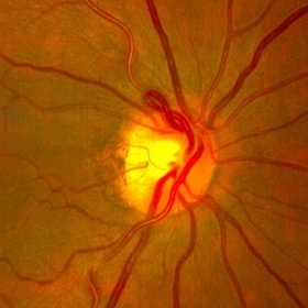
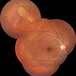
This is the fundus picture of right eye of a young 32 years female depicting pre papillary vascular loop. A prepapillary vascular loop is a congenital anomaly of the optic disc that presents as an elevated and twisted bundle of vessels projecting into the vitreous cavity. It is a benign condition, usually unilateral but can be bilateral. It is asymptomatic and discovered during routine eye examination. This anomaly can sometimes cause complications like branch retinal artery occlusion, vitreous hemorrhage, or sub retinal hemorrhage.
Photographer: Dr. Kanwaljeet Harjot Madan, Thind Eye Hospital, Jalandhar City (Punjab) INDIA.
Imaging device: Zeiss Fundus Camera
Condition/keywords: branch retinal artery occlusion (BRAO), optic disc, Prepapillary Vascular Loop, SUB RETINAL HEMORRHAGE, Vitreous hemorrhage
-
 Pigmented Paravenous Retinochoroidal Atrophy (PPRCA)
Jun 30 2025 by Maria Letícia Costa Holanda
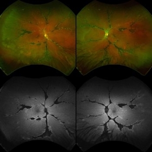
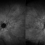
Fundoscopy of a 42-year-old asymptomatic man with pigmented paravenous chorioretinal atrophy. Pigmented paravenous retinochoroidal atrophy (PPRCA) is a rare disorder of unknown etiology. The disease is characterized by pigment accumulation along the distribution of retinal veins. The findings are usually incidental with minimal effect on vision.
Photographer: Guilherme da Cruz Reis, CLINOS Eye Hospital - Feira de Santana (BA),Brazil
Condition/keywords: pigmented paravenous chorioretinal atrophy (PPCRA)
-
 Pigmented Paravenous Retinochoroidal Atrophy (PPRCA)
Jun 27 2025 by Maria Letícia Costa Holanda
Fundoscopy of a 42-year-old asymptomatic man with pigmented paravenous chorioretinal atrophy. Pigmented paravenous retinochoroidal atrophy (PPRCA) is a rare disorder of unknown etiology. The disease is characterized by pigment accumulation along the distribution of retinal veins. The findings are usually incidental with minimal effect on vision.
Photographer: Guilherme da Cruz Reis, CLINOS Eye Hospital - Feira de Santana (BA),Brazil
Condition/keywords: Pigmented Paravenous Retinochoroidal Atrophy
-
 Pigmented Paravenous Retinochoroidal Atrophy (PPRCA)
Jun 27 2025 by Maria Letícia Costa Holanda
Fundoscopy of a 42-year-old asymptomatic man with pigmented paravenous chorioretinal atrophy. Pigmented paravenous retinochoroidal atrophy (PPRCA) is a rare disorder of unknown etiology. The disease is characterized by pigment accumulation along the distribution of retinal veins. The findings are usually incidental with minimal effect on vision.
Photographer: Guilherme da Cruz Reis, CLINOS Eye Hospital - Feira de Santana (BA),Brazil
Condition/keywords: pigmented paravenous chorioretinal atrophy (PPCRA)
-
 Pigmented Paravenous Chorioretinal Atrophy (PPCRA)
Jun 27 2025 by Maria Letícia Costa Holanda
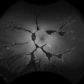
Fundoscopy of a 42-year-old asymptomatic man with pigmented paravenous chorioretinal atrophy. Pigmented paravenous retinochoroidal atrophy (PPRCA) is a rare disorder of unknown etiology. The disease is characterized by pigment accumulation along the distribution of retinal veins. The findings are usually incidental with minimal effect on vision.
Photographer: Guilherme da Cruz Reis, CLINOS Eye Hospital - Feira de Santana (BA),Brazil
Condition/keywords: pigmented paravenous chorioretinal atrophy (PPCRA)
-
 Myelinated Nerve Fibers
Apr 18 2025 by DR Rohit Gupta
The **myelinated nerve fibers of the optic disc** (also known as **medullated nerve fibers**) are retinal nerve fibers that retain their myelin sheath as they pass through the optic nerve head. Normally, retinal nerve fibers are unmyelinated to allow for light transparency, but in some cases, myelination extends anteriorly into the retina, appearing as a striking white, feathery patch on the optic disc or peripapillary retina. ### **Key Features:** 1. **Appearance:** - Dense, white, striated patches with feathery edges. - Typically located at the superior or inferior pole of the optic disc. - May obscure retinal vessels underneath. 2. **Clinical Significance:** - Usually **benign** and asymptomatic. - **Congenital** (present at birth or early childhood). - Rarely associated with **visual field defects** (e.g., scotomas corresponding to the area of myelination). - Occasionally linked with **high myopia** or **amblyopia** if extensive. 3. **Pathophysiology:** - Failure of oligodendrocytes or Schwann cells to stop myelination at the lamina cribrosa. - Normally, myelination stops at the optic nerve head, but in this condition, it extends into the retina. 4. **Diagnosis:** - **Fundoscopy:** Classic white, feathery appearance. - **Optical Coherence Tomography (OCT):** Shows thickened retinal nerve fiber layer (RNFL). - **Visual Field Testing:** May detect defects if large. 5. **Differential Diagnosis:** - Optic disc edema - Cotton wool spots - Retinoblastoma (rarely, but must be ruled out in children) 6. **Management:** - No treatment required if asymptomatic. - Monitor for amblyopia in children. - Rare cases with significant visual impairment may need further evaluation. ### **Fun Fact:** Myelinated nerve fibers are seen in **~0.5-1%** of the population and are usually an incidental finding.
Photographer: Dr Rohit gupta
Imaging device: Samsung S21
Condition/keywords: Medulated Nerve fibre, Medullated Nerve fibres, myelinated nerve fibers, Myelinated Nerve Fibres, optic disc drusen
-
 Hosreshoe Tears on Posterior Pole
Mar 22 2025 by Deepak Bhojwani, MS
A fundus image of an asymptomatic 64 year old male with large horseshoe shaped breaks in inferonasal quadrant on posterior pole, an unusual location for retinal breaks.
Photographer: DR DEEPAK BHOJWANI
Condition/keywords: horseshoe tear, posterior pole break, retinal break
-
 Multimodal Imaging in CHRPE
Mar 6 2025 by Gerardo - Montante Montelongo, MD
Fundus photograph of an 83-year-old male with a history of Diabetes, smoking, cataract surgery on the right eye in 2022, and open-angle glaucoma. Asymptomatic. Indirect ophthalmoscopy revealed 80% excavation, peripapillary atrophy, and a hyperpigmented perifoveal lesion with 35% atrophy, 10% drusen, and 5.1 mm diameter, corresponding to a CHRPE. At multimodal imaging, FFA shows hypoautofluorescence of the lesion, OCT shows preservation of internal retinal layers, atrophy of external retinal layer, with an RPE disruption, and posterior shadowing. USG shows a flat hyperechoic lesion 5.1 mm in diameter and 1.32 mm in thickness, solid and with high internal reflectance.
Photographer: Gerardo Montante-Montelongo, MD, Mexican Institute of Ophthalmology
Imaging device: Clarus 700
Condition/keywords: congenital hypertrophy of the retinal pigment epithelium (CHRPE), multimodal imaging
-
 Choroidal Osteoma
Feb 10 2025 by Virginia Gebhart
10 year old female referred for amelanotic lesion in the inferior macula. Clinical exam and ultrasound consistent with choroidal osteoma. Pt is asymptomatic, will observe.
Photographer: Virginia Gebhart, Retina Consultants of Carolina
Imaging device: Topcon 50DX
Condition/keywords: choroidal osteoma
-
 Indocyanine Green (ICG) of Circumscribed Choroidal Hemangioma (CCH)
Feb 6 2025 by Jack B Margines, MD, MHCI
Peripheral patchy hyperfluorescence is seen on this early image of ICG-A on a 53-year-old asymptomatic with an extramacular circumscribed choroidal hemangioma.
Photographer: W Ryan Miliam, CRA, OCT-C, University of California, Irvine Gavin Herbert Eye Institute
Imaging device: Optos
Condition/keywords: choroidal hemangioma, indocyanine green (ICG) angiography
-
 CCH
CCH
Feb 6 2025 by Jack B Margines, MD, MHCI
SD-OCT of an extramacular Circumscribed Choroidal Hemangioma in an asymptomatic 53 year-old female
Photographer: Ryan Milam, University of California, Irvine Gavin Herbert Eye Institute
Imaging device: Zeiss Cirrus
Condition/keywords: Circumscribed Choroidal Hemangioma, OCT
-
 Melanocytoma of Optic Disc
Jan 13 2025 by Virginia Gebhart
25 year-old female referred for melanocytoma of optic disc. Lesion is benign, no treatment necessary. Pt asymptomatic.
Photographer: Virginia Gebhart, Retina Consultants of Carolina
Imaging device: Topcon 50DX
Condition/keywords: benign melanocytoma, melanocytoma, optic disc melanocytoma
-
 Retinal Telangiectasis
Nov 13 2024 by Virginia Gebhart
42 year old female with telangiectatic vessels and vascular sheathing. FA showed mild leakage with areas of peripheral non-perfusion. Pt is asymptomatic without inflammation in the vitreous. No history of systemic inflammatory disease. Will observe.
Photographer: Virginia Gebhart, Retina Consultants of Carolina
Imaging device: Optos California
Condition/keywords: retinal telangiectasia, telangiectatic vessels, vascular sheathing of retina
-
 Stargardt's Disease
Oct 23 2024 by Virginia Gebhart
62 year old female with bullseye RPE changes and flecks, mottled FAF, and silent choroid on FA consistent with late onset Stargardt's Disease. Pt is asymptomatic with 20/20 vision OU at this time
Photographer: Virginia Gebhart, Retina Consultants of Carolina
Imaging device: Optos California
Condition/keywords: Stargardt disease, Stargardts Disease
-
 Retinitis Pigmentosa
Oct 16 2024 by Virginia Gebhart
74 year old female with bone spicule pigmentation associated with Retinitis Pigmentosa. Pt diagnosed at age 53, relatively asymptomatic prior to diagnosis. Pt reports gradual vision loss over 10+ years. BCVA 20/40
Photographer: Virginia Gebhart, Retina Consultants of Carolina
Imaging device: Optos California
Condition/keywords: bone spicule, retinitis pigmentosa, retinitis pigmentosa (RP) dystrophy
-
 IOFB
IOFB
Oct 11 2024 by Virginia Gebhart
55 year old male s/p RD repair with SB and SO in Mexico June 2023. Questionable foreign body inferior vitreous base. Pt asymptomatic, had no previous knowledge of IOFB.
Photographer: Virginia Gebhart, Retina Consultants of Carolina
Imaging device: Optos California
Condition/keywords: intraocular foreign body, IOFB, scleral buckle
-
 Pattern dystrophies – Asymptomatic middle-aged man with normal vision and a multifocal PD
Sep 17 2024 by Nicolas A Yannuzzi, MD
The PD simulates Stargardt disease/fundus flavimaculatus with irregular yellow-white flecks scattered throughout the posterior pole. Some lesions extend beyond the retinal vascular arcades.
Condition/keywords: inherited retinal disease, pattern dystrophy
-
 Torpedo Retinopathy
Sep 16 2024 by Sriharanathan Poopalaratnam, MD,FRCS
Fundus photograph of a 13-year-old boy asymptomatic accidental finding of unilateral, extramacular oval hypopigmented lesion, with its tip directed toward the central macula, suggestive of a "torpedo" lesion in the nonclassical location.
Photographer: Ms.Samitha
Condition/keywords: torpedo Retinopathy
-
 Vasoproliferative Tumor
Aug 29 2024 by César Adrián Gómez Valdivia, MD
Inferior retinal vasoproliferative tumor found in a 66 year-old female patient. Asymptomatic.
Photographer: @eyemissu2
Imaging device: California ICG OPTOS
Condition/keywords: Vasoproliferative Tumor
-
 Fundus Flavimaculatus
Aug 12 2024 by Virginia Gebhart
77 year old female with symmetrical retinal flecks consistent with hereditary dystrophy. Unable to complete genetic testing today, will consider in the future. Pt Asymptomatic, Dcc 20/20 OU
Photographer: Virginia Gebhart
Imaging device: Optos California
Condition/keywords: fundus flavimaculatus
-
 Ruptured Retinal Artery Macroaneurysm
Jun 18 2024 by KANWALJEET HARJOT MADAN, M.S. (Ophthalmology); FAICO (Vitreous - Retina)
This is a fundus photo depicting ruptured Retinal Artery Macroaneurysm (RAM) in the left eye of a 63 years old female. RAM is an acquired saccular or fusiform dilatation of the retinal arterioles that usually occur within the first three orders of bifurcation. The Superotemporal artery is the most common location. RAM may be asymptomatic or cause a number of complications such as macular edema, serous macular detachment, and hemorrhages.
Photographer: Dr Kanwaljeet Harjot Madan
Condition/keywords: Haemorrhage, macroaneurysm, retinal arteriole
-
 Choroidal Osteoma
May 30 2024 by Virginia Gebhart
33 year old female with regressed osteoma OS s/p focal laser, TTT and PDT (first treatment in 2013). Vision 20/20, pt remains asymptomatic.
Photographer: Virginia Gebhart
Imaging device: Topcon 50DX
Condition/keywords: choroidal osteoma
 Loading…