Search results (281 results)
-
 White Retinal Tuft
Nov 9 2012 by Norman Byer
After six years, the previous lesion looked like this. The former flap has been completely avulsed and is now a free operculum. The white zone around the tear represents the small area of detachment and subretinal fluid. It is still asymptomatic and does not require treatment.
Condition/keywords: does not require treatment, free operculum, operculated retinal hole, subretinal fluid, white retinal tuft
-
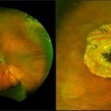
 Inferior Rhegmatogenous Retinal Detachment with Subretinal Fibrosis
Aug 23 2012 by Gabriela Lopezcarasa Hernandez, MD
Asymptomatic 25-year-old woman with high myopia.
Photographer: Gabriela Lopezcarasa Hernandez, Hospital Angeles Lomas
Imaging device: FF4
Condition/keywords: high myopia, subretinal fibrosis
-
 Operculated Hole and CHRPE
Jan 16 2018 by Carolyn Daley
58-year-old woman with an operculated hole and CHRPE in the right eye. Patient is asymptomatic so no treatment was recommended at this time.
Photographer: Carolyn Daley
Imaging device: Optos ultra wide field image
Condition/keywords: congenital hypertrophy of the retinal pigment epithelium (CHRPE), operculated retinal hole, Optos, ultra-wide field imaging
-
 Pigmented Peripheral Retinal Degeneration
Jun 27 2013 by Jason S. Calhoun
42-year-old male came in for routine eye exam and to follow up on peripheral retinal degeneration in both eyes. VA is 20/20, right eye and 20/25, left eye. Patient is asymptomatic with no visual complaints.
Photographer: Jason S. Calhoun, Mayo Clinic Jacksonville, Florida
Imaging device: TOPCON TRC 50-EX
Condition/keywords: peripheral retinal degeneration
-
 Asymptomatic Rhegmatogenous Retinal Detachment
Sep 14 2012 by Sharon Fekrat, MD FACS FASRS
Fundus photograph of a 25-year-old emmetropic male graduate student with an inferotemporal phakic chronic asymptomatic rhegmatogenous retinal detachment with a demarcation line in the right eye. His sister who is an ophthalmology resident discovered this incidental finding. Vision 20/20.
Photographer: Brian Lutman CRA, Duke University Eye Center, Durham, NC
Condition/keywords: asymptomatic, demarcation line
-
 Retinal Break at Site of Lattice Degeneration with Scleral Indentation
Nov 9 2012 by Norman Byer
This is the same case as the previous photograph. With scleral indentation slightly more posterior, the flap is seen to be associated with a large retinal tear. This is a tractional tear and it is possible that in this case the cryotherapy itself may have increased the vitreoretinal traction at this site and in this way led to this new tear. The age of the tear is unknown because it was asymptomatic, and even though the eye is aphakic the tear has not caused a clinical retinal detachment.
Condition/keywords: retinal flap, scleral indentation, tractional retinal tear, vitreoretinal traction
-
 Asymptomatic Lesion
Nov 9 2012 by Norman Byer
This asymptomatic lesion in a 27-year-old woman is a very interesting example of a cystic retinal tuft. Note the discrete white nubbin, which is the chief characteristic of this lesion. In this case, it is surrounded by a small area of subretinal fluid. The next slide pair will reveal the reason for this.
Condition/keywords: asymptomatic, cystic retinal tuft, subretinal fluid
-
 OCT Myopic Staphyloma With Schisis and ERM
Apr 24 2014 by Scott E. Pautler, MD
OCT of high myope with asymptomatic macular schisis.
Imaging device: Heidelberg Spectralis
Condition/keywords: foveal schisis, maculopathy, maculoschisis, optical coherence tomography (OCT), pathologic myopia, staphyloma
-
 White Retinal Tuft
Nov 9 2012 by Norman Byer
This white retinal tuft was seen in a 20-year-old man. It is associated with an asymptomatic retinal tear posterior to the tuft and with a tiny adjacent amount of subretinal fluid. It remained just like this for six years and then underwent the change shown in the next slide pair.
Condition/keywords: asymptomatic, subretinal fluid, white retinal tuft
-
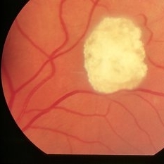 Tuberous Sclerosis
Oct 9 2012 by Alan D. Letson, MD
Small astrocytic hamartoma in asymptomatic 65-year-old woman with Tuberous sclerosis.
Photographer: Beverly Radcliffe
Condition/keywords: astrocytoma, hamartoma, tuberous sclerosis
-
 Retinoschisis
Nov 9 2012 by Norman Byer
This 51-year-old woman has retinoschisis with a large outer layer hole which has a white posterior rolled border. The left side of the posterior border of this hole can be seen to lie quite close to the inner layer showing that the outer layer is detached. This, therefore, is actually a combined schisis detachment which may safely be observed without treatment. This is an asymptomatic process, and the detachment of the outer layer is almost always localized and self limited.
Condition/keywords: intact inner layer, localized detachment of outer layer, outer layer hole, retinoschisis, rolled edges of retina, schisis detachment, white posterior
-
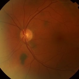 Multiple Choroidal Nevi
Jul 9 2014 by Susanna S. Park, MD, PhD
Asymptomatic 45-year-old woman with multiple small pigmented choroidal lesions in the posterior pole consistent with nevi.
Photographer: Ellen Redenbo
Condition/keywords: choroidal nevus
-
 Asymptomatic Lesion
Nov 9 2012 by Norman Byer
This is the same lesion as seen in the previous slide pair. Here the scleral indentation is carried more posterior revealing a tiny, round, full thickness retinal hole. This is not a tear produced by traction even though vitreous is always attached to these flaps. You will note that the hole is round and is separated by a slight distance from the flap itself. It is probably the result of long continued atrophy and devitalization of the retina. A posterior vitreous was not detached. This lesion has not changed its appearance for more than a year of observation, but the age of the hole is actually unknown.
Condition/keywords: asymptomatic, atrophy, full thickness retinal hole, posterior scleral indentation, retinal hole, round hole
-
 Remnant of Hyaloidal Artery
Feb 5 2014 by Gerardo Garcia-Aguirre, MD
Fundus photograph of the left eye of a 14-year-old asymptomatic female. The photograph is focused on the retina, and a prepapillary vitreous opacity is observed (white arrows). The opacity is attached to the origin of the retinal vessels in the optic nerve head.
Photographer: Gerardo Garcia-Aguirre, MD
Condition/keywords: persistence of the hyaloid artery
-
 Retinal Cavernous Hemangioma
Oct 30 2012 by Roy D. Brod, MD
Color fundus photograph of right eye in a 32-year-old asymptomatic female showing typical "cluster of grapes" appearence. Normal MRI of brain. No skin lesions.
Photographer: Julia Walker
Condition/keywords: cavernous hemangioma of the retina, cluster of grapes
-
 Optic Nerve Head Drusen
Feb 12 2015 by Timothy S Fuller, MD
Fundus photograph of a 34-year-old woman with striking, asymptomatic optic nerve head drusen.
Photographer: Nice Hesse, Texas Retina Associates
Condition/keywords: drusen of optic disc
-
 Acute Retinal Detachment
Nov 9 2012 by Norman Byer
This 54-year-old man was referred because of sudden symptoms in his opposite eye in which he had suffered an acute retinal detachment secondary to a horseshoe tear around lattice degeneration. During the examination, the fellow eye shown here was also found to have this large horseshoe tear about 1 o’clock hour (4 disc diameters) in size. A tear occurred around a lattice lesion which is present on the flap but is out of focus. This tear had been asymptomatic even though it was caused by a posterior vitreous detachment and illustrates that even very large tears may produce no symptoms or mild symptoms that are easily overlooked.
Condition/keywords: lattice degeneration, posterior vitreous detachment
-
 Remnant of Hyaloidal Artery
Feb 5 2014 by Gerardo Garcia-Aguirre, MD
Fundus photograph of the left eye of a 14-year-old asymptomatic female. The photograph is focused on the posterior vitreous where a prepapillary vitreous opacity is observed (white arrows). The opacity is attached to the origin of the retinal vessels in the optic nerve head.
Photographer: Gerardo Garcia-Aguirre, MD
Condition/keywords: persistence of the hyaloid artery
-
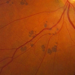 Bear Tracks Pigmentation
Mar 1 2014 by Homayoun Tabandeh, MD, FASRS
Bear tracks pigmentation in a 58-year-old asymptomatic female.
Condition/keywords: bear tracks
-
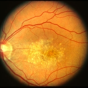
 Pattern Dystrophy slide 1
Oct 22 2012 by Ronald C. Gentile, MD
Asymptomatic middle-age man with normal vision and a multifocal pattern dystrophy. The pattern dystrophy simulates Stargardt disease/fundus flavimaculatus with irregular yellow-white flecks scattered throughout the posterior pole. Some lesions extend beyond the retinal vascular arcades.
Photographer: The New York Eye & Ear Infirmary Department of Medical Imaging
Condition/keywords: pattern macular dystrophy
-
9(Z001---thumb.BMP/image-square;max$300,300.ImageHandler) Torpedo Maculopathy
Torpedo Maculopathy
Jul 25 2013 by Ian C Reddie, LLB (QUT), MBBS (Qld), FRANZCO, FASRS
Color picture of asymptomatic 8-year-old girl with incidental retinal finding at posterior pole of left eye.
Condition/keywords: torpedo maculopathy
-
 Malattia Leventinese 2 - color LE
Jan 11 2013 by Alex P. Hunyor, MD
Malattia levantinese - left eye. Asymptomatic patient with 20/25 vision OU.
Condition/keywords: Malattia Leventinese
-
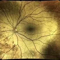 Oguchi Disease OS
Dec 30 2017 by David Lam
Fundus photo of asymptomatic 42-year-old male with BCVA 20/20 OD and OS. Note golden yellow metallic sheen of posterior pole and mid-periphery.
Photographer: David Lam OD
Imaging device: Eidon
Condition/keywords: congenital nyctalopia, Oguchi's disease
-
 Crystalline Retinopathy
Jun 27 2020 by Thirumalesh Mochi Basavaraj, MD
23-year-old asymptomatic female, came for routine examination. Inset shows OCT image with hyperreflective lesion scattered throughout the retina, choroidal sclerosis can also be noted.
Photographer: Ravikrishna, Puttaswamy
Imaging device: Heidelberg Spectralis
Condition/keywords: crystalline retinopathy
-
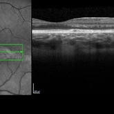 SD-OCT - Torpedo Maculopathy
Jul 25 2013 by Ian C Reddie, LLB (QUT), MBBS (Qld), FRANZCO, FASRS
SD-OCT through lesion at posterior pole of asymptomatic 8-year-old female.
Condition/keywords: torpedo maculopathy
 Loading…