Search results (66 results)
-
 Advanced Angioid Streak-Associated Choroidal Neovasclar Membranes
Dec 27 2016 by Young Hee Yoon, MD, PhD
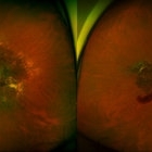
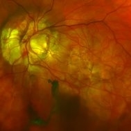
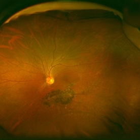
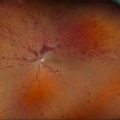
UWF fundus photographs of an 74-year-old woman who received several anti-VEGF injections due to CNV associated with angioid streak in both eyes. Note diffuse scar change and pigmentary degenerations aroud disc in both eyes. There is some retinal hemorrhage due to CNV in her left eye.
Photographer: Young Hee Yoon, University of Ulsan, Asan Medical Center, Seoul, Korea
Imaging device: Optomap
Condition/keywords: angioid streaks, choroidal neovascularization (CNV)
-
 Central Retinal Vein Occlusion With Waldenstroms macroglobulinemia
Jun 18 2025 by Korey Starkey
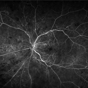
64-year-old patient presents with CRVO with secondary macular edema in both eyes. Venous beading present in 2/4 quadrants OU. Patient diagnosed with Waldenstroms macroglobulinemia, found on SPEP and bone marrow biopsy. Treatment recommended of anti-vegF intravitreal injections OU.
Photographer: Korey Starkey
Imaging device: Optos
Condition/keywords: attenuated vessels, central retinal vein occlusion (CRVO), CRVO, FA early phase, FLUORESCEIN ANGIOGRAPHY, macular edema, Optomap, OPTOS CALIFORNIA, severe NPDR, venous beading, Waldenstroms macroglobulinemia
-
 Congenital Hypertrophy of the Retinal Pigment Epithelium Autofluorescence Optomap
Sep 24 2019 by Sophia El Hamichi, MD
A 52-year-old female followed for 2 temporal lesions of CHRPE OD and white without pressure.
Photographer: Sophia El Hamichi, MD, Murray Ocular Oncology and Retina, Miami
Condition/keywords: autofluorescence imaging, congenital hypertrophy of the retinal pigment epithelium (CHRPE), Optomap, ultra-wide field imaging, white without pressure
-
 Congenital Hypertrophy of the Retinal Pigment Epithelium Wide Field Optomap
Sep 24 2019 by Sophia El Hamichi, MD
A 52-year-old female followed for 2 temporal lesions of CHRPE OD and white without pressure.
Photographer: Sophia El Hamichi,MD, Murray Ocular Oncology and Retina, Miami
Condition/keywords: congenital hypertrophy of the retinal pigment epithelium (CHRPE), Optomap, ultra-wide field imaging, white without pressure
-
 Giant Retinal Tear
Mar 29 2014 by Min Kim, MD, PhD, MBA, FASRS
Wide field fundus photograph of a 25 year-old male shows giant retinal tear with inverted retinal flap.
Photographer: Young Duk Bae, Yonsei University, Gangnam Severance Hospital
Imaging device: Optomap
Condition/keywords: giant retinal tear
-
 Gyrate Atrophy
Sep 23 2020 by Hashim Ali Khan, OD, FAAO
Widefield red-free image from a young male with gyrate atrophy.
Imaging device: Optomap
Condition/keywords: gyrate atrophy
-
 Gyrate Atrophy
Sep 23 2020 by Hashim Ali Khan, OD, FAAO
Widefield choroidal image from a young male with gyrate atrophy.
Imaging device: Optomap
Condition/keywords: gyrate atrophy
-
 Gyrate Atrophy
Sep 23 2020 by Hashim Ali Khan, OD, FAAO
Widefield color fundus image of a young male with gyrate atrophy.
Imaging device: Optomap
Condition/keywords: gyrate atrophy
-
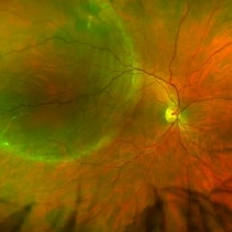
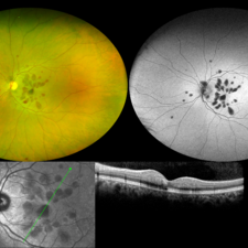
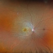
 Morning Glory Disc Anomaly
Feb 12 2024 by NIDHI PANWAR, MD FNB FICO
Fundus photograph of 43 year old male, hypertensive on medication, came for routine check up, and has been diagnosed to have poor vision left eye since childhood, denies any history of trauma. Vision left eye 6/18, Anterior segment normal, Fundus left eye shows excavated ,funnel-shaped optic nerve head, with central tuft of glial tissue obscuring the cup . The retinal vessels were seen emanating from the edge of disc in radial manner. In addition, the sectoral nasal retina shows localized area of hyperpigmented bony spicules like lesions. However, no history of nyctalopia or any other neurological disorder could be obtained.
Photographer: Nidhi Panwar, NMC Royal hospital, Sharjah , UAE
Imaging device: OPTOMAP
Condition/keywords: Morning Glory Anomaly, optic disc excavation
-
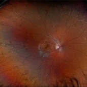
 Morning-Glory-Syndrome
Dec 22 2017 by James B. Soque, CRA, OCT-C, COA, FOPS
68-year-old WM with Morning Glory Syndrome with PVD OS with Staphyloma surrounding optic nerve and extending into the macula. Also, esotropia OS from V1 nerve paresis from birth, with amblyopia.
Photographer: James B Soque, CRA OCT-C COA FOPS
Imaging device: Optos Daytona
Condition/keywords: color photo, esotropia, fundus photograph, Optomap, Optos, peripheral vascular disease (PVD), posterior vitreous detachment, staphyloma, ultra-wide field imaging, wide angle imaging
-
 Retinal Detachment Right Eye Optomap
Mar 31 2014 by James B. Soque, CRA, OCT-C, COA, FOPS
36-year-old white male presented with non traumatic retinal detachment OD, with six very distinct demarcation lines and isolated tear, and detachment parameters. Patient underwent PPV OD on 12/3/13 with 20% SF6 gas placement and face down in his first 1 month post op period.
Photographer: James Soque, CRA, COA
Imaging device: Optos Daytona
Condition/keywords: Cryopexy, demarcation line, gas pneumatic displacement, Optomap, Optos, pars plana vitrectomy (PPV), retinal tear, scanning laser ophthalmoscope
-
 Serpiginous Choroidal Atrophy
Mar 29 2019 by Jessica Norkus
Optos ultra wide field color image of 20-year-old female presenting with serpiginous choroidal atrophy. Patient was unaware of vision loss OD, until accidentally covering OS and noticing the change. Acuity of 20/200 OD and 20/15 OS at time of imaging.
Photographer: Jessica Norkus
Imaging device: Optos Wide Field Camera
Condition/keywords: abnormal fundus, color fundus photograph, fundus photograph, macula serpiginous choroidopathy, Optomap, Optos, ultra-wide field imaging
-
 X-Linked Juvenile Retinoschisis
Dec 16 2016 by Young Hee Yoon, MD, PhD
Ultrawide field fundus photograph of a 27-year-old male with x-linked juvenile retinoschisis shows peripheral retinoshisis in his right eye
Photographer: Yoo Jin Jang, University of Ulsan, Asan Medical Center, Seoul, Korea
Imaging device: Optomap
Condition/keywords: juvenile retinoschisis
-
 X-Linked Juvenile Retinoschisis
Dec 16 2016 by Young Hee Yoon, MD, PhD
Ultrawide field fundus photograph of a 27-year-old male with x-linked juvenile retinoschisis shows peripheral retinoshisis in his left eye
Photographer: Yoo Jin Jang, University of Ulsan, Asan Medical Center, Seoul, Korea
Imaging device: Optomap
Condition/keywords: juvenile retinoschisis
-
 Central Retinal Vein Occlusion With Macular Edema
Feb 27 2025 by Kimberly Wakester
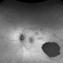
Optomap RGB image of a 34-year-old woman with central retinal vein occlusion with macular edema in the left eye. Patient has had a fairly acute onset central retinal vein occlusion in her left eye with dense superior IRH and macular edema. Modest ischemic changes are seen on exam and fundus photos. Patient was educated on the etiology of CRVOs and the relationship to systemic risk factors. Recommended hypercoagulable work-up with her PCP and bloodwork was ordered. Treatment with intravitreal injections was recommended to reduce the macular edema. Patient is to continue monthly follow ups with repeat OCT.
Photographer: Kimberly Wakester, COA
Imaging device: Optos California
Condition/keywords: CRVO with macular edema
-
 Central Retinal Vein Occlusion with Macular Edema
Feb 11 2025 by Kimberly Wakester
Optomap RGB image of an 61-year-old man with a central retinal vein occlusion with macular edema in the left eye. Will continue monthly follow up care with repeat OCT and treatment with intravitreal anti-VEGF injections as needed.
Photographer: Kimberly Wakester, COA
Imaging device: Optos California
Condition/keywords: CRVO with macular edema, DBH
-
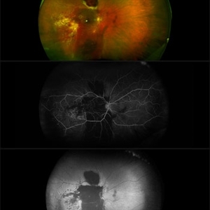 Coats' Disease
Nov 30 2018 by Darin R. Goldman, MD
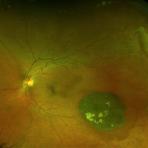
58-year-old male with a Coats’-like process in his right eye. The patient has undergone both laser photocoagulation and anti-VEGF therapy.
Photographer: Harold Rodriguez, CMA, Retina Group of Florida
Imaging device: Optomap color image, fluorescein angiogram, and fundus autofluorescence
Condition/keywords: Coats' disease, macroaneurysm, serous retinal detachment, subretinal hemorrhage
-
000---thumb.jpg/image-square;max$300,300.ImageHandler) Dropped IOL into the Vitreous Cavity
Dropped IOL into the Vitreous Cavity
Oct 7 2012 by Young Hee Yoon, MD, PhD
Fundus photograph of an 70-year-old man with a history of cataract operation 20 years ago. He visited our clinic with decreased visual acuity for 2 days.
Photographer: Yoon-hwa Kim, Asan Medical Center
Imaging device: Optomap, optos
Condition/keywords: intraocular lens dislocation
-
 Extensive Macular Atrophy with Pseudodrusen-Like Appearance
Dec 10 2020 by Cláudia Farinha
71-year-old male presented with progressive vision loss OD, now reduced to CF, without nyctalopia. The SD-OCT scans are one year apart and show extensive and progressive macular atrophy with marked disruption of the outer retinal layers and slightly larger vertical diameter, plus choroidal thinning. Reticular pseudodrusen-like deposits are heavily present in the posterior pole and are better depicted in the infra-red. The widefield imaging shows extensive paving stone peripheral degeneration. The patient denies any systemic medication or known disease. No family history of similar findings.
Photographer: Claudia Farinha
Imaging device: Optomap ultra-widefield imaging, Optos
Condition/keywords: macular atrophy
-
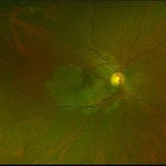
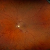
 Healthy Retina
Jul 6 2021 by Vipin Kumar
Optomap of healthy retina.
Photographer: Vipin Kumar
Imaging device: Optos Daytona
Condition/keywords: retina
-
 Henle Fiber Layer Hemorrhage
Jan 11 2021 by Cláudia Farinha
Henle fiber layer hemorrhage in a 56-year-old woman, who presented with a sudden decrease of visual acuity after sleeping. HBP, no other known systemic comorbidities.
Photographer: Claudia Farinha
Imaging device: Optomap, Optos
Condition/keywords: Henle fiber layer hemorrhage
-
 Hereditary Retinal Dystrophy
Feb 27 2025 by Kimberly Wakester
Optomap RGB image of a 7-year-old girl with Hereditary retinal dystrophy. Biological mother is a CHM gene carrier and biological father is diagnosed with RP. Patient had genetic testing and was also confirmed to be a CHM gene carrier and also has the TTC21B gene. There is linear pigmentary changes on clinical exam and fundus photos. Atypical appearance of Retinitis Pigmentosa. Patient will continue follow up care with repeat imaging.
Photographer: Kimberly Wakester, COA
Imaging device: Optos California
Condition/keywords: CHM gene, hereditary retinal dystrophy, linear pigmentary changes
-
 Macroaneurysm
Jun 23 2021 by Cláudia Farinha
Color Optomap from a middle-aged man with preretinal hemorrhage due to a macroaneurysm.
Photographer: Claudia Farinha, MD
Imaging device: Optomap, Optos
Condition/keywords: macroaneurysm, preretinal hemorrhage
-
 Multifocal Choroiditis and Panuveitis- Schlaegel lines
Nov 16 2021 by Manuel Ángel Alcántara Delgado, MD
Optomap ultra-widefield retinal imaging of an 52-year-old woman showed multiple punched-out chorioretinal lesions and 2 rows of peripheral curvilinear pigmented chorioretinal streaks (Schlaegel lines).
Photographer: Manuel Ángel Alcántara Delgado. Conde de Valenciana.
Condition/keywords: multifocal choroiditis, myopia, retina, uveitis
-
 Multifocal Pattern Dystrophy
Feb 5 2025 by Kimberly Wakester
Optomap RGB and AF photograph of an 37-year-old woman with multifocal pattern dystrophy in both eyes. Previously believed to be Stargardts, but genetic testing returned positive for PRPH2 mutation. Likely Multifocal Pattern Dystrophy given phenotypical appearance of SGD. There is stable NVE in the left eye. Will continue to monitor both eyes and consider treatment with PRP laser if needed for NVE in the left eye.
Photographer: Kimberly Wakester, COA
Imaging device: Optos California
Condition/keywords: multifocal pattern dystrophy, NVE, PRPH2 Positive
 Loading…