Search results (66 results)
-
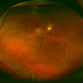
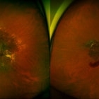
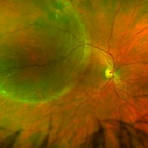 Retinal Detachment Right Eye Optomap
Mar 31 2014 by James B. Soque, CRA, OCT-C, COA, FOPS
36-year-old white male presented with non traumatic retinal detachment OD, with six very distinct demarcation lines and isolated tear, and detachment parameters. Patient underwent PPV OD on 12/3/13 with 20% SF6 gas placement and face down in his first 1 month post op period.
Photographer: James Soque, CRA, COA
Imaging device: Optos Daytona
Condition/keywords: Cryopexy, demarcation line, gas pneumatic displacement, Optomap, Optos, pars plana vitrectomy (PPV), retinal tear, scanning laser ophthalmoscope
-
000---thumb.jpg/image-square;max$300,300.ImageHandler) Dropped IOL into the Vitreous Cavity
Dropped IOL into the Vitreous Cavity
Oct 7 2012 by Young Hee Yoon, MD, PhD
Fundus photograph of an 70-year-old man with a history of cataract operation 20 years ago. He visited our clinic with decreased visual acuity for 2 days.
Photographer: Yoon-hwa Kim, Asan Medical Center
Imaging device: Optomap, optos
Condition/keywords: intraocular lens dislocation
-
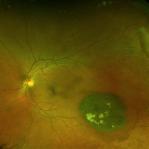
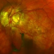
 Giant Retinal Tear
Mar 29 2014 by Min Kim, MD, PhD, MBA, FASRS
Wide field fundus photograph of a 25 year-old male shows giant retinal tear with inverted retinal flap.
Photographer: Young Duk Bae, Yonsei University, Gangnam Severance Hospital
Imaging device: Optomap
Condition/keywords: giant retinal tear
-
 Retinal Detachment with Proliferative Vitreoretinopathy
Mar 20 2014 by Min Kim, MD, PhD, MBA, FASRS
Wide field fundus photograph of a 59-year-old male with chronic total RD and PVR, with multiple retinal breaks that developed a few months after LASIK surgery.
Photographer: Young Duk Bae, Yonsei University, Gangnam Severance Hospital
Imaging device: Wide field fundus photography, Optomap
Condition/keywords: proliferative vitreoretinopathy (PVR), retinal detachment with retinal defect
-
 Congenital Hypertrophy of the Retinal Pigment Epithelium Wide Field Optomap
Sep 24 2019 by Sophia El Hamichi, MD
A 52-year-old female followed for 2 temporal lesions of CHRPE OD and white without pressure.
Photographer: Sophia El Hamichi,MD, Murray Ocular Oncology and Retina, Miami
Condition/keywords: congenital hypertrophy of the retinal pigment epithelium (CHRPE), Optomap, ultra-wide field imaging, white without pressure
-
 Multifocal Choroiditis and Panuveitis- Schlaegel lines
Nov 16 2021 by Manuel Ángel Alcántara Delgado, MD
Optomap ultra-widefield retinal imaging of an 52-year-old woman showed multiple punched-out chorioretinal lesions and 2 rows of peripheral curvilinear pigmented chorioretinal streaks (Schlaegel lines).
Photographer: Manuel Ángel Alcántara Delgado. Conde de Valenciana.
Condition/keywords: multifocal choroiditis, myopia, retina, uveitis
-
 Congenital Hypertrophy of the Retinal Pigment Epithelium Autofluorescence Optomap
Sep 24 2019 by Sophia El Hamichi, MD
A 52-year-old female followed for 2 temporal lesions of CHRPE OD and white without pressure.
Photographer: Sophia El Hamichi, MD, Murray Ocular Oncology and Retina, Miami
Condition/keywords: autofluorescence imaging, congenital hypertrophy of the retinal pigment epithelium (CHRPE), Optomap, ultra-wide field imaging, white without pressure
-
---thumb.jpg/image-square;max$300,300.ImageHandler) Ocular Lymphoma
Ocular Lymphoma
Jan 3 2014 by Young Hee Yoon, MD, PhD
Wide field fundus photograph of a 51-year-old female in a complete remission status after treatment for diffuse large B cell lymphoma (DLBCL) (Stage IV). Her best-corrected visual acuity was 20/80. Cytology for vitreous fluid revealed malignant lymphocytes, suggesting vitreoretinal relapse.
Photographer: Soo Hyun Cho, Asan Medical Center
Imaging device: Fundus photography using Optomap, optos
Condition/keywords: ocular lymphoma
-
 X-Linked Juvenile Retinoschisis
Dec 16 2016 by Young Hee Yoon, MD, PhD
Ultrawide field fundus photograph of a 27-year-old male with x-linked juvenile retinoschisis shows peripheral retinoshisis in his right eye
Photographer: Yoo Jin Jang, University of Ulsan, Asan Medical Center, Seoul, Korea
Imaging device: Optomap
Condition/keywords: juvenile retinoschisis
-
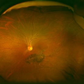
 Morning-Glory-Syndrome
Dec 22 2017 by James B. Soque, CRA, OCT-C, COA, FOPS
68-year-old WM with Morning Glory Syndrome with PVD OS with Staphyloma surrounding optic nerve and extending into the macula. Also, esotropia OS from V1 nerve paresis from birth, with amblyopia.
Photographer: James B Soque, CRA OCT-C COA FOPS
Imaging device: Optos Daytona
Condition/keywords: color photo, esotropia, fundus photograph, Optomap, Optos, peripheral vascular disease (PVD), posterior vitreous detachment, staphyloma, ultra-wide field imaging, wide angle imaging
-
-2000---thumb.jpg/image-square;max$300,300.ImageHandler) Retinoschisis in ROP
Retinoschisis in ROP
Jan 22 2013 by Young Hee Yoon, MD, PhD
Images of an 18-year-old male with a history of premature birth (30 weeks).
Photographer: Soohyun Cho, Asan Medical Center, Seoul, Korea
Imaging device: Fundus photography using Optomap, optos
Condition/keywords: retinoschisis
-
000---thumb.jpg/image-square;max$300,300.ImageHandler) Retinoschisis in ROP
Retinoschisis in ROP
Jan 22 2013 by Young Hee Yoon, MD, PhD
Images of an 18-year-old male with a history of premature birth (30 weeks).
Photographer: Soohyun Cho, Asan Medical Center, Seoul, Korea
Imaging device: Fluorescein angiography using Optomap, optos
Condition/keywords: retinoschisis
-
 Gyrate Atrophy
Sep 23 2020 by Hashim Ali Khan, OD, FAAO
Widefield color fundus image of a young male with gyrate atrophy.
Imaging device: Optomap
Condition/keywords: gyrate atrophy
-
 X-Linked Juvenile Retinoschisis
Dec 16 2016 by Young Hee Yoon, MD, PhD
Ultrawide field fundus photograph of a 27-year-old male with x-linked juvenile retinoschisis shows peripheral retinoshisis in his left eye
Photographer: Yoo Jin Jang, University of Ulsan, Asan Medical Center, Seoul, Korea
Imaging device: Optomap
Condition/keywords: juvenile retinoschisis
-
---thumb.jpg/image-square;max$300,300.ImageHandler) Choroidal Metastasis
Choroidal Metastasis
Jan 3 2014 by Young Hee Yoon, MD, PhD
Wide field fundus photograph of a 68-year-old male with a history of nonsmall cell lung cancer (Stage: T3 N3 M1). His best-corrected visual acuity was counting fingers at 50cm in the left eye.
Photographer: Soo Hyun Cho, Asan Medical Center
Imaging device: Fundus photography using Optomap, optos
Condition/keywords: choroidal metastasis
-
 Proliferative Diabetic Retinopathy
Aug 27 2013 by Carmen L Gonzalez, MD
Ultra-wide-field fundus photograph of a diabetic patient with a proliferative diabetic retinopathy.
Photographer: Regina Victoria, Denver Health Medical Center, Denver, Colorado
Imaging device: Optomap, Panoramic 200; Optos PLC, Scotland , UK
Condition/keywords: vitreous hemorrhage
-
 Pan-retinal Photocoagulation in a Myopic Eye
Aug 27 2013 by Carmen L Gonzalez, MD
Ultra-wide-field fundus photograph of a myopic patient.
Photographer: Regina Victoria, Denver Health Medical Center, Denver, Colorado
Imaging device: Optomap, Panoramic 200; Optos PLC, Scotland , UK
Condition/keywords: pan-retinal photocoagulation (PRP)
-
 Advanced Angioid Streak-Associated Choroidal Neovasclar Membranes
Dec 27 2016 by Young Hee Yoon, MD, PhD
UWF fundus photographs of an 74-year-old woman who received several anti-VEGF injections due to CNV associated with angioid streak in both eyes. Note diffuse scar change and pigmentary degenerations aroud disc in both eyes. There is some retinal hemorrhage due to CNV in her left eye.
Photographer: Young Hee Yoon, University of Ulsan, Asan Medical Center, Seoul, Korea
Imaging device: Optomap
Condition/keywords: angioid streaks, choroidal neovascularization (CNV)
-
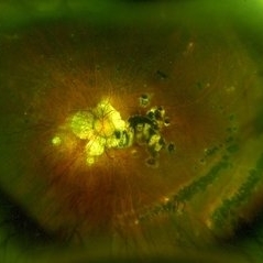
 Coats' Disease
Nov 30 2018 by Darin R. Goldman, MD
58-year-old male with a Coats’-like process in his right eye. The patient has undergone both laser photocoagulation and anti-VEGF therapy.
Photographer: Harold Rodriguez, CMA, Retina Group of Florida
Imaging device: Optomap color image, fluorescein angiogram, and fundus autofluorescence
Condition/keywords: Coats' disease, macroaneurysm, serous retinal detachment, subretinal hemorrhage
-
 Sunset Glow Fundus
May 15 2022 by Manuel Ángel Alcántara Delgado, MD
Optomap ultra-widefield retinal imaging of an 35-year-old woman showed sunset glow fundus, multiple nummular chorioretinal atrophic lesions, macular subretinal fibrosis and pigment clumping in chronic recurrent stage of Vogt-Koyanagi-Harada disease.
Photographer: Manuel Ángel Alcántara Delgado. Conde de Valenciana.
Condition/keywords: abnormal retina, benign pigmented lesions, pigment clumps, retinal fibrosis, uveitis, Vogt-Koyanagi-Harada
-
---thumb.jpg/image-square;max$300,300.ImageHandler) Choroidal Metastasis
Choroidal Metastasis
Jan 3 2014 by Young Hee Yoon, MD, PhD
Wide field fundus photograph of a 68-year-old male with a history of nonsmall cell lung cancer (Stage: T3 N3 M1). His best-corrected visual acuity remained at 20/20 in the right eye.
Photographer: Soo Hyun Cho, Asan Medical Center
Imaging device: Fundus photography using Optomap, optos
Condition/keywords: choroidal metastasis
-
---thumb.jpg/image-square;max$300,300.ImageHandler) Choroidal Metastasis
Choroidal Metastasis
Jan 3 2014 by Young Hee Yoon, MD, PhD
Wide field fluorescein angiography (FA) image of a 68-year-old male with a history of nonsmall cell lung cancer (Stage: T3 N3 M1). His best-corrected visual acuity remained at 20/20 in the right eye.
Photographer: Soo Hyun Cho, Asan Medical Center
Imaging device: Fluorescein angiography using Optomap, optos
Condition/keywords: choroidal metastasis
-
 Serpiginous Choroidal Atrophy
Mar 29 2019 by Jessica Norkus
Optos ultra wide field color image of 20-year-old female presenting with serpiginous choroidal atrophy. Patient was unaware of vision loss OD, until accidentally covering OS and noticing the change. Acuity of 20/200 OD and 20/15 OS at time of imaging.
Photographer: Jessica Norkus
Imaging device: Optos Wide Field Camera
Condition/keywords: abnormal fundus, color fundus photograph, fundus photograph, macula serpiginous choroidopathy, Optomap, Optos, ultra-wide field imaging
-
 Retinal Tear
Sep 4 2025 by Kimberly Wakester
Optomap RBG of a 55-year-old woman with a retinal tear at 12 with bridging vessel and some fluid. Treatment with prophylaxis laser was recommended. Patient is to continue follow up care post operatively.
Photographer: Kimberly Wakester, COA, OCT-C
Imaging device: Optos California
Condition/keywords: left eye, PVD, Retinal tear
-
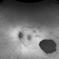
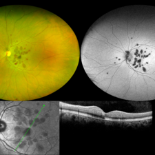 Henle Fiber Layer Hemorrhage
Jan 11 2021 by Cláudia Farinha
Henle fiber layer hemorrhage in a 56-year-old woman, who presented with a sudden decrease of visual acuity after sleeping. HBP, no other known systemic comorbidities.
Photographer: Claudia Farinha
Imaging device: Optomap, Optos
Condition/keywords: Henle fiber layer hemorrhage
 Loading…