Search results (66 results)
-
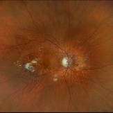
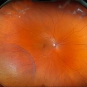
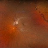
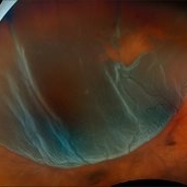 Retinal Detachment with Multiple Breaks
Mar 5 2025 by Kimberly Wakester
Optomap RGB image of an 44-year-old man with a retinal detachment with a complex lattice break in the right eye. Surgery was recommended. Patient is to continue follow up care post operatively.
Photographer: Kimberly Wakester, COA
Imaging device: Optos California
Condition/keywords: Retinal Detachment, retinal tear
-
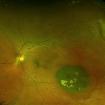
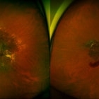
 Sunset Glow Fundus
May 15 2022 by Manuel Ángel Alcántara Delgado, MD
Optomap ultra-widefield retinal imaging of an 35-year-old woman showed sunset glow fundus, multiple nummular chorioretinal atrophic lesions, macular subretinal fibrosis and pigment clumping in chronic recurrent stage of Vogt-Koyanagi-Harada disease.
Photographer: Manuel Ángel Alcántara Delgado. Conde de Valenciana.
Condition/keywords: abnormal retina, benign pigmented lesions, pigment clumps, retinal fibrosis, uveitis, Vogt-Koyanagi-Harada
-
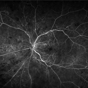 Central Retinal Vein Occlusion With Waldenstroms macroglobulinemia
Jun 18 2025 by Korey Starkey
64-year-old patient presents with CRVO with secondary macular edema in both eyes. Venous beading present in 2/4 quadrants OU. Patient diagnosed with Waldenstroms macroglobulinemia, found on SPEP and bone marrow biopsy. Treatment recommended of anti-vegF intravitreal injections OU.
Photographer: Korey Starkey
Imaging device: Optos
Condition/keywords: attenuated vessels, central retinal vein occlusion (CRVO), CRVO, FA early phase, FLUORESCEIN ANGIOGRAPHY, macular edema, Optomap, OPTOS CALIFORNIA, severe NPDR, venous beading, Waldenstroms macroglobulinemia
-
 Henle Fiber Layer Hemorrhage
Jan 11 2021 by Cláudia Farinha
Henle fiber layer hemorrhage in a 56-year-old woman, who presented with a sudden decrease of visual acuity after sleeping. HBP, no other known systemic comorbidities.
Photographer: Claudia Farinha
Imaging device: Optomap, Optos
Condition/keywords: Henle fiber layer hemorrhage
-
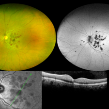
 Multifocal Pattern Dystrophy
Feb 5 2025 by Kimberly Wakester
Optomap RGB and AF photograph of an 37-year-old woman with multifocal pattern dystrophy in both eyes. Previously believed to be Stargardts, but genetic testing returned positive for PRPH2 mutation. Likely Multifocal Pattern Dystrophy given phenotypical appearance of SGD. There is stable NVE in the left eye. Will continue to monitor both eyes and consider treatment with PRP laser if needed for NVE in the left eye.
Photographer: Kimberly Wakester, COA
Imaging device: Optos California
Condition/keywords: multifocal pattern dystrophy, NVE, PRPH2 Positive
-
 Neovascular AMD with Active CNV
May 22 2025 by Kimberly Wakester
Optomap RGB of an 82-year-old man with Neovascular AMD with Active CNV and Dry AMD in the right eye. There is advanced atrophic changes without subfoveal involvement located temporally to the fovea. Patient is to continue follow up care with dilated exam, repeat OCT, and treatment of intravitreal injection of Vabysmo every 5 weeks at this time.
Photographer: Kimberly Wakester, COA, OCT-C
Imaging device: Optos California
Condition/keywords: advanced geographic atrophy, dry age-related macular degeneration (dry AMD), neovascular age-related macular degeneration (AMD)
-
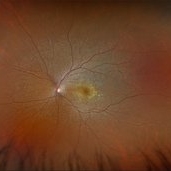
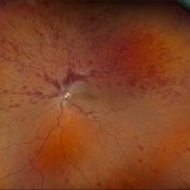
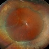 Repaired Retinal Detachment
Jun 24 2025 by Kimberly Wakester
Optomap RGB of an 45-year-old woman with a repaired retinal detachment in the right eye. The operative eye is doing well three-month s/p surgery. Retina is attached 360 on SB. There is resolving residual SRF at 6:00. Discussed the possible need for added laser. Will continue to observe and will return in 3 months for follow up exam.
Photographer: Kimberly Wakester, COA, OCT-C
Imaging device: Optos California
Condition/keywords: repaired RD, scleral buckle
-
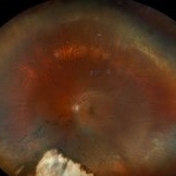 Repaired Retinal Detachment
May 7 2025 by Kimberly Wakester
Optomap RGB montage of an 56-year-old woman with a repaired retinal detachment with scleral buckle and cryotherapy in the left eye. Patient remains stable s/p Vitreo-retinal surgery in 2007. Patient is to return in 1 year for follow up exam with repeat imaging.
Photographer: Kimberly Wakester, COA, OCT-C
Imaging device: Optos California
Condition/keywords: cryotherapy, repaired RD, scleral buckle
-
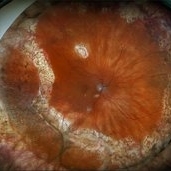
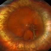 Repaired Retinal Detachment with Scleral Buckle
Mar 25 2025 by Kimberly Wakester
Optomap RGB montage of an 64-year-old woman with a repaired retinal detachment with scleral buckle in the right eye. There is nasal and inferior pre-retinal membranes with traction. PPV was recommended but patient defers to proceed with sx at this time. Will continue to follow patient closely for worsening traction. Patient was educated on how to monitor their peripheral vision and was advised to report any changes immediately.
Photographer: Kimberly Wakester, COA, OCT-C
Imaging device: Optos California
Condition/keywords: pre-retinal membrane with traction, repaired RD, scleral buckle
-
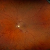
 Retinoschisis
Feb 26 2025 by Kimberly Wakester
Optomap RGB of a 56-year-old woman with bullous retinoschisis in the right eye. The patient remains stable with very mild progression. Patient is to continue follow up care at 6 month intervals to monitor for worsening progression.
Photographer: Kimberly Wakester, COA
Imaging device: Optos California
Condition/keywords: bullous retinoschisis
-
000---thumb.jpg/image-square;max$300,300.ImageHandler) Retinoschisis in ROP
Retinoschisis in ROP
Jan 22 2013 by Young Hee Yoon, MD, PhD
Images of an 18-year-old male with a history of premature birth (30 weeks).
Photographer: Soohyun Cho, Asan Medical Center, Seoul, Korea
Imaging device: Fluorescein angiography using Optomap, optos
Condition/keywords: retinoschisis
-
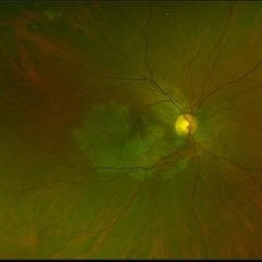
 Gyrate Atrophy
Sep 23 2020 by Hashim Ali Khan, OD, FAAO
Widefield color fundus image of a young male with gyrate atrophy.
Imaging device: Optomap
Condition/keywords: gyrate atrophy
-
 Congenital Hypertrophy of the Retinal Pigment Epithelium Wide Field Optomap
Sep 24 2019 by Sophia El Hamichi, MD
A 52-year-old female followed for 2 temporal lesions of CHRPE OD and white without pressure.
Photographer: Sophia El Hamichi,MD, Murray Ocular Oncology and Retina, Miami
Condition/keywords: congenital hypertrophy of the retinal pigment epithelium (CHRPE), Optomap, ultra-wide field imaging, white without pressure
-
000---thumb.jpg/image-square;max$300,300.ImageHandler) Dropped IOL into the Vitreous Cavity
Dropped IOL into the Vitreous Cavity
Oct 7 2012 by Young Hee Yoon, MD, PhD
Fundus photograph of an 70-year-old man with a history of cataract operation 20 years ago. He visited our clinic with decreased visual acuity for 2 days.
Photographer: Yoon-hwa Kim, Asan Medical Center
Imaging device: Optomap, optos
Condition/keywords: intraocular lens dislocation
-
-2000---thumb.jpg/image-square;max$300,300.ImageHandler) Retinoschisis in ROP
Retinoschisis in ROP
Jan 22 2013 by Young Hee Yoon, MD, PhD
Images of an 18-year-old male with a history of premature birth (30 weeks).
Photographer: Soohyun Cho, Asan Medical Center, Seoul, Korea
Imaging device: Fundus photography using Optomap, optos
Condition/keywords: retinoschisis
-
 Serpiginous Choroidal Atrophy
Mar 29 2019 by Jessica Norkus
Optos ultra wide field color image of 20-year-old female presenting with serpiginous choroidal atrophy. Patient was unaware of vision loss OD, until accidentally covering OS and noticing the change. Acuity of 20/200 OD and 20/15 OS at time of imaging.
Photographer: Jessica Norkus
Imaging device: Optos Wide Field Camera
Condition/keywords: abnormal fundus, color fundus photograph, fundus photograph, macula serpiginous choroidopathy, Optomap, Optos, ultra-wide field imaging
-
 Repaired Retinal Detachment with PVR
Mar 25 2025 by Kimberly Wakester
Optomap RGB of a 79-year-old-woman with a repaired retinal detachment with PVR in the right eye. Patient is doing well over 7 months s/p vitrectomy with silicone oil and scleral buckle placement. Retina remains attached on the buckle under oil. Patient is to return in 6 months for follow up exam with repeat imaging.
Photographer: Kimberly Wakester, COA, OCT-C
Imaging device: Optos California
Condition/keywords: PVR, repaired RD, Retinal detachment under Silicon Oil, scleral buckle
-
 Retinoschisis with Outer Layer Holes
Jul 18 2025 by Kimberly Wakester
Optomap RGB of an 56-year-old woman with retinoschisis with outer layer holes s/p laser in the left eye. Patient remains stable. Will continue follow up care with dilated exam and optos imaging.
Photographer: Kimberly Wakester, COA, OCT-C
Imaging device: Optos California
Condition/keywords: outer layer hole, retinoschisis
-
 Advanced Angioid Streak-Associated Choroidal Neovasclar Membranes
Dec 27 2016 by Young Hee Yoon, MD, PhD
UWF fundus photographs of an 74-year-old woman who received several anti-VEGF injections due to CNV associated with angioid streak in both eyes. Note diffuse scar change and pigmentary degenerations aroud disc in both eyes. There is some retinal hemorrhage due to CNV in her left eye.
Photographer: Young Hee Yoon, University of Ulsan, Asan Medical Center, Seoul, Korea
Imaging device: Optomap
Condition/keywords: angioid streaks, choroidal neovascularization (CNV)
-
 Central Retinal Vein Occlusion With Macular Edema
Feb 27 2025 by Kimberly Wakester
Optomap RGB image of a 34-year-old woman with central retinal vein occlusion with macular edema in the left eye. Patient has had a fairly acute onset central retinal vein occlusion in her left eye with dense superior IRH and macular edema. Modest ischemic changes are seen on exam and fundus photos. Patient was educated on the etiology of CRVOs and the relationship to systemic risk factors. Recommended hypercoagulable work-up with her PCP and bloodwork was ordered. Treatment with intravitreal injections was recommended to reduce the macular edema. Patient is to continue monthly follow ups with repeat OCT.
Photographer: Kimberly Wakester, COA
Imaging device: Optos California
Condition/keywords: CRVO with macular edema
-
 Central Retinal Vein Occlusion with Macular Edema
Feb 11 2025 by Kimberly Wakester
Optomap RGB image of an 61-year-old man with a central retinal vein occlusion with macular edema in the left eye. Will continue monthly follow up care with repeat OCT and treatment with intravitreal anti-VEGF injections as needed.
Photographer: Kimberly Wakester, COA
Imaging device: Optos California
Condition/keywords: CRVO with macular edema, DBH
-
---thumb.jpg/image-square;max$300,300.ImageHandler) Choroidal Metastasis
Choroidal Metastasis
Jan 3 2014 by Young Hee Yoon, MD, PhD
Wide field fundus photograph of a 68-year-old male with a history of nonsmall cell lung cancer (Stage: T3 N3 M1). His best-corrected visual acuity was counting fingers at 50cm in the left eye.
Photographer: Soo Hyun Cho, Asan Medical Center
Imaging device: Fundus photography using Optomap, optos
Condition/keywords: choroidal metastasis
-
---thumb.jpg/image-square;max$300,300.ImageHandler) Choroidal Metastasis
Choroidal Metastasis
Jan 3 2014 by Young Hee Yoon, MD, PhD
Wide field fluorescein angiography (FA) image of a 68-year-old male with a history of nonsmall cell lung cancer (Stage: T3 N3 M1). His best-corrected visual acuity remained at 20/20 in the right eye.
Photographer: Soo Hyun Cho, Asan Medical Center
Imaging device: Fluorescein angiography using Optomap, optos
Condition/keywords: choroidal metastasis
-
---thumb.jpg/image-square;max$300,300.ImageHandler) Choroidal Metastasis
Choroidal Metastasis
Jan 3 2014 by Young Hee Yoon, MD, PhD
Wide field fundus photograph of a 68-year-old male with a history of nonsmall cell lung cancer (Stage: T3 N3 M1). His best-corrected visual acuity remained at 20/20 in the right eye.
Photographer: Soo Hyun Cho, Asan Medical Center
Imaging device: Fundus photography using Optomap, optos
Condition/keywords: choroidal metastasis
-
---thumb.jpg/image-square;max$300,300.ImageHandler) Choroidal Metastasis
Choroidal Metastasis
Jan 3 2014 by Young Hee Yoon, MD, PhD
Wide field fluorescein angiography (FA) image of a 68-year-old male with a history of nonsmall cell lung cancer (Stage: T3 N3 M1). His best-corrected visual acuity was counting fingers at 50cm in the left eye.
Photographer: Soo Hyun Cho, Asan Medical Center
Imaging device: Fluorescein angiography using Optomap, optos
Condition/keywords: choroidal metastasis
 Loading…