Search results (6 results)
-
 Retinoschisis
Feb 26 2025 by Kimberly Wakester
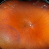
Optomap RGB of a 56-year-old woman with bullous retinoschisis in the right eye. The patient remains stable with very mild progression. Patient is to continue follow up care at 6 month intervals to monitor for worsening progression.
Photographer: Kimberly Wakester, COA
Imaging device: Optos California
Condition/keywords: bullous retinoschisis
-
 Repaired Retinal Detachment with Scleral Buckle
Mar 25 2025 by Kimberly Wakester
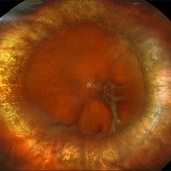
Optomap RGB montage of an 64-year-old woman with a repaired retinal detachment with scleral buckle in the right eye. There is nasal and inferior pre-retinal membranes with traction. PPV was recommended but patient defers to proceed with sx at this time. Will continue to follow patient closely for worsening traction. Patient was educated on how to monitor their peripheral vision and was advised to report any changes immediately.
Photographer: Kimberly Wakester, COA, OCT-C
Imaging device: Optos California
Condition/keywords: pre-retinal membrane with traction, repaired RD, scleral buckle
-
 Multifocal Choroiditis and Panuveitis- Schlaegel lines
Nov 16 2021 by Manuel Ángel Alcántara Delgado, MD
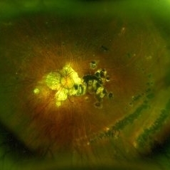
Optomap ultra-widefield retinal imaging of an 52-year-old woman showed multiple punched-out chorioretinal lesions and 2 rows of peripheral curvilinear pigmented chorioretinal streaks (Schlaegel lines).
Photographer: Manuel Ángel Alcántara Delgado. Conde de Valenciana.
Condition/keywords: multifocal choroiditis, myopia, retina, uveitis
-
 Gyrate Atrophy
Sep 23 2020 by Hashim Ali Khan, OD, FAAO
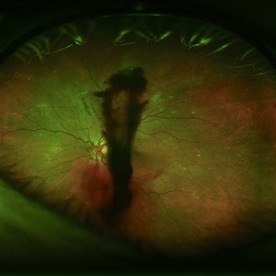
Widefield color fundus image of a young male with gyrate atrophy.
Imaging device: Optomap
Condition/keywords: gyrate atrophy
-
---thumb.jpg/image-square;max$300,300.ImageHandler) Choroidal Metastasis
Choroidal Metastasis
Jan 3 2014 by Young Hee Yoon, MD, PhD
Wide field fundus photograph of a 68-year-old male with a history of nonsmall cell lung cancer (Stage: T3 N3 M1). His best-corrected visual acuity was counting fingers at 50cm in the left eye.
Photographer: Soo Hyun Cho, Asan Medical Center
Imaging device: Fundus photography using Optomap, optos
Condition/keywords: choroidal metastasis
-
 Proliferative Diabetic Retinopathy
Aug 27 2013 by Carmen L Gonzalez, MD
Ultra-wide-field fundus photograph of a diabetic patient with a proliferative diabetic retinopathy.
Photographer: Regina Victoria, Denver Health Medical Center, Denver, Colorado
Imaging device: Optomap, Panoramic 200; Optos PLC, Scotland , UK
Condition/keywords: vitreous hemorrhage
 Loading…