 Initializing download.
-
 By Gary R. Cook, MD, FACS
- Uploaded on Mar 27, 2019.
- Last modified by Caroline Bozell on Aug 2, 2019.
- Rating
- Appears in
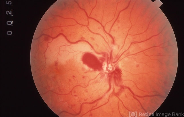
- COMBINED BRAO AND CRVO
- Condition/keywords
- central retinal vein occlusion (CRVO), branch retinal artery occlusion (BRAO)
- Imaging device
-
Fundus camera
Topcon VT-50 - Description
- Right eye of a 56-year-old white male with a combined perfused CRVO (venous dilation and dot & blot hemorrhages in all 4 quadrants) and a superotemporal BRAO with peripapillary hemorrhages and cotton wool spots, and an area of retinal whitening inside of the ST arcade. V.A.= 20/70.


