Search results (195 results)
-
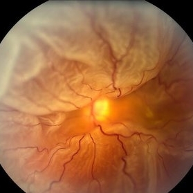
 Total Rhegmatogenous Retinal Detachment With Severe PVR
May 27 2015 by Darin R. Goldman, MD
63-year-old pseudophakic male with hand motion vision in the left eye due to a total retinal detachment with severe proliferative vitreoretinopathy.
Condition/keywords: proliferative vitreoretinopathy (PVR), retinal tear
-
 360 Degree Retinal Detachment
Jun 29 2013 by Jason S. Calhoun
Total retinal detachment in the left eye.
Photographer: Jason S. Calhoun, Mayo Clinic Jacksonville, Florida
Imaging device: TOPCON TRC 50-EX
-
 Traumatic Macular Hole with Retinal Detachment and PVR
Sep 27 2012 by Pauline T Merrill, MD, FASRS
Fundus photo of a 13-year-old boy s/p soccer ball injury 1 month previously. In addition to full-thickness macular hole and total retinal detachment with grade C PVR, note pigment granules visible in vitreous over optic nerve.
Photographer: Karen Parque, Illinois Retina Associates, Chicago, IL
Condition/keywords: proliferative vitreoretinopathy (PVR), traumatic macular hole
-
 PVR Retinal Detachment following Laser Retinopexy Slide 1
Oct 22 2012 by Ronald C. Gentile, MD
Acute onset total retinal detachment with PVR 10 weeks following laser retinopexy.
Photographer: The New York Eye & Ear Infirmary Department of Medical Imaging
Condition/keywords: laser retinopexy, proliferative vitreoretinopathy (PVR)
-
---thumb.jpg/image-square;max$300,300.ImageHandler) Sturge-Weber Diffuse Hemangioma and Retinal Detachment on B-scan
Sturge-Weber Diffuse Hemangioma and Retinal Detachment on B-scan
Apr 18 2014 by Susanna S. Park, MD, PhD
B-scan ultrasonogram of the right eye of an 8 year old Hispanic boy with Sturge -Weber Syndrome showing diffuse choroidal thickening from diffuse choroidal hemangioma and associated total exudative retinal detachment.
Photographer: Ellen Redenbo, University of California Davis Eye Center
Condition/keywords: B scan ultrasound, diffuse choroidal hemangioma, Sturge-Weber syndrome
-
 Hyphema
-
 Massive Subretinal Hemorrhage With Near Total Retina Detachment
Nov 27 2013 by David W. Faber, MD
Fundus photo of an 71-year-old male with massive subretinal hemorrhage. Had been given 6 week Avastin treatments. Was put on coumadin for 6 weeks following knee surgery.
Photographer: Donna Knight, Rocky Mountain Retina Consultants, Salt Lake City, Utah
-
 Venous Stasis Retinopathy
Feb 20 2015 by H. Michael Lambert, MD
Scattered hemorrhages associated with venous stasis retinopathy in eye with ipsilateral total internal carotid artery occlusion. Vision is 20/40.
Condition/keywords: arterial occlusion, venous stasis retinopathy
-
 PVR Retinal Detachment with subretinal bands Slide 1
Oct 22 2012 by Ronald C. Gentile, MD
Total retinal detachment with pre-retinal and sub-retinal proliferation. The subretinal bands have a napkin ring configuration posteriorly with the macula folded and dragged above the optic nerve.
Photographer: The New York Eye & Ear Infirmary Department of Medical Imaging
Condition/keywords: proliferative vitreoretinopathy (PVR), subretinal bands
-
 Chronic Total Retinal Detachment
Oct 12 2012 by Jeffrey G. Gross, MD, FASRS
Chronic, total RD, with shifting inferior subretinal fluid.
Condition/keywords: chronic, inferior subretinal fluid
-
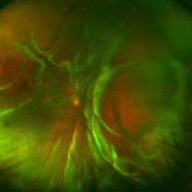
 Retinal Detachment with Proliferative Vitreoretinopathy
Mar 20 2014 by Min Kim, MD, PhD, MBA, FASRS
Wide field fundus photograph of a 59-year-old male with chronic total RD and PVR, with multiple retinal breaks that developed a few months after LASIK surgery.
Photographer: Young Duk Bae, Yonsei University, Gangnam Severance Hospital
Imaging device: Wide field fundus photography, Optomap
Condition/keywords: proliferative vitreoretinopathy (PVR), retinal detachment with retinal defect
-
 Funnel Retinal Detachment
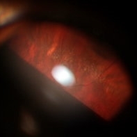
Feb 2 2015 by Matt Poe, COA
The patient presented with total vision loss for >2months. Patient had history of exudative ARMD with intravitreal injections. No surgical intervention was done due to the long standing detachment and patient health.
Photographer: Matt Poe, COA. Northwest Arkansas Retina Associates, Springdale, AR.
Condition/keywords: retinal defect
-
 CNV due to AMPPE
Oct 16 2012 by Ratimir Lazic, MD, PhD
OCT image of 58-year- old male. Total resolution of fluid one and a half month after treatment can be seen. The patient was treated with intravitreal bevacizumab.
Photographer: Marko Lukic, MD
Imaging device: OCT Copernicus
Condition/keywords: acute posterior multifocal placoid pigment epitheliopathy (APMPPE), anti-VEGF, choroidal neovascularization (CNV)
-
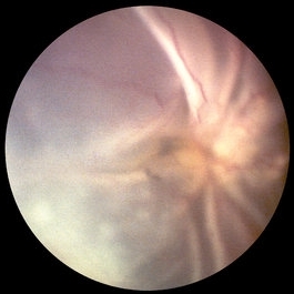
 Funnel Retinal Detachment With Proliferative Vitreoretinopathy
Oct 2 2013 by Jerald A. Bovino, MD
There is a total retinal detachment. The proliferative vitreoretinopathy has caused the retina to assume a funnel shape.
Condition/keywords: funnel, proliferative vitreoretinopathy (PVR), re-attached retinal detachment (RRD)
-
---thumb.jpg/image-square;max$300,300.ImageHandler) Retina Flower 1
Retina Flower 1
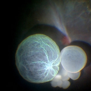
Mar 22 2013 by Cesare Forlini, MD
Post-traumatic Total Choroidal and Retinal Detachment: "like a rose on the rock."
Photographer: Matteo Forlini MD, University of Modena, Institute of Ophthalmology, Modena, Italy
Condition/keywords: choroidal detachment
-
 Retinal Tear in Aphakic Fellow Eye
Nov 9 2012 by Norman Byer
This 59-year-old man presented with sudden symptoms of retinal detachment in his opposite aphakic eye secondary to a tiny retinal tear about 1/8th disc diameter in size. During the examination, the fellow eye shown here was found to have this much larger tractional tear approximately 2 disc diameters in total length. If you look carefully, you will see that this is really a series of three separate tears with a common flap. The tears are separated by tiny bridges of remaining tissue which cause the edges of the apparent large tear to be serrated. This was also an aphakic eye with a posterior vitreous detachment but the lesion had produced no symptoms.
Condition/keywords: asymptomatic, bridge of tissue between tears, posterior vitreous detachment, tractional retinal tear
-
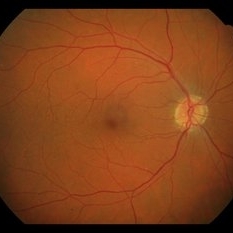 Example of AREDS Category 1 (Small Drusen But Not Considered AMD)
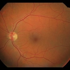
Feb 11 2013 by Neil M. Bressler, MD
Person in AREDS Category 1 were essentially free of age-related macular abnormalities, with a total drusen area less than 5 small drusen (<63 microns) within 3,000 microns of the center of the macula, and visual acuity of 20/32 or better in both eyes1. These are fundus photographs of a 53-year-old man, with visual acuity 20/20 OD and 20/32 OS presenting for evaluation of any diabetic retinopathy. Reference: 1 Age-Related Eye Disease Study Research Group. A randomized, placebo controlled clinical trial of high-dose supplementation with vitamins C and E, beta carotene, and zinc for age-related macular degeneration and vision loss: AREDS report No. 8. Arch Ophthalmol. 2001;119(10):1417-1436.
Condition/keywords: age-related macular degeneration (AMD)
-
 Retrohyaloid Hemorrhage
May 24 2016 by Hazem Alaskar, MD, FEBO
35-year-old pregnant woman with spontaneous retrohyaloid premacular hemorrhage. Treated by observation and total resolution without any treatment.
Photographer: Hazem Alaskar, Hospital de Poniente de Almeria, SPAIN
Imaging device: Topcon
Condition/keywords: premacular hemorrhage, retrohyaloid hemorrhage
-
 Intraocular Multiple Cysticercus
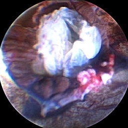
Oct 10 2018 by Vishal Agrawal, MD, FRCS,FACS,FASRS
Intraoperative fundus picture of right eye of a 18-year-old boy with complaints of DOV for the past 2 months. There were 12 intravitreal cysts in total with vitritis sclerosis retinal vessels and TRD. To note here, the largest cyst has a flimsy wall and no scolex (possibly ruptured) and the rest of the smaller cysts have a scolex and a taut wall.
Photographer: Vishal Agrawal MD,FRCS
Imaging device: SONY PMW-10 MD HD
Condition/keywords: cysticercosis, scolex
-
 Snowflake Vitreoretinal Degeneration
Nov 29 2018 by Hashim Ali Khan, OD, FAAO
Peripheral snowflake in a 16-year-old female. The fellow eye had chronic total retinal detachment.
Imaging device: Goldman triple mirror lens
Condition/keywords: peripheral fundus lesion, snowflake hereditary degeneration
-
 Retinal Detachment
Nov 9 2012 by Norman Byer
This 18-year-old girl gave the history of having been hit in this eye three years before with a fist and of having retinal surgery nine months previously, which was temporarily successful. When the photograph was taken, she had a total left retinal detachment with a small nasal dialysis which had not been treated. She also had two prominent intraretinal cysts, one of which is shown here. The retina promptly reattached following further surgery and the next slide shows an interesting change in this cyst.
Condition/keywords: intraretinal cyst, small nasal dialysis
-
 Funnel Retinal Detachment With Proliferative Vitreoretinopathy
Oct 2 2013 by Jerald A. Bovino, MD
There is a total retinal detachment. The proliferative vitreoretinopathy has caused the retina to assume a funnel shape.
Condition/keywords: funnel, proliferative vitreoretinopathy (PVR), re-attached retinal detachment (RRD)
-
 Hyphema, Total
-
 Example of AREDS Category 1 (Small Drusen But Not Considered AMD)
Feb 11 2013 by Neil M. Bressler, MD
Person in AREDS Category 1 were essentially free of age-related macular abnormalities, with a total drusen area less than 5 small drusen (<63 microns) within 3,000 microns of the center of the macula, and visual acuity of 20/32 or better in both eyes1. These are fundus photographs of a 53-year-old man, with visual acuity 20/20 OD and 20/32 OS presenting for evaluation of any diabetic retinopathy. Reference: 1 Age-Related Eye Disease Study Research Group. A randomized, placebo controlled clinical trial of high-dose supplementation with vitamins C and E, beta carotene, and zinc for age-related macular degeneration and vision loss: AREDS report No. 8. Arch Ophthalmol. 2001;119(10):1417-1436.
Condition/keywords: age-related macular degeneration (AMD)
-
 Retina Flower 3
Mar 22 2013 by Cesare Forlini, MD
Post-traumatic Total Choroidal and Retinal Detachment: "like a rose on the rock."
Photographer: Matteo Forlini MD, University of Modena, Institute of Ophthalmology, Modena, Italy
Condition/keywords: choroidal detachment
 Loading…