Search results (195 results)
-
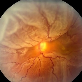
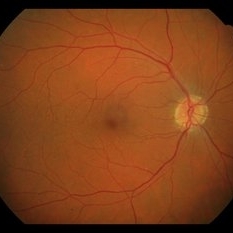
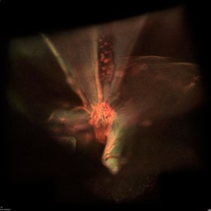
 Total Rhegmatogenous Retinal Detachment With Severe PVR
May 27 2015 by Darin R. Goldman, MD
63-year-old pseudophakic male with hand motion vision in the left eye due to a total retinal detachment with severe proliferative vitreoretinopathy.
Condition/keywords: proliferative vitreoretinopathy (PVR), retinal tear
-
 Massive Subretinal Hemorrhage With Near Total Retina Detachment
Nov 27 2013 by David W. Faber, MD
Fundus photo of an 71-year-old male with massive subretinal hemorrhage. Had been given 6 week Avastin treatments. Was put on coumadin for 6 weeks following knee surgery.
Photographer: Donna Knight, Rocky Mountain Retina Consultants, Salt Lake City, Utah
-
 Giant Retinal Tear
Feb 20 2024 by Soobien Lee
Optos color fundus photograph of a 40-year-old caucasian male who is a UFC fighter with a total retinal detachment in his right eye secondary to a giant retinal tear from 10 o'clock to 2 o'clock.
Photographer: Trinity Wolf, Elman Retina Group
Imaging device: Optos Ultra-Widefield Imaging
Condition/keywords: giant retinal tear, optos, Retinal Detachment, Retinal tear with detachment, trauma
-
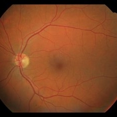
 360 Degree Retinal Detachment
Jun 29 2013 by Jason S. Calhoun
Total retinal detachment in the left eye.
Photographer: Jason S. Calhoun, Mayo Clinic Jacksonville, Florida
Imaging device: TOPCON TRC 50-EX
-
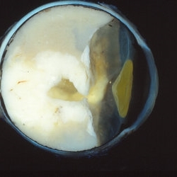
 Choroidal Melanoma
Nov 3 2022 by pedro fernandes souza neto
Transillumination of Enucleation specimen of Choroidal Melanoma: anterior chamber is closed. Total secondary retinal detachment with subretinal serous fluid and some subretinal hemorrhages are present.
Photographer: Eduardo Marback, Federal University of Bahia, Brazil
Condition/keywords: enucleation, melanoma
-
 Spontaneously Dropped Lens in a Congenital Rubella Syndrome
Apr 30 2022 by NEIFFER RABELO
Intraoperative photograph of a 68-year-old patient with congenital rubella syndrome and light perception visual acuity since childhood. The image shows a pigmentary retinopathy and the lens spontaneously displaced into the vitreous cavity. The patient sought care complaining of a total and sporadic loss of vision that was hindering her in daily tasks. Surgery was indicated to remove the lens.
Photographer: Rodrigo Dos Anjos Versiani - Retina Institute - Belo Horizonte - Brazil
Imaging device: ZEISS OPMI LUMERA 700
Condition/keywords: dropped nucleus, retina surgery, rubella retinopathy
-
 Horseshoe Retinal Break
Apr 3 2018 by Wesam Safwat
Fundus photograph of an 40-year-old woman with lower temporal horseshoe retinal tear associated with lower sub total retinal detachment not involving macula.
Photographer: Wesam Safwat, Elferdaws eye hospital , Zagazig, Egypt.
Imaging device: Topcon
-
 Coats' disease
Sep 7 2022 by Niloofar Piri, MD
Total exudative RD with extensive subretinal exudates and peripheral telangiectatic vascular anomalies in stage 4 Coats's disease. Patient is a 12 yo who presented with severe eye pain and neovascular glaucoma secondary to the above.
Photographer: Jacob Grodsky, MD
Condition/keywords: Coats' disease
-
 Dislocated Intraocular Lens (IOL)
Aug 2 2019 by JEFFERSON R SOUSA, Tecg.º (Biomedical Systems Technology)
A 53-year-old male patient suffered blunt trauma 15 days after cataract surgery. Note total dislocation of the intraocular lens. No glass reaction.
Photographer: JEFFERSON R SOUSA - Study Center and Ophthalmological Research Dr. Andre M V Gomes, Institute Dr. Suel Abujamra São Paulo-Brazil
Imaging device: Topcon TRC-50 DX, Imaginet 4.0, angle de 50 graus. Flash 18w-s
Condition/keywords: dislocated intraocular lens (IOL)
-
 Example of AREDS Category 1 (Small Drusen But Not Considered AMD)
Feb 11 2013 by Neil M. Bressler, MD
Person in AREDS Category 1 were essentially free of age-related macular abnormalities, with a total drusen area less than 5 small drusen (<63 microns) within 3,000 microns of the center of the macula, and visual acuity of 20/32 or better in both eyes1. These are fundus photographs of a 53-year-old man, with visual acuity 20/20 OD and 20/32 OS presenting for evaluation of any diabetic retinopathy. Reference: 1 Age-Related Eye Disease Study Research Group. A randomized, placebo controlled clinical trial of high-dose supplementation with vitamins C and E, beta carotene, and zinc for age-related macular degeneration and vision loss: AREDS report No. 8. Arch Ophthalmol. 2001;119(10):1417-1436.
Condition/keywords: age-related macular degeneration (AMD)
-
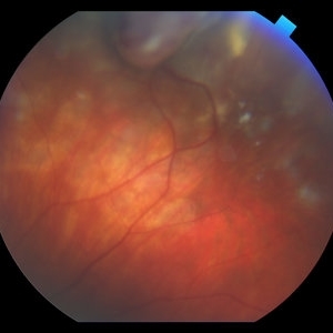
 Inferior retinal detachment with lattice and holes
May 31 2023 by Aditya S Kelkar, MS, FRCS, FASRS,FRCOphth
Importance of dilated retina check up before Lasik surgery can't be better demonstrated...patient totally asymptomatic came for Lasik opinion and has inferior retinal detachment with lattice and holes, sparing the macula
Photographer: Dr. Sahil Wagh , National Institute of Opthalmology, Pune , India
Imaging device: Zeiss Clarus 500
Condition/keywords: inferior retinal detachment
-
 Macrocyst in the Fovea
Feb 2 2021 by Peter J Belin, MD
36-year-old male with a white cataract and a chronic total retinal detachment for 1 year presented with a recurrent PVR detachment after primary repair 2 weeks prior. This OCT- EDI demonstrates a large retinal cyst through the fovea.
Photographer: Holly Cheshier, CRA, OCT-C, COT
Imaging device: Heidelberg Spectralis
Condition/keywords: chronic retinal detachment, proliferative vitreoretinopathy (PVR), retinal cyst, retinal macrocyst
-
 Retinal Detachment
Nov 9 2012 by Norman Byer
This 18-year-old girl gave the history of having been hit in this eye three years before with a fist and of having retinal surgery nine months previously, which was temporarily successful. When the photograph was taken, she had a total left retinal detachment with a small nasal dialysis which had not been treated. She also had two prominent intraretinal cysts, one of which is shown here. The retina promptly reattached following further surgery and the next slide shows an interesting change in this cyst.
Condition/keywords: intraretinal cyst, small nasal dialysis
-
 Retinoblastoma
Oct 9 2019 by McGill University Health Centre
Enucleated eye of a pediatric patient showing a total retinal detachment and a large exophytic retinoblastoma.
Photographer: Miguel N. Burnier, McGill University Health Center-McGill University Ocular Pathology & Translational Research Laboratory
Condition/keywords: exophytic, gross pathology, retinoblastoma
-
 Silicone oil in traumatic aniridia
Apr 19 2022 by Thais Bastos
A 27-year-old patient who developed aniridia, aphakia and retinal detachment after ocular trauma in the left eye. She underwent vitrectomy with silicone oil. Photo of the anterior segment 3 months after surgery showing a double meniscus made of silicone oil. Note red reflex, the retina is totally attached.
Photographer: Thaís Azeredo Bastos, CBCO Hospital de Olhos, Goiânia - Brazil
Imaging device: Zeiss Clarus 700
Condition/keywords: aniridia, ocular trauma, silicone oil
-
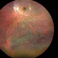
---thumb.jpg/image-square;max$300,300.ImageHandler) Sturge-Weber Diffuse Hemangioma and Retinal Detachment on B-scan
Sturge-Weber Diffuse Hemangioma and Retinal Detachment on B-scan
Apr 18 2014 by Susanna S. Park, MD, PhD
B-scan ultrasonogram of the right eye of an 8 year old Hispanic boy with Sturge -Weber Syndrome showing diffuse choroidal thickening from diffuse choroidal hemangioma and associated total exudative retinal detachment.
Photographer: Ellen Redenbo, University of California Davis Eye Center
Condition/keywords: B scan ultrasound, diffuse choroidal hemangioma, Sturge-Weber syndrome
-
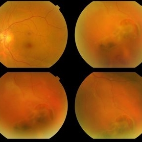
 Total Retinal Detachment With PVR Changes
Nov 5 2018 by awaneesh m upadhyay, MBBS, DNB
Fundus images of a male pseudophakic patient with total rhegmatogenous retinal detachment and PVR changes.
Photographer: Hiteshwar Saikia
Condition/keywords: proliferative vitreoretinopathy (PVR)
-
 Total Rhegmatogenous and Tractional Retinal Detachment Following Choroidal Melanoma Laser Ablation Treatment
Sep 22 2020 by Sophia El Hamichi, MD
A 69-year-old female, with a history of choroidal melanoma in her left eye with exudative detachment, underwent tumor laser ablation. She then developed a complex combined tractional and rhegmatogenous retinal detachment with a giant retinal tear.
Photographer: Belinda Rodriguez, Murray Ocular Oncology and Retina, Miami
Condition/keywords: tractional retinal detachment
-
 Traumatic Macular Hole with Retinal Detachment and PVR
Sep 27 2012 by Pauline T Merrill, MD, FASRS
Fundus photo of a 13-year-old boy s/p soccer ball injury 1 month previously. In addition to full-thickness macular hole and total retinal detachment with grade C PVR, note pigment granules visible in vitreous over optic nerve.
Photographer: Karen Parque, Illinois Retina Associates, Chicago, IL
Condition/keywords: proliferative vitreoretinopathy (PVR), traumatic macular hole
-
 Vasoproliferative Tumor (VPT)
Apr 25 2017 by Christopher G Fuller, MD
Fundus photograph of a presumptive vasoproliferative tumor (with resultant total exudative retinal detachment) in a 54-year-old white truck driver. Image is taken on post-operative day 4, after 25/27 gauge vitrectomy with drainage retinotomy, air-fluid exchange, endoscopic laser blanching of VPT, oil, and Ozurdex.
Photographer: Ray Garner, Texas Retina Associates [Lubbock, TX]
Condition/keywords: vasoproliferative retinopathy
-
 Xanthocoria in Coats' disease
Sep 7 2022 by Niloofar Piri, MD
Xanthocoria secondary to total exudative RD in Coats' disease. Yellow color is secondary to extensive subretinal exudates.
Photographer: Jacob Grodsky, MD
Condition/keywords: Coats' disease, Xanthocoria
-
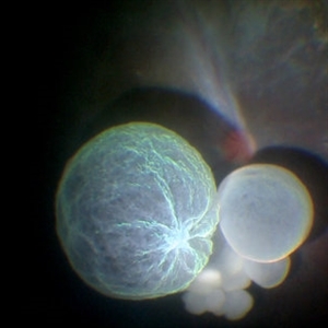 Intraocular Multiple Cysticercus
Oct 10 2018 by Vishal Agrawal, MD, FRCS,FACS,FASRS
Intraoperative fundus picture of right eye of a 18-year-old boy with complaints of DOV for the past 2 months. There were 12 intravitreal cysts in total with vitritis sclerosis retinal vessels and TRD. To note here, the largest cyst has a flimsy wall and no scolex (possibly ruptured) and the rest of the smaller cysts have a scolex and a taut wall.
Photographer: Vishal Agrawal MD,FRCS
Imaging device: SONY PMW-10 MD HD
Condition/keywords: cysticercosis, scolex
-
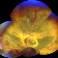
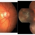
 Total retinal Detachment multiple holes
Sep 26 2022 by Denica Rodriguez
60 year old Male presented with two week old Macula off Retinal detachment with multiple tears.
Photographer: Denica Rodriguez
Imaging device: Optos California
Condition/keywords: color fundus photograph, color photo, macula-off, optos, pseudocolor, Retinal detachment, retinal holes, retinal tear, Retinal tear with detachment, superior arcade, superior field, superior retina, total retinal detachment
-
 Subconjuntival IOL After Blunt Trauma
Jun 27 2018 by Gabriel Costa Andrade, PhD
A 73-year-old male patient was referred to our ophthalmic emergency department with complaints of redness, pain, and diminution of vision in his left eye, after fall from height. The patient underwent small incision cataract surgery with polymethylmethacrylate (PMMA) IOL implantation in both the eyes 15 years back through superior sclerocorneal incision under local anesthesia. His best-corrected visual acuity was perception of light in the left eye. Ophthalmic examination using slit lamp biomicroscopy of the left eye revealed diffuse subconjunctival hemorrhage with no conjunctival laceration and inferior bulbar conjunctiva showed traumatic pseudophacocele with a sign “golden half ring,” suggesting the presence of PCIOL in subconjunctival space.There was total hyphema obscuring the view of rest of the ocular structures in his left eye.
Photographer: Gabriel Andrade, RETINA CLINIC, São Paulo, BRAZIL
Condition/keywords: dislocated intraocular lens (IOL), trauma
 Loading…