Search results (195 results)
-
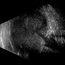
 Advanced Proliferative Diabetic Retinopathy
Apr 9 2025 by Gustavo Uriel Fonseca Aguirre
B-mode ultrasound of a patient with long-standing poorly controlled diabetes demonstrates characteristic findings of advanced proliferative diabetic retinopathy. The examination reveals moderate vitreous hemorrhage appearing as diffuse hyperechoic opacities throughout the vitreous cavity, along with a posterior hyaloid membrane densely infiltrated by hemorrhagic material, showing irregular thickening and increased reflectivity. A mild subhyaloid hemorrhage is visible as a subtle hyphema-like space anterior to the retinal surface. The study documents a total tractional retinal detachment, evidenced by rigid retinal folds with clear insertion points of vitreous strands, accompanied by a significant subretinal hemorrhage seen as a prominent hyperechoic collection beneath the elevated retina. These findings collectively illustrate the severe vitreoretinal interface pathology characteristic of end-stage diabetic eye disease, with predominant tractional components and distinct echographic stratification of hemorrhagic layers - from anterior vitreous involvement to deeper subretinal blood accumulation.
Photographer: Gustavo U. Fonseca Aguirre, Hospital Conde de Valenciana, Ciudad de México
Condition/keywords: diabetic retinopathy, tractional retinal detachment, Vitreous hemorrhage
-
 Retinopathy of Prematurity
Apr 7 2025 by Gustavo Uriel Fonseca Aguirre
B-mode ultrasound of a 7-month-old premature infant with a history of perinatal supplemental oxygen therapy reveals a total funnel-shaped retinal detachment with significant vasoproliferative tissue causing retinal traction.
Photographer: Gustavo U. Fonseca Aguirre, Hospital Conde de Valenciana, Ciudad de México
Condition/keywords: retinopathy of prematurity
-
 Negative Retinal Detachment
Apr 7 2025 by Gustavo Uriel Fonseca Aguirre
B-mode ultrasound of a 7-month-old premature infant with a history of perinatal supplemental oxygen therapy reveals a total funnel-shaped retinal detachment appearing as a hypoechoic structure, accompanied by significant hyperechoic subretinal hemorrhage. This distinctive echographic pattern creates the characteristic appearance of a "negative retinal detachment."
Photographer: Gustavo U. Fonseca Aguirre, Hospital Conde de Valenciana, Ciudad de México
Condition/keywords: retinopathy of prematurity
-
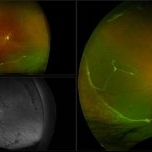
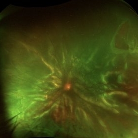 Rhegmatogenous Retinal Detachment with Gd C PVR Changes
Mar 28 2025 by Shrishti mishra
Fundus photograph of a 58 year old male who had undergone a pneumatic retinopexy elsewhere presented to us with a total retinal detachment with a retinal tear in the superotemporal quadrant and grade c pvr changes.
Photographer: Mrs Vinutha
Imaging device: Optos nikon
Condition/keywords: proliferative vitreoretinopathy (PVR), retinal tear with detachment, rhegmatogenous retinal detachment
-
 Repaired Retinal Detachment with Grade C PVR
Dec 23 2024 by Virginia Gebhart
61 year old male 1 day s/p retinectomy/SO exchange. Retina is attached under SO with good laser to retinectomy edge.
Photographer: Virginia Gebhart, Retina Consultants of Carolina
Imaging device: Optos California
Condition/keywords: gas bubble, proliferative vitreoretinopathy (PVR), retinectomy, silicone oil, total retinal detachment
-
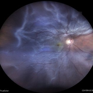
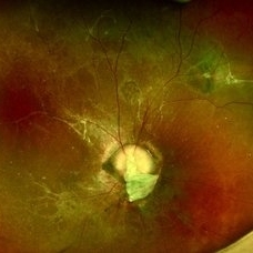 Morning Glory Anomaly With Retinal Detachment Managed With Amniotic Membrane Graft
Oct 15 2024 by Hemanth Murthy, MBBS, MD, FASRS
10 year-old boy presented with noticed blurring of vision. He had total retinal detachment with complicated cataract. He underwent lensectomy with 240 band and vitrectomy with silicone oil. The retina failed to settle due to minute breaks in the inferior part of the disc. Repeat surgery with AMG was done to cover the inferior part of disc. The retina settled under silicone oil. Silicone oil was removed and he is presently undergoing amblyopia treatment. Vision is 2/60 with +14.5 diopter lens.
Photographer: Mr Veda Vyas
Condition/keywords: amniotic membrane graft, Morning Glory Anomaly
-
 Emulsified Silicone Oil in Macular Hole
Jun 7 2024 by Vaidehi Jethwa
Fundus photograph of 72 year old man was having a/h/o Left Eye trauma by a cow horn, 8 years Ago, and developed Left Eye total Retinal detachment and was operated for Left Eye vitrectomy with FAX, SOI, Endolaser on 11 /04/2015 and was advised Left Eye silicone Oil removal.
Photographer: Dr. Vaidehi Jethwa, M and J institute of Ophthalmology, Ahmedabad, Gujarat.
Imaging device: Zeiss Visucam lite
Condition/keywords: macular hole, silicon oil emulsification in vitreous cavity
-
 Retinal Detachment Status Post Trauma
May 7 2024 by Akansha Sharma
Color fundus photograph of a 47 year old male with total retinal detachment.
Photographer: Dr. Akansha Sharma, Bharati Eye Hospital
Condition/keywords: RD, Retinal Detachment
-
 Vogt Koyanagi Harada
May 1 2024 by Marco Antonio Sauza
Fundus photograph of an 30-year-old woman with total serour detachment, the photo was taken after the SRF resolution with topical and systemic treatment.
Photographer: MARCO SAUZA CASTILLEJOS
Imaging device: VISUCAM ZEISS
Condition/keywords: vkh, VOGT KOYANAGI HARADA
-
 Total Serour Detachment
May 1 2024 by Marco Antonio Sauza
Fundus photograph of an 30-year-old woman with total serour detachment, the photo was taken after the srf resolution with topical and systemic treatment.
Photographer: MARCO SAUZA CASTILLEJOS
Imaging device: VISUCAM ZEISS
Condition/keywords: vkh, VOGT KOYANAGI HARADA
-
 Belt Buckle- Post RD Surgery
Apr 30 2024 by Eesh Nigam, MS
Fundus photograph of 50 years old lady of left eye superior horse shoe tear break and subtotal rhegmatogenous retinal detachment underwent left eye vitrectomy with belt buckling with endotamponade with SF6 . This is the post op image of attached retina with laser barraged breaks.
Photographer: Susmita Ghosh , Adity Birla SankaraNethralaya Mukundapur Kolkata WB India
Imaging device: Clarus 700
Condition/keywords: Attached retina with Belt buckle indent all over 360 Degrees after Vitrectomy for retinal detachment
-
 Giant Retinal Tear
Feb 20 2024 by Soobien Lee
Optos color fundus photograph of a 40-year-old caucasian male who is a UFC fighter with a total retinal detachment in his right eye secondary to a giant retinal tear from 10 o'clock to 2 o'clock.
Photographer: Trinity Wolf, Elman Retina Group
Imaging device: Optos Ultra-Widefield Imaging
Condition/keywords: giant retinal tear, optos, Retinal Detachment, Retinal tear with detachment, trauma
-
 Sub-total Retinal Detachment
Jan 30 2024 by Akansha Sharma
Color fundus photograph of a 53 year old male patient with sub-total retinal detachment.
Photographer: Dr. Akansha Sharma, Bharati Eye Hospital
Condition/keywords: RD, Retinal Detachment, sub-total retinal detachment
-
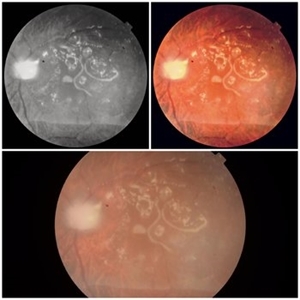
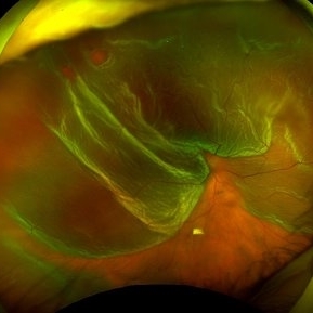
 Chronic Retinal Detachment with Proliferative Vitreoretinopathy
Jan 25 2024 by Isaac Agranoff
Widefield fundus photography of a 24 year old male presenting with subtotal retinal detachment with circumferential anterior proliferative vitreoretinopathy. The detachment is bullous inferiorly with atrophic retina and subretinal bands. There are also scattered patches of lattice with atrophic holes and associated detachment in the periphery. Patient presented with flashes for 2 years with worsening vision over the past 6-8 months, measured at 20/100 ph 20/60 OS.
Photographer: Isaac Agranoff, Ashley Rigdon
Imaging device: Optos California
Condition/keywords: atrophic hole, chronic retinal detachment, lattice degeneration, proliferative vitreoretinopathy (PVR), subretinal bands
-
 Macula off Retinal Detachment
Jan 23 2024 by Annaka Gooding
Ultra-widefield fundus photograph of an 81-year-old male with a Macula Off Retinal Detachment affecting his right eye. Patient presented at office with complaints of flashes of light for about 2 weeks accompanied by a curtain veil covering inferior visual field. Patient had total vision loss 24 hours prior to visit. His vision was scHM. The physician recommended Retinal Detachment Repair with PPV.
Photographer: Annaka Gooding, CPO
Imaging device: Optos California
Condition/keywords: detachment, fundus photography, macula off retinal detachment, Optos, retinal detachment of the macula, right eye, ultra-wide field imaging
-
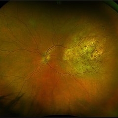 Choroidal Metastasis
Dec 6 2023 by Virginia Gebhart
60 year old female with totally regressed tumor in temporal macula s/p external beam radiation and chemo. Pt diagnosed with stage IV metastatic lung cancer.
Photographer: Virginia Gebhart
Imaging device: Optos
Condition/keywords: choroidal metastasis, choroidal tumor
-
 Mac off Retinal Detachment with Horseshoe Tear
Dec 5 2023 by Virginia Gebhart
68 year old male presented with HM vision in OD. Near total detachment with multiple breaks. Scheduled PPV with GFE. Visual prognosis guarded
Photographer: Virginia Gebhart
Imaging device: Topcon
Condition/keywords: Retinal Detachment, retinal detachment of the macula, Retinal Detachment with multiple breaks
-
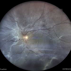
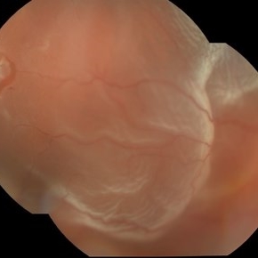
 Restoring the glow: Retinochoroidal Coloboma associated Retinal detachment - Post-op
Aug 21 2023 by Harsh Vardhan Singh, MS
17 year-old female with RC coloboma with total bullous retinal detachment - operated for the same
Photographer: Dr Harsh Vardhan Singh
Condition/keywords: coloboma of choroid, post-op
-
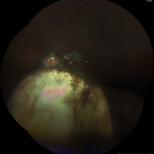
 Restoring the glow : Retinochoroidal Coloboma associated Retinal detachment - Pre-op
Aug 21 2023 by Harsh Vardhan Singh, MS
17 year-old female with RC coloboma with total bullous retinal detachment
Photographer: Dr Harsh Vardhan Singh
Condition/keywords: coloboma of choroid, pre-op
-
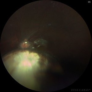
 Restoring the glow : Retinochoroidal Coloboma associated Retinal detachment - Pre-op
Aug 21 2023 by Harsh Vardhan Singh, MS
17-year-old female with RC coloboma with total bullous retinal detachment
Photographer: Dr Harsh Vardhan Singh
Condition/keywords: coloboma of choroid, pre-op
-
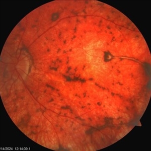
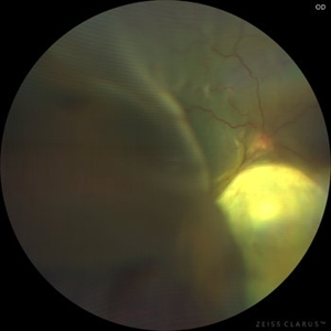
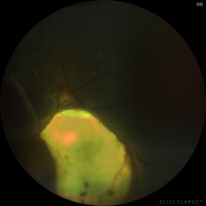
 Total Rhegmatogenous retinal detachment with lattice degeneration & Vitreous haemorrhage
Jul 31 2023 by Harsh Vardhan Singh, MS
72-year male presented PVD induced total retinal detachment with vitreous hemorrhage
Photographer: Dr Harsh Vardhan Singh, AIIMS, Guwahati
Imaging device: Zeiss Clarus 700
Condition/keywords: chronic retinal detachment, hemorrhage, rrd
-
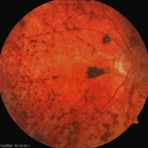
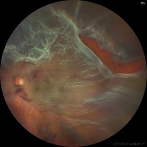
 Total Rhegmatogenous retinal detachment with opened posterior margin of lattice degeneration
Jul 18 2023 by Harsh Vardhan Singh, MS
78-year-old man with history of defective following cataract surgery showed total retinal detachment on examination
Photographer: Harsh Vardhan Singh, AIIMS, Guwahati
Imaging device: Zeiss Clarus 700
Condition/keywords: chronic retinal detachment, peripheral lattice degeneration, rrd
-
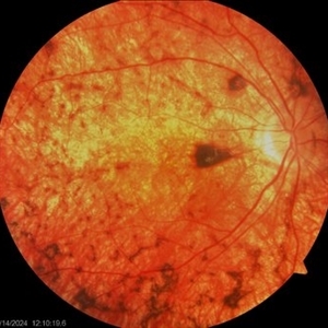
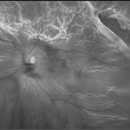
 Total Rhegmatogenous retinal detachment with opened posterior margin of lattice degeneration
Jul 18 2023 by Harsh Vardhan Singh, MS
78-year-old man with history of defective following cataract surgery showed total retinal detachment on examination
Photographer: Harsh Vardhan Singh, AIIMS, Guwahati
Imaging device: Zeiss Clarus 700
Condition/keywords: chronic retinal detachment, peripheral lattice degeneration, rrd
 Loading…