Search results (222 results)
-
 Retinoblastoma Ultrasound
Oct 2 2015 by Aparna Ramasubramanian
Ultrasonography of a retinoblastoma tumor shows hyperreflective echoes suggestive of calcification. It is seen in 90% of retinoblastoma patients and is an important diagnostic sign.
Photographer: Aparna Ramasubramanian
Condition/keywords: A-scan ultrasound, B scan ultrasound, calcification, retinoblastoma
-
 Retinoblastoma
Oct 5 2012 by Ronald C. Gentile, MD
B scan ultrasonography of the large endophytic retinoblastoma revealing internal hyper-reflective spots consistent with internal calcification.
Photographer: The New York Eye & Ear Infirmary Department of Medical Imaging
Condition/keywords: B scan ultrasound, retinoblastoma
-
 Retinoblastoma with Multiple Vitreous Seeding
Oct 9 2012 by Audina M. Berrocal, MD FASRS
Retinoblastoma with vitreous seeding.
Photographer: Ditte Hess CRA, BPEI
Imaging device: RETCAM
Condition/keywords: retinoblastoma, vitreous seeding
-
 Retinoblastoma
Jul 4 2012 by John T. Thompson, MD
Retinoblastoma filling enucleated eye
Condition/keywords: enucleation, pediatric tumor, retinoblastoma
-
---thumb.jpg/image-square;max$300,300.ImageHandler) Retinoblastoma To Chemothermotherapy
Retinoblastoma To Chemothermotherapy
Oct 4 2013 by Maurice F. Rabb
A 7 week old girl with a family history of retinoblastoma was found to have a small retinoblastoma in each eye. In the right eye the tumor was adjacent to the optic disc in the papillomacular bundle and measured 2 X 2 X 2 mm. Its temporal margin was 1.0 mm from the foveola and it overhung 20% of the optic disc surface. There was not clinical or ultrasonographic evidence of vitreous seeking or optic nerve invation. In the left eye there was a solitary tumor 1mm superonasal to the optic disc. The tumor measured 1 X 1 X 1 mm. The foveal reflex was normal in both eyes. Both tumors showed a fluorescein angiographic pattern compatible with retinoblastoma with rapid filling and late hyperfluorescence.
Condition/keywords: retina
-
 Retinoblastoma - Regressed
May 3 2013 by Suber S. Huang, MD, MBA, FASRS
24-year-old male status post radiation for retinoblastoma with secondary metastatic carcinoma.
Imaging device: Retina Diseases Imaging Analysis Reading Center
Condition/keywords: endophytic tumor growth, intraocular tumor, macular lesion, radiotherapy, retinoblastoma
-
---thumb.jpg/image-square;max$300,300.ImageHandler) Retinoblastoma To Chemothermotherapy
Retinoblastoma To Chemothermotherapy
Oct 4 2013 by Maurice F. Rabb
A 7 week old girl with a family history of retinoblastoma was found to have a small retinoblastoma in each eye. In the right eye the tumor was adjacent to the optic disc in the papillomacular bundle and measured 2 X 2 X 2 mm. Its temporal margin was 1.0 mm from the foveola and it overhung 20% of the optic disc surface. There was not clinical or ultrasonographic evidence of vitreous seeking or optic nerve invation. In the left eye there was a solitary tumor 1mm superonasal to the optic disc. The tumor measured 1 X 1 X 1 mm. The foveal reflex was normal in both eyes. Both tumors showed a fluorescein angiographic pattern compatible with retinoblastoma with rapid filling and late hyperfluorescence.
Condition/keywords: retina
-
 Retinoblastoma Type 2 Regression After Chemo and Laser
Apr 17 2014 by Susanna S. Park, MD, PhD
Retcam fundus photograph of a 2-year-old boy with history of bilateral Group D retinoblastoma completing 6 cycles of systemic chemotherapy and retinal laser and cryotherapy with residual regressing posterior pole tumor showing predominantly type 2 regression. Pigmented rim shows scarring from prior diode and argon laser treatments.
Photographer: Ellen Redenbo, University of California Davis Eye Center
Condition/keywords: retina, retinoblastoma, type 2 regression
-
 Retinoblastoma
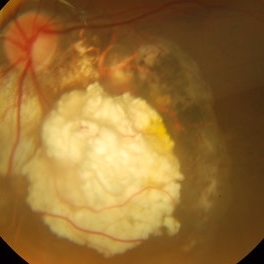
Jul 11 2013 by Jerald A. Bovino, MD
No history, probably treated, cottage cheese.
Condition/keywords: retinoblastoma
-
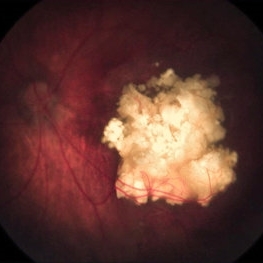
 Retinoblastoma
Oct 5 2012 by Ronald C. Gentile, MD
A white, large, highly vascularized retinoblastoma with endophytic tumor growth in a child presenting with leukocoria (white pupil reflex) and strabismus (turned eye).
Photographer: The New York Eye & Ear Infirmary Department of Medical Imaging
Condition/keywords: leukocoria, retinoblastoma, strabismus
-
 Retinoblastoma
Sep 13 2013 by Maria Ana Martinez-Castellanos, MD
Fundus photograph, fluorescein angiography and OCT of the macula and of the tumor of a 2-years-old boy with retinoblastoma.
Photographer: Maria A. Martinez-Castellanos. Asociacion para Evitar la Ceguera en Mexico
Imaging device: RetCAm II
Condition/keywords: leakage, optical coherence tomography (OCT), pediatric tumor, retinoblastoma
-
 Retinoblastoma
Apr 27 2018 by Brenda Fallas
2-year-old boy with stage D+ retinoblastoma of the right eye.
Photographer: Brenda Fallas, Bascom Palmer Eye Institute, Miami, FL
Imaging device: RETCAM III 130 degree lens montage
Condition/keywords: tumor, tumor seeding
-
 Retinoblastoma
Jul 11 2013 by Jerald A. Bovino, MD
Sectioned globe, calcium visible.
Condition/keywords: retinoblastoma, sectioned globe
-
 Retinoblastoma
Jul 11 2013 by Jerald A. Bovino, MD
No history, probably treated, cottage cheese overlying atrophy.
Condition/keywords: retinoblastoma
-
 Retinoblastoma
Nov 7 2013 by Maria Ana Martinez-Castellanos, MD
Retinoblastoma in a 2-year-old boy.
Photographer: Maria A. Martinez-Castellanos. Asociacion para Evitar la Ceguera en Mexico
Imaging device: RetCamII
Condition/keywords: retinoblastoma
-
 Group D Retinoblastoma After Chemo and Laser
Apr 17 2014 by Susanna S. Park, MD, PhD
Retcam fundus photograph of a 2 year old boy with history of bilateral Group D retinoblastoma completing 6 cycles of systemic chemotherapy and retinal laser and cryotherapy with residual regressing posterior pole tumor showing type 3 (type 1 and 2) regression pattern. Some pigmented scarring and hemorrhage are also noted nasal to the disc from prior laser treatment of tumor.
Photographer: Ellen Redenbo
Condition/keywords: retina
-
 MRI: The Preferred Method to Image the Brain and Orbit of Children with Retinoblastoma
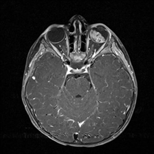
Oct 2 2015 by Paul T. Finger, MD, FACS
MRI used to evaluate a child with retinoblastoma. Note the white tumor on the T1 weighted image. There is no evidence of extrascleral or intramural invasion nor PNET.
Imaging device: MRI
Condition/keywords: MRI, retinoblastoma
-
 Retinoblastom Group E
Apr 17 2014 by Susanna S. Park, MD, PhD
Retcam Fundus photograph of a 3-year-old girl with no family history of retinoblastoma noted with a large retinal tumor with calcification filling 70% of globe with diffuse vitreous and subretinal seeding and exudative retinal detachment--unilateral Group E.
Photographer: Ellen Redenbo, University of California Davis Eye Center
Condition/keywords: retinoblastoma, tumor seeding
-
 Retinoblastoma Recurrent Seeding After Chemotherapy
Apr 17 2014 by Susanna S. Park, MD, PhD
Retcam fundus photograph of a 2-year-old boy with history of bilateral Group D retinoblastoma completing 6 cycles of systemic chemotherapy and retinal laser and cryotherapy who was noted with recurrent peripheral seeding of tumor.
Photographer: Ellen Redenbo, University of California Davis Eye Center
Condition/keywords: retinoblastoma, tumor seeding
-
 Retinoblastoma with Vitreous Seeding
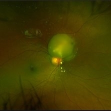
Oct 2 2015 by Paul T. Finger, MD, FACS
Exophytic retinoblastoma shedding clumps of tumor (seeds).
Photographer: Anonymous
Condition/keywords: retinoblastoma, vitreous, vitreous seeding
-
 Retinoblastoma Regressed
Dec 31 2015 by P. Mahesh Shanmugam, MBBS, DO, FRCSEd, PhD, FAICO
Regressed Retinoblastoma S/P chemotherapy and multiple sessions of TTT. Central calcific residue with surrounding chorio-retinal atrophy is well noted.
Condition/keywords: retinoblastoma
-
 Retinoblastoma Group-B (International Classification)
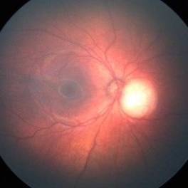
Oct 2 2015 by Aparna Ramasubramanian
Retcam fundus photography of a child with bilateral sporadic retinoblastoma showing an elevated tumor adjacent to the optic nerve. By the international classification this would be graded as a Group B retinoblasoma (Rb size >3 mm or =3 mm to foveola or =1.5 mm to disc or srf =3 mm from margin).
Photographer: Aparna Ramasubramanian
Condition/keywords: retinoblastoma
-
 Retinocytoma
Jul 13 2018 by Olivia Rainey
Ultra-wide field pseudocolor image of a 5-year-old male with a retinocytoma affecting his right eye. The retinal tumor has associated calcium which looks suspicious for retinoblastoma. However, there are a number of atypical features which raise the possibility of a masquerade tumor.
Photographer: Olivia Rainey
Imaging device: Optos
Condition/keywords: Optos, pseudocolor, retinocytoma, ultra-wide field imaging
-
 Retinoblastoma
Dec 22 2014 by H. Michael Lambert, MD
Cartoon of histopath features.
Condition/keywords: retinoblastoma
-
 Regressed Endophytic Retinoblastoma
Oct 7 2018 by Victor M Villegas, MD
Color fundus photograph of a 2-year-old child with germline retinoblastoma. Type 1 regression over the macula is shown.
Photographer: Brenda Fallas, Bascom Palmer Eye Institute, Miami FL
Imaging device: RetCam3
Condition/keywords: endophytic tumor growth, retinoblastoma, tumor
 Loading…