Search results (222 results)
-
 Bilateral Retinoblastoma
Apr 2 2019 by Gary R. Cook, MD, FACS
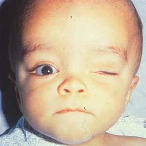
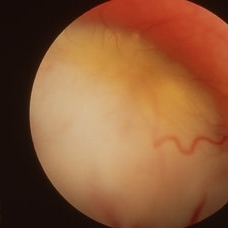
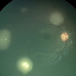
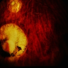
Right eye of a 13-month old white male infant with bilateral retinoblastoma.
Condition/keywords: retinoblastoma
-
 Bilateral Retinoblastoma
Nov 20 2019 by McGill University Health Centre
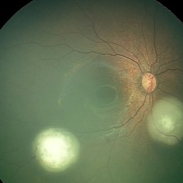
A 3 month-old boy was noticed to have a pupillary reflex on his left eye. A diagnosis of bilateral retinoblastoma was made and the left eye was enucleated. The small tumor on the right eye was treated with radiation. Imaging of the brain revealed a tumor in the region of the choroid plexus consistent with a choroid plexus carcinoma. A removal of the brain tumor was attempted.
Condition/keywords: retinoblastoma
-
 Calcifications in Retinoblastoma
Apr 2 2019 by Gary R. Cook, MD, FACS
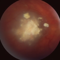
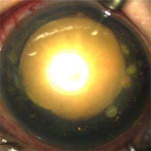
A calcified, necrotic retinoblastoma lesion OD in a 13-month old white male infant with bilateral retinoblastoma.
Condition/keywords: retinoblastoma
-
 Calcified Retinoblastoma after intra-arterial chemotherapy
Jan 19 2024 by Hector Gabriel Moreno Solano, MD, MHA
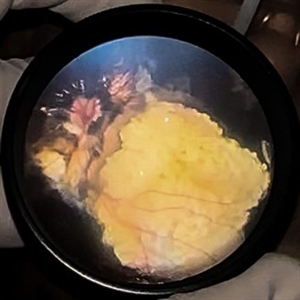
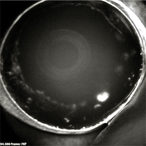
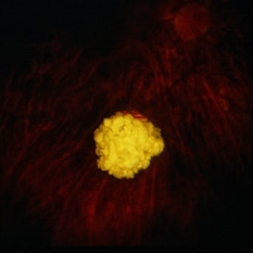
Fundus photography of a 5- Year-old Mexican child with bilateral retinoblastoma following unilateral enucleation and 4 cycles of intra-arterial chemotherapy in her only remaining eye. The image shows a succesfully treated tumor with a completely calcificied regression.
Photographer: Hector Solano, Hospital General de Zona #20 IMSS, Puebla
Imaging device: SmartPhone (IPhone 11 ProMax)
Condition/keywords: pediatic retina, pediatric tumor, retinoblastoma
-
 Calcified Retinoblastoma After Intra-arterial Chemotherapy
Apr 6 2024 by Hector Gabriel Moreno Solano, MD, MHA
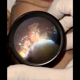
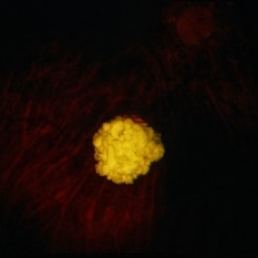
Fundus photography of a 5 yea-old Mexican child with bilateral retinoblastoma following unilateral enucleation and 4 cycles of intra-arterial chemotherapy in her only remaining eye. The image shows a successfully treated tumor with a completely calcificied regression.
Photographer: Héctor Gabriel Moreno-Solano, MD, MHA
Imaging device: SmartPhone (IPhone 11 pro Max)
Condition/keywords: pediatric retina, pediatric tumor, retinoblastoma
-
 Endophytic Retinoblastoma
May 18 2020 by McGill University Health Centre
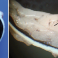
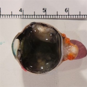
Image (A) shows an endophytic grayish tumor located on the retina of this enucleation specimen (arrow). Higher magnification of the same specimen (B) shows small hemorrhagic areas (arrow). The choroidal layer is not compromised.
Condition/keywords: enucleation, retinoblastoma
-
 Exo-Endophytic Retinoblastoma
May 18 2020 by McGill University Health Centre
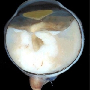
An exo-endophytic tumor is located on the optic nerve head and has produced a retinal detachment. Note the proteinaceous subretinal fluid. The optic nerve is thickened due to retinoblastoma infiltration.
Condition/keywords: enucleation, retinoblastoma
-
 Exophytic Retinoblastoma
May 18 2020 by McGill University Health Centre
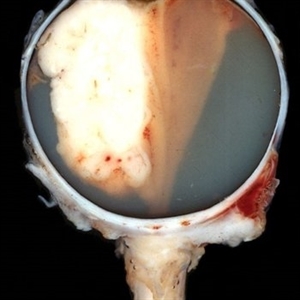
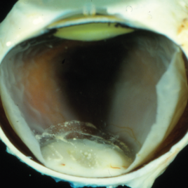
This type of tumor grows from the retina toward the choroid. In this enucleation specimen, the retina is completely detached, and the tumor is growing inside the subretinal proteinaceous fluid. Note the distance between the tumor and the optic nerve head.
Condition/keywords: enucleation, retinoblastoma
-
 Exophytic Retinoblastoma
Apr 27 2020 by Sophia El Hamichi, MD
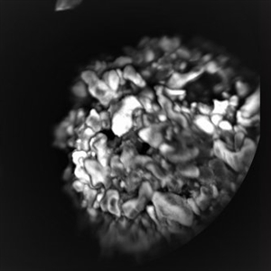
Exophytic retinoblastoma.
Photographer: Belinda Rodriguez, Murray Ocular Oncology and Retina, Miami
Imaging device: Heidelberg Engineering
Condition/keywords: exophytic, retinoblastoma
-
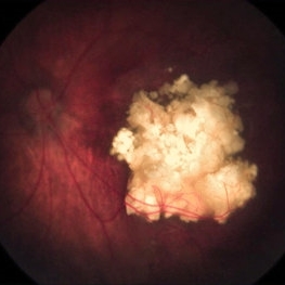
 Exophytic Retinoblastoma
Apr 2 2019 by Gary R. Cook, MD, FACS
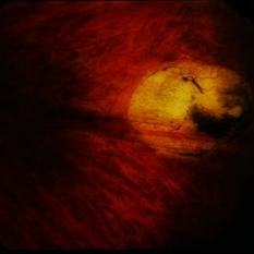
Solid white-appearing exophytic retinoblastoma with dilated tortuous vessels visible on its surface in the left eye of a WF infant.
Condition/keywords: retinoblastoma
-
 Group E Retinoblastoma Specimen
Jan 10 2019 by Rahul Komati, MD
Enucleation specimen of a 3-year-old boy with Group E unilateral sporadic retinoblastoma.
Condition/keywords: retinoblastoma
-
 Mixed Retinoblastoma
May 18 2020 by McGill University Health Centre
This enucleation specimen shows a whitish, thickened area of the neurosensory retina over the head of the optic nerve, corresponding to a mixed retinoblastoma. Snowballlike structures are present in the vitreous chamber overlying the tumor, corresponding to vitreous seeding. A retinal detachment artifact is present.
Condition/keywords: mixed, retinoblastoma
-
 MRI: The Preferred Method to Image the Brain and Orbit of Children with Retinoblastoma
Oct 2 2015 by Paul T. Finger, MD, FACS
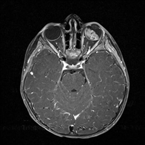
MRI used to evaluate a child with retinoblastoma. Note the white tumor on the T1 weighted image. There is no evidence of extrascleral or intramural invasion nor PNET.
Imaging device: MRI
Condition/keywords: MRI, retinoblastoma
-
 Multifocal Retinoblastoma
Jun 9 2021 by Thirumalesh Mochi Basavaraj, MD
3-year-old kid with multifocal retinoblastoma.
Photographer: Puttaswamy , Narayana Nethralaya , Bangalore
Imaging device: Retcam
Condition/keywords: retinoblastoma
-
 Multifocal Retinoblastoma
Jun 9 2021 by Thirumalesh Mochi Basavaraj, MD
Inferotemporal tumor is blanched immediately post TTT, in comparison to the nasal tumor which has not received TTT.
Photographer: Puttaswamy, Narayana Nethralaya, Bangalore
Imaging device: Retcam
Condition/keywords: retinoblastoma, tumor
-
 Necrotic Multifocal Retinoblastoma Group E (ICRB) / cT3e (AJCC)
Jul 7 2021 by Linda A Cernichiaro- Espinosa, MD
A 3-year, 9-month-old male presented with unilateral advanced group E multifocal retinoblastoma cT3e (AJCC). Anterior seeding vascularized over the iris surface. Fluorescein angiogram fills the vascularized tumors. Aseptic orbital cellulitis, birefringent anterior segment crystals, cataract and dense vitritis are secondary to necrosis.
Photographer: Jose Oyervides-Alvarado MD
Imaging device: RetCam3
Condition/keywords: retinoblastoma
-
 Necrotic Multifocal Retinoblastoma Group E (ICRB) / cT3e (AJCC)
Jul 7 2021 by Linda A Cernichiaro- Espinosa, MD
A 3-year, 9-month-old male presented with unilateral advanced group E multifocal retinoblastoma cT3e (AJCC). Anterior seeding vascularized over the iris surface. Fluorescein angiogram fills the vascularized tumors. Aseptic orbital cellulitis, birefringent anterior segment crystals, cataract and dense vitritis are secondary to necrosis.
Photographer: Jose Oyervides-Alvarado MD
Imaging device: RetCam3
Condition/keywords: fluorescein angiogram (FA), pediatric tumor, retinoblastoma
-
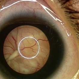 Pupillary View of Retinoblastoma
Jun 9 2021 by Thirumalesh Mochi Basavaraj, MD
Group D retinoblastoma.
Photographer: Puttaswamy, Narayana Nethralaya
Imaging device: Retcam Shuttle
Condition/keywords: retinoblastoma
-
 Regressed Endophytic Retinoblastoma
Oct 7 2018 by Victor M Villegas, MD
Color fundus photograph of a 2-year-old child with germline retinoblastoma. Type 1 regression over the macula is shown.
Photographer: Brenda Fallas, Bascom Palmer Eye Institute, Miami FL
Imaging device: RetCam3
Condition/keywords: endophytic tumor growth, retinoblastoma, tumor
-
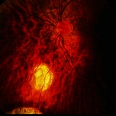 Regressed RB / Retinoblastoma
Sep 18 2015 by David Callanan, MD
20-year-old Hispanic female, regressed RB / retinoblastoma.
Condition/keywords: regressed, retinoblastoma
-
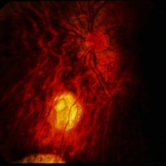 Regressed RB / Retinoblastoma
Sep 18 2015 by David Callanan, MD
20-year-old Hispanic female, regressed RB / retinoblastoma.
Condition/keywords: regressed, retinoblastoma
-
 Regressed RB / Retinoblastoma
Sep 18 2015 by David Callanan, MD
20-year-old Hispanic female, regressed RB / retinoblastoma.
Condition/keywords: regressed, retinoblastoma
-
 Regressed RB / Retinoblastoma
Sep 18 2015 by David Callanan, MD
20-year-old Hispanic female, regressed RB / retinoblastoma.
Condition/keywords: regressed, retinoblastoma
-
 Regressed RB / Retinoblastoma
Sep 18 2015 by David Callanan, MD
20-year-old Hispanic female, regressed RB / retinoblastoma.
Condition/keywords: regressed, retinoblastoma
-
 Regressed RB / Retinoblastoma
Sep 18 2015 by David Callanan, MD
20-year-old Hispanic female, regressed RB / retinoblastoma.
Condition/keywords: regressed, retinoblastoma
 Loading…