Search results (222 results)
-
 Retinoblastoma
Apr 27 2018 by Brenda Fallas
2-year-old boy with stage D+ retinoblastoma of the right eye.
Photographer: Brenda Fallas, Bascom Palmer Eye Institute, Miami, FL
Imaging device: RETCAM III 130 degree lens montage
Condition/keywords: tumor, tumor seeding
-
 Retinoblastoma
Apr 23 2021 by Giselle DeOliveira
5 week old baby girl born with retinoblastoma.
Photographer: Giselle DeOliveira, Bascom Palmer Eye Institute, Miami ,Fl
Imaging device: Retcam
Condition/keywords: retinoblastoma
-
 Retinoblastoma Regressed
Dec 31 2015 by P. Mahesh Shanmugam, MBBS, DO, FRCSEd, PhD, FAICO
Regressed Retinoblastoma S/P chemotherapy and multiple sessions of TTT. Central calcific residue with surrounding chorio-retinal atrophy is well noted.
Condition/keywords: retinoblastoma
-
 Retinoblastoma
Jul 4 2012 by John T. Thompson, MD
Retinoblastoma filling enucleated eye
Condition/keywords: enucleation, pediatric tumor, retinoblastoma
-
 Myelinated Nerve Fibers
Apr 18 2025 by DR Rohit Gupta
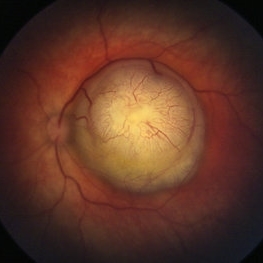
The **myelinated nerve fibers of the optic disc** (also known as **medullated nerve fibers**) are retinal nerve fibers that retain their myelin sheath as they pass through the optic nerve head. Normally, retinal nerve fibers are unmyelinated to allow for light transparency, but in some cases, myelination extends anteriorly into the retina, appearing as a striking white, feathery patch on the optic disc or peripapillary retina. ### **Key Features:** 1. **Appearance:** - Dense, white, striated patches with feathery edges. - Typically located at the superior or inferior pole of the optic disc. - May obscure retinal vessels underneath. 2. **Clinical Significance:** - Usually **benign** and asymptomatic. - **Congenital** (present at birth or early childhood). - Rarely associated with **visual field defects** (e.g., scotomas corresponding to the area of myelination). - Occasionally linked with **high myopia** or **amblyopia** if extensive. 3. **Pathophysiology:** - Failure of oligodendrocytes or Schwann cells to stop myelination at the lamina cribrosa. - Normally, myelination stops at the optic nerve head, but in this condition, it extends into the retina. 4. **Diagnosis:** - **Fundoscopy:** Classic white, feathery appearance. - **Optical Coherence Tomography (OCT):** Shows thickened retinal nerve fiber layer (RNFL). - **Visual Field Testing:** May detect defects if large. 5. **Differential Diagnosis:** - Optic disc edema - Cotton wool spots - Retinoblastoma (rarely, but must be ruled out in children) 6. **Management:** - No treatment required if asymptomatic. - Monitor for amblyopia in children. - Rare cases with significant visual impairment may need further evaluation. ### **Fun Fact:** Myelinated nerve fibers are seen in **~0.5-1%** of the population and are usually an incidental finding.
Photographer: Dr Rohit gupta
Imaging device: Samsung S21
Condition/keywords: Medulated Nerve fibre, Medullated Nerve fibres, myelinated nerve fibers, Myelinated Nerve Fibres, optic disc drusen
-
 Retinal Detachment After Retinoblastoma Treatment
Mar 10 2024 by Alexandre Grandinetti, MD, PhD
Inferior retinal detachment occurring 6 years after treatment with intraarterial chemotherapy and laser in an 8-year-old boy.
Photographer: Corina Szrek
Condition/keywords: pediatric, retinoblastoma
-
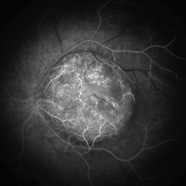
 Retinoblastoma
Sep 13 2013 by Maria Ana Martinez-Castellanos, MD
Fundus photograph, fluorescein angiography and OCT of the macula and of the tumor of a 2-years-old boy with retinoblastoma.
Photographer: Maria A. Martinez-Castellanos. Asociacion para Evitar la Ceguera en Mexico
Imaging device: RetCAm II
Condition/keywords: leakage, optical coherence tomography (OCT), pediatric tumor, retinoblastoma
-
 Retinoblastoma
Apr 23 2021 by Giselle DeOliveira
Angiography of retinoblastoma in 5 week old baby girl.
Photographer: Giselle DeOliveira, Bascom Palmer Eye Institute, Miami ,Fl
Imaging device: Retcam
Condition/keywords: retinoblastoma
-
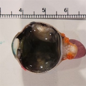
 Retinoblastoma
Oct 9 2019 by McGill University Health Centre
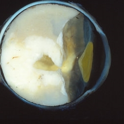
Enucleated eye of a pediatric patient showing a total retinal detachment and a large exophytic retinoblastoma.
Photographer: Miguel N. Burnier, McGill University Health Center-McGill University Ocular Pathology & Translational Research Laboratory
Condition/keywords: exophytic, gross pathology, retinoblastoma
-
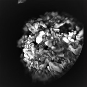
 Retinoblastoma
Oct 9 2019 by McGill University Health Centre
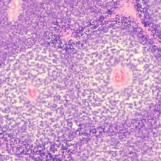
Histopathology of retinoblastoma displaying the typical sleeve pattern of the neoplastic cells around blood vessels.
Photographer: Miguel N. Burnier, McGill University Health Center-McGill University Ocular Pathology & Translational Research Laboratory
Condition/keywords: histopathology, retinoblastoma
-
 Retinoblastoma - Regressed
May 3 2013 by Suber S. Huang, MD, MBA, FASRS
24-year-old male status post radiation for retinoblastoma with secondary metastatic carcinoma.
Imaging device: Retina Diseases Imaging Analysis Reading Center
Condition/keywords: endophytic tumor growth, intraocular tumor, macular lesion, radiotherapy, retinoblastoma
-
 Retinoblastoma Pathology
Jan 10 2019 by Rahul Komati, MD
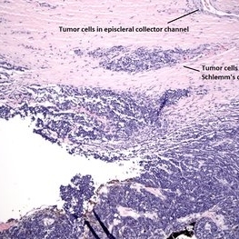
Histopathology showing tumor cell invasion of trabecular meshwork, Schlemm's canal, episcleral collector channels. Patient was treated with enucleation & adjuvant chemotherapy.
Condition/keywords: retinoblastoma
-
 Retinoblastoma Type 2 Regression After Chemo and Laser
Apr 17 2014 by Susanna S. Park, MD, PhD
Retcam fundus photograph of a 2-year-old boy with history of bilateral Group D retinoblastoma completing 6 cycles of systemic chemotherapy and retinal laser and cryotherapy with residual regressing posterior pole tumor showing predominantly type 2 regression. Pigmented rim shows scarring from prior diode and argon laser treatments.
Photographer: Ellen Redenbo, University of California Davis Eye Center
Condition/keywords: retina, retinoblastoma, type 2 regression
-
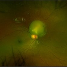
 Retinoblastoma
Apr 30 2020 by Giselle DeOliveira
Fundus photograph of an 17-month-old female infant with retinoblastoma over optic nerve.
Photographer: Giselle DeOliveira, University of Miami, Bascom Palmer Eye Institute
Imaging device: Retcam III
Condition/keywords: retinoblastoma
-
 Group E Retinoblastoma Specimen
Jan 10 2019 by Rahul Komati, MD
Enucleation specimen of a 3-year-old boy with Group E unilateral sporadic retinoblastoma.
Condition/keywords: retinoblastoma
-
 Retinoblastoma Stage 5 After One Cycle of Systemic Chemotherapy and Laser Ablation
Sep 17 2019 by Sophia El Hamichi, MD
A 1-year-old patient with stage 5B retinoblastoma, fundus after one cycle of systemic chemotherapy and laser ablation.
Photographer: Abby Orcutt-Hayes, Murray Ocular Oncology and Retina
Condition/keywords: chemoreduction, laser photocoagulation, montage, retinoblastoma, stage 5
-
 Retinocytoma
Jul 13 2018 by Olivia Rainey
Ultra-wide field pseudocolor image of a 5-year-old male with a retinocytoma affecting his right eye. The retinal tumor has associated calcium which looks suspicious for retinoblastoma. However, there are a number of atypical features which raise the possibility of a masquerade tumor.
Photographer: Olivia Rainey
Imaging device: Optos
Condition/keywords: Optos, pseudocolor, retinocytoma, ultra-wide field imaging
-
 Retinoblastoma
Jun 4 2018 by Diva Kant Misra, MBBS, DO, DNB, MNAMS, FVRS
Fundus photograph of a 2-year-old male child with retinoblastoma.
Photographer: DIVA KANT MISRA
Imaging device: RETCAM
Condition/keywords: retinoblastoma
-
 Retinoblastoma OD FA 6-2-2015-12-15-42 PM Proof
Jun 4 2015 by Kathy Karsten, COT
15-year-old male born with retinoblastoma. S/P enucleation OS at 3 months of age. S/P chemo/radiation lesions OD
Photographer: Kathy Karsten, COT
Imaging device: Topcon TRC 50-DX
Condition/keywords: retinoblastoma
-
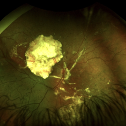
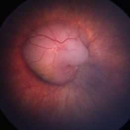
 Exophytic Retinoblastoma
Apr 27 2020 by Sophia El Hamichi, MD
Exophytic retinoblastoma.
Photographer: Belinda Rodriguez, Murray Ocular Oncology and Retina, Miami
Imaging device: Heidelberg Engineering
Condition/keywords: exophytic, retinoblastoma
-
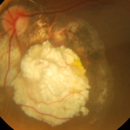
 Retinoblastoma
Oct 5 2012 by Ronald C. Gentile, MD
A white, large, highly vascularized retinoblastoma with endophytic tumor growth in a child presenting with leukocoria (white pupil reflex) and strabismus (turned eye).
Photographer: The New York Eye & Ear Infirmary Department of Medical Imaging
Condition/keywords: leukocoria, retinoblastoma, strabismus
-
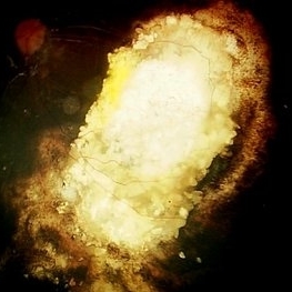
 Retinoblastoma With Calcifications
Dec 16 2019 by Sophia El Hamichi, MD
A 4-year-old male patient with germline retinoblastoma, treated with intraarterial chemotherapy and thermal transpupillary laser.
Photographer: Abby Orcutt-Hayes, Murray Ocular Oncology and Retina
Condition/keywords: calcification, chemoreduction, retinoblastoma
-
 Retinoblastoma with Multiple Vitreous Seeding
Oct 9 2012 by Audina M. Berrocal, MD FASRS
Retinoblastoma with vitreous seeding.
Photographer: Ditte Hess CRA, BPEI
Imaging device: RETCAM
Condition/keywords: retinoblastoma, vitreous seeding
-
 Retinoblastoma: Color Picture
Nov 10 2017 by Linda A Cernichiaro- Espinosa, MD
Color image of the right eye from a 19-month-old male. Treatment naïve bilateral germinal retinoblastoma ICRB Group C (R-E: Vb) with multiple tumors. A Coats-like response is observed.
Photographer: Abby Orcutt
Imaging device: RetCam III
Condition/keywords: pediatric retina, retinoblastoma
-
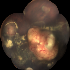
 Retrolental Bullous Retinal Detachment
Mar 22 2019 by Abdulaziz A. Alshamrani, MD
External photo for a 2-year-old child with exophytic retinoblastoma.
Condition/keywords: bullous retinal detachment, retinoblastoma
 Loading…