Search results (222 results)
-
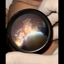
 Myelinated Nerve Fibers
Apr 18 2025 by DR Rohit Gupta
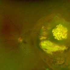
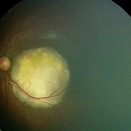
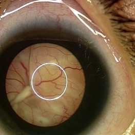
The **myelinated nerve fibers of the optic disc** (also known as **medullated nerve fibers**) are retinal nerve fibers that retain their myelin sheath as they pass through the optic nerve head. Normally, retinal nerve fibers are unmyelinated to allow for light transparency, but in some cases, myelination extends anteriorly into the retina, appearing as a striking white, feathery patch on the optic disc or peripapillary retina. ### **Key Features:** 1. **Appearance:** - Dense, white, striated patches with feathery edges. - Typically located at the superior or inferior pole of the optic disc. - May obscure retinal vessels underneath. 2. **Clinical Significance:** - Usually **benign** and asymptomatic. - **Congenital** (present at birth or early childhood). - Rarely associated with **visual field defects** (e.g., scotomas corresponding to the area of myelination). - Occasionally linked with **high myopia** or **amblyopia** if extensive. 3. **Pathophysiology:** - Failure of oligodendrocytes or Schwann cells to stop myelination at the lamina cribrosa. - Normally, myelination stops at the optic nerve head, but in this condition, it extends into the retina. 4. **Diagnosis:** - **Fundoscopy:** Classic white, feathery appearance. - **Optical Coherence Tomography (OCT):** Shows thickened retinal nerve fiber layer (RNFL). - **Visual Field Testing:** May detect defects if large. 5. **Differential Diagnosis:** - Optic disc edema - Cotton wool spots - Retinoblastoma (rarely, but must be ruled out in children) 6. **Management:** - No treatment required if asymptomatic. - Monitor for amblyopia in children. - Rare cases with significant visual impairment may need further evaluation. ### **Fun Fact:** Myelinated nerve fibers are seen in **~0.5-1%** of the population and are usually an incidental finding.
Photographer: Dr Rohit gupta
Imaging device: Samsung S21
Condition/keywords: Medulated Nerve fibre, Medullated Nerve fibres, myelinated nerve fibers, Myelinated Nerve Fibres, optic disc drusen
-
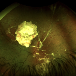
 Calcified Retinoblastoma After Intra-arterial Chemotherapy
Apr 6 2024 by Hector Gabriel Moreno Solano, MD, MHA
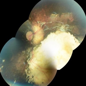
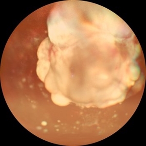
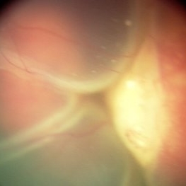
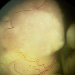
Fundus photography of a 5 yea-old Mexican child with bilateral retinoblastoma following unilateral enucleation and 4 cycles of intra-arterial chemotherapy in her only remaining eye. The image shows a successfully treated tumor with a completely calcificied regression.
Photographer: Héctor Gabriel Moreno-Solano, MD, MHA
Imaging device: SmartPhone (IPhone 11 pro Max)
Condition/keywords: pediatric retina, pediatric tumor, retinoblastoma
-
 Retinal Detachment After Retinoblastoma Treatment
Mar 10 2024 by Alexandre Grandinetti, MD, PhD
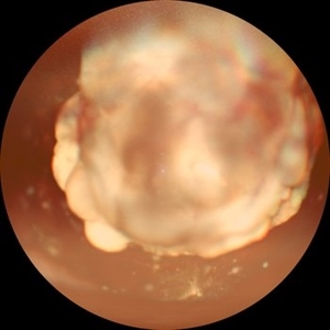
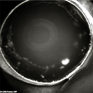
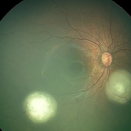
Inferior retinal detachment occurring 6 years after treatment with intraarterial chemotherapy and laser in an 8-year-old boy.
Photographer: Corina Szrek
Condition/keywords: pediatric, retinoblastoma
-
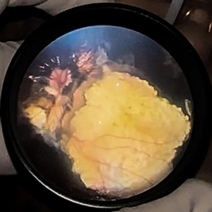 Calcified Retinoblastoma after intra-arterial chemotherapy
Jan 19 2024 by Hector Gabriel Moreno Solano, MD, MHA
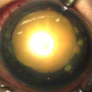
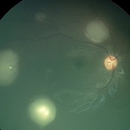
Fundus photography of a 5- Year-old Mexican child with bilateral retinoblastoma following unilateral enucleation and 4 cycles of intra-arterial chemotherapy in her only remaining eye. The image shows a succesfully treated tumor with a completely calcificied regression.
Photographer: Hector Solano, Hospital General de Zona #20 IMSS, Puebla
Imaging device: SmartPhone (IPhone 11 ProMax)
Condition/keywords: pediatic retina, pediatric tumor, retinoblastoma
-
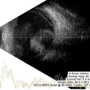 Retinoblastoma
Nov 1 2023 by ANKIT JAIN
USG B SCAN image showing hyperechogenic mass lesion with moderate spikes with restricted after movements on dynamic scan. In between high spikes noted suggestive of calcification in a case of Retinoblastoma
Photographer: DR ANKIT JAIN
Condition/keywords: B scan ultrasound, retinoblastoma, ultrasound
-
 Retinoma
Sep 11 2023 by Naveenam Srinivasa Muralidhar, MD
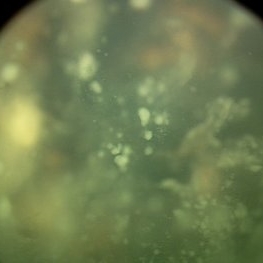
Optos widefield image of left eye of a 31 year old male c/o defective vision in left eye since 6 years with esotropia of 30 degree. Fundus shows translucent greyish mass temporal to macula surrounded by zone of atrophy with pigmentation. Right eye fundus within normal limits.
Photographer: Mr. Vedavyasa N K
Imaging device: Optos
Condition/keywords: optos, Retinoma, spontaneously regressed retinoblastoma
-
 Retinoblastoma Pseudohypopyon
Dec 10 2022 by Jordan D Deaner, MD
6-year-old female referred for left eye pain with elevated intraocular pressure, neovascularization of the iris, and a hypopyon concerned for uveitis eventually diagnosed with pseudohypopyon secondary to diffuse infiltrating retinoblastoma.
Condition/keywords: Retinoblastoma Pseudohypopyon
-
 Retinoblastoma
Nov 8 2022 by Heitor Nogueira
Calcified retinoblastoma after chemotherapy therapy
Photographer: Heitor Nogueira, Penido Burnier Institute, Campinas-SP, Brazil
Condition/keywords: retinoblastoma
-
 Retinoblastoma
Nov 6 2022 by Akansha Sharma
WIDE-FIELD COLOUR FUNDUS PHOTOGRAPH OF A 2 MONTH OLD FEMALE WITH RETINOBLASTOMA
Photographer: Dr. Akansha Sharma-Retina Foundation, Ahmedabad
Condition/keywords: RB gene mutation, retinoblastoma
-
 Retinoblastoma
Nov 6 2022 by Akansha Sharma
Wide-field color fundus photograph of a 2-month old female with retinoblastoma.
Photographer: Dr. Akansha Sharma-Retina Foundation, Ahmedabad
Condition/keywords: RB gene mutation, retinoblastoma
-
 Necrotic Multifocal Retinoblastoma Group E (ICRB) / cT3e (AJCC)
Jul 7 2021 by Linda A Cernichiaro- Espinosa, MD
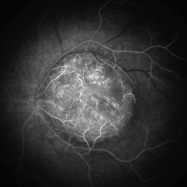
A 3-year, 9-month-old male presented with unilateral advanced group E multifocal retinoblastoma cT3e (AJCC). Anterior seeding vascularized over the iris surface. Fluorescein angiogram fills the vascularized tumors. Aseptic orbital cellulitis, birefringent anterior segment crystals, cataract and dense vitritis are secondary to necrosis.
Photographer: Jose Oyervides-Alvarado MD
Imaging device: RetCam3
Condition/keywords: fluorescein angiogram (FA), pediatric tumor, retinoblastoma
-
 Necrotic Multifocal Retinoblastoma Group E (ICRB) / cT3e (AJCC)
Jul 7 2021 by Linda A Cernichiaro- Espinosa, MD
A 3-year, 9-month-old male presented with unilateral advanced group E multifocal retinoblastoma cT3e (AJCC). Anterior seeding vascularized over the iris surface. Fluorescein angiogram fills the vascularized tumors. Aseptic orbital cellulitis, birefringent anterior segment crystals, cataract and dense vitritis are secondary to necrosis.
Photographer: Jose Oyervides-Alvarado MD
Imaging device: RetCam3
Condition/keywords: retinoblastoma
-
 Vitreous Seeds
Jun 9 2021 by Thirumalesh Mochi Basavaraj, MD
Vitreous seeds in a case of retinoblastoma.
Photographer: Puttaswamy, Narayana Nethralaya, Bangalore
Imaging device: retcam
Condition/keywords: retinoblastoma
-
 Macular Retinoblastoma
Jun 9 2021 by Thirumalesh Mochi Basavaraj, MD
Macular retinoblastoma.
Photographer: Puttaswamy, Narayana Nethralaya
Imaging device: Retcam
Condition/keywords: tumor
-
 Retinoblastoma with Exudative Retinal Detachment
Jun 9 2021 by Thirumalesh Mochi Basavaraj, MD
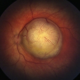
A case of retinoblastoma with exudative retinal detachment and subretinal seeding.
Photographer: Puttaswamy, Narayan Nethralaya, Bangalore
Imaging device: Retcam
Condition/keywords: exudative retinal detachment
-
 Multifocal Retinoblastoma
Jun 9 2021 by Thirumalesh Mochi Basavaraj, MD
Inferotemporal tumor is blanched immediately post TTT, in comparison to the nasal tumor which has not received TTT.
Photographer: Puttaswamy, Narayana Nethralaya, Bangalore
Imaging device: Retcam
Condition/keywords: retinoblastoma, tumor
-
 Multifocal Retinoblastoma
Jun 9 2021 by Thirumalesh Mochi Basavaraj, MD
3-year-old kid with multifocal retinoblastoma.
Photographer: Puttaswamy , Narayana Nethralaya , Bangalore
Imaging device: Retcam
Condition/keywords: retinoblastoma
-
 Pupillary View of Retinoblastoma
Jun 9 2021 by Thirumalesh Mochi Basavaraj, MD
Group D retinoblastoma.
Photographer: Puttaswamy, Narayana Nethralaya
Imaging device: Retcam Shuttle
Condition/keywords: retinoblastoma
-
 Retinoblastoma
Jun 9 2021 by Thirumalesh Mochi Basavaraj, MD
Large retinoblastoma.
Photographer: Puttaswamy ,Narayana Nethralaya
Imaging device: Retcam shuttle
Condition/keywords: retinoblastoma
-
 Retinoblastoma
Apr 23 2021 by Giselle DeOliveira
Angiography of retinoblastoma in 5 week old baby girl.
Photographer: Giselle DeOliveira, Bascom Palmer Eye Institute, Miami ,Fl
Imaging device: Retcam
Condition/keywords: retinoblastoma
-
 Retinoblastoma
Apr 23 2021 by Giselle DeOliveira
5 week old baby girl born with retinoblastoma.
Photographer: Giselle DeOliveira, Bascom Palmer Eye Institute, Miami ,Fl
Imaging device: Retcam
Condition/keywords: retinoblastoma
-
 Endophytic Retinoblastoma
May 18 2020 by McGill University Health Centre
Image (A) shows an endophytic grayish tumor located on the retina of this enucleation specimen (arrow). Higher magnification of the same specimen (B) shows small hemorrhagic areas (arrow). The choroidal layer is not compromised.
Condition/keywords: enucleation, retinoblastoma
-
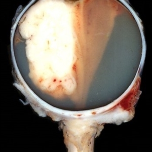 Exophytic Retinoblastoma
May 18 2020 by McGill University Health Centre
This type of tumor grows from the retina toward the choroid. In this enucleation specimen, the retina is completely detached, and the tumor is growing inside the subretinal proteinaceous fluid. Note the distance between the tumor and the optic nerve head.
Condition/keywords: enucleation, retinoblastoma
-
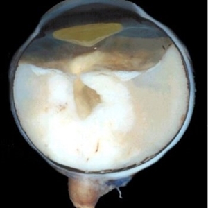 Exo-Endophytic Retinoblastoma
May 18 2020 by McGill University Health Centre
An exo-endophytic tumor is located on the optic nerve head and has produced a retinal detachment. Note the proteinaceous subretinal fluid. The optic nerve is thickened due to retinoblastoma infiltration.
Condition/keywords: enucleation, retinoblastoma
-
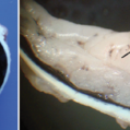
 Infiltration of the Optic Nerve
May 18 2020 by McGill University Health Centre
The main morphological prognostic factor for retinoblastoma is infiltration of the optic nerve. The invasion of the optic nerve has to be assessed by a pathologist in all cases. The superior calotte is removed in this enucleation specimen to show an extensively exophytic necrotic tumor occupying the vitreous chamber with severe infiltration of the optic nerve (arrowhead). Intratumoral calcification is a hallmark of this tumor and can be seen in the chalky white areas (arrow).
Condition/keywords: infiltration of the optic nerve
 Loading…