Search results (625 results)
-
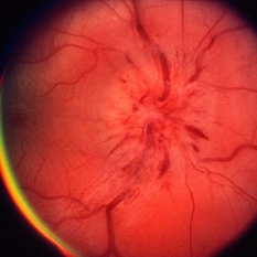
 Bergmeister's Papillae
Mar 29 2013 by Henry J. Kaplan, MD
Remnants of fetal hyaloid artery as fibrous tuft called Bergmeister`s papillae on the optic disc.
Condition/keywords: Bergmeister's Papillae, hyaloid artery
-
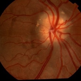
 Papillitis
May 2 2013 by Henry J. Kaplan, MD
Anterior optic neuropathy or papillitis in the right eye; notice the blurred optic disc margin, engorged capillaries and flame shaped hemorrhages at the margin.
Condition/keywords: optic disc edema, optic disc swelling, papillitis
-
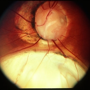
 Optic Disc Edema and Hemorrhages with Subdural Hematoma
Oct 1 2012 by Jeffrey G. Gross, MD, FASRS
Optic disc edema and hemorrhages with subdural hematoma.
Condition/keywords: optic disc edema, subdural hematoma
-
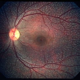
 Optic Disc Drusen
Jul 31 2016 by Mitzy E Torres Soriano, MD
Optic Disc Drusen (Right eye)
Photographer: Mitzy E. Torres Soriano. Retina Department. Hospital Provincial del Centenario. Rosario, Argentina
Imaging device: TOPCON
Condition/keywords: optic disc drusen, optic nerve drusen
-
 Coloboma
Mar 29 2013 by Henry J. Kaplan, MD
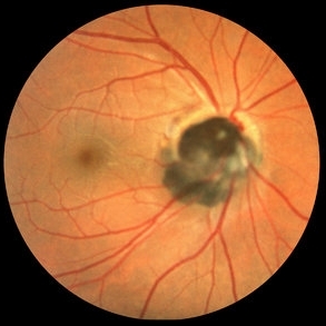
Optic disc and inferonasal choroidal coloboma in the same patient #2.
Condition/keywords: coloboma, coloboma of choroid, coloboma of optic disc
-
 Fundus Albipunctatus
Mar 29 2013 by Henry J. Kaplan, MD
Fundus albipunctatus (one of the stationary night blindness syndromes with multiple white dots in the periphery and normal optic disc and vessels).
Condition/keywords: fundus albipunctatus
-
---thumb.jpg/image-square;max$300,300.ImageHandler) Anterior Ischemic Optic Neuropathy
Anterior Ischemic Optic Neuropathy
Mar 29 2013 by Henry J. Kaplan, MD
Anterior Ischemic Optic Neuropathy; notice the typical pale optic disc swelling and faint splinter hemorrhages.
Condition/keywords: anterior ischemic optic neuropathy
-
 Coloboma of Disc & Choroid
Oct 6 2012 by Hamid Ahmadieh, MD
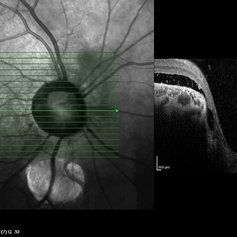
OCT image of a 25-year-old woman with serous retinal detachment secondary to coloboma of disc associated with coloboma of choroid.
Photographer: Hamid Ahmadieh, MD, Ophthalmic Research Center, Labbafinejad Medical Center, Shahid Beheshti University of Medical Sciences
Imaging device: Heidelberg Spectralis
Condition/keywords: coloboma of choroid, coloboma of optic disc, optical coherence tomography (OCT), serous retinal detachment
-
 Central Retinal Vein Occlusion
Sep 2 2012 by Hyung-Woo Kwak, MD
Multiple dense, dark, blotchy hemorrhages, cotton-wool spots, and pale optic disc are signs suggestive of retinal ischemia in CRVO.
Imaging device: Zeiss F450 plus
Condition/keywords: central retinal vein occlusion (CRVO)
-
 Central Retinal Artery Occlusion
Aug 23 2012 by Gerardo Garcia-Aguirre, MD
Fundus photograph of a left eye with central retinal artery occlusion. Note the paleness of the retina (except for a very small area adjacent to the optic disc, probably irrigated by a very small cillioretinal artery), and the cherry red spot. Visual acuity is light perception.
Photographer: Noemí Hernández, Asociación para Evitar la Ceguera en México
Condition/keywords: central retinal artery occlusion (CRAO), cherry red spot, cilioretinal artery occlusion
-
---thumb.jpg/image-square;max$300,300.ImageHandler) Possible CMV Retinitis with Frosted Branch Angiitis
Possible CMV Retinitis with Frosted Branch Angiitis
Feb 14 2013 by From the Collections of Thomas M. Aaberg, MD and Thomas M. Aaberg Jr., MD
Possible CMV Retinitis with frosted branch angiitis appearance and disc edema---late macular star appearance, but diagnosis is not certain.
Condition/keywords: frosted branch angiitis, late macular star, optic disc edema
-
 Hypertensive Retinopathy
Aug 24 2012 by Geoffrey G. Emerson, MD, PhD, FASRS
A 35-year-old man has headaches and decreased vision. The right eye measures 20/25 and the left eye measures 3/200. The blood pressure measures 180/110.This fluorescein angiogram shows leakage of dye from the optic disc (papilledema), ischemia, and dilated capillaries around the foveal avascular zone
Photographer: Geoffrey Emerson, MD, PhD, Retina Center, Minneapolis
Condition/keywords: hypertensive retinopathy, ischemia, papilledema
-
 Myelinated nerve fibres at optic disc
Jan 11 2013 by Alex P. Hunyor, MD
Myelinated nerve fibres at optic disc, left eye.
Condition/keywords: myelinated nerve fibers
-
 Myelinated nerve fibres
Jan 11 2013 by Alex P. Hunyor, MD
Myelinated nerve fibres at optic disc, left eye.
Condition/keywords: myelinated nerve fibers
-
 Hypertensive Retinopathy
Feb 25 2013 by Suber S. Huang, MD, MBA, FASRS
32-year-old African American male with Grade IV hypertensive retinopathy and acute renal failure. Vision OD 20/70, OS 20/25. Creatine 7.1. BP: 250/150.
Photographer: Geoffrey Pankhurst, University Hospitals, Eye Institute/Dept. Ophthalmology and Visual Sciences Case Western Reserve University Cleveland, OH
Imaging device: Topcon TRC 50x
Condition/keywords: acute renal failure, disc edema, exudate, hypertension, hypertensive retinopathy, ischemia, macular edema, macular ischemia, optic disc edema
-
 Myopic Shift With Tilted Optic Disc.
Jul 11 2013 by Jason S. Calhoun
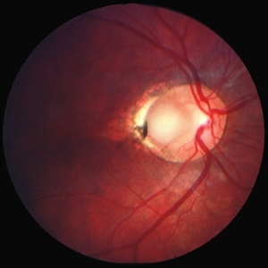
Black female who has a myopic shift shows tilted optic disc in the right eye.
Photographer: Jason S. Calhoun, Department of Ophthalmology, Mayo Clinic Jacksonville, Florida
Condition/keywords: high myopia
-
 Behcet's Disease
Nov 25 2012 by Mallika Goyal, MD
Fundus photograph of left eye of a 23-year-old gentleman with Behcet's Disease shows occlusive retinal vasculitis with optic disc pallor and macular ischemia. This eye has no light perception; other eye has similar fundus appearance.
Photographer: Mallika Goyal, MD, Apollo Health City, Hyderabad, India
Condition/keywords: macular ischemia, occlusive vasculitis, optic disc pallor
-
 Hypertensive Retinopathy
Feb 25 2013 by Suber S. Huang, MD, MBA, FASRS
32-year-old African American male with Grade IV hypertensive retinopathy and acute renal failure. Vision OD 20/70, OS 20/25. Creatine 7.1. BP: 250/150.
Photographer: Geoffrey Pankhurst, University Hospitals, Eye Institute/Dept. Ophthalmology and Visual Sciences Case Western Reserve University Cleveland, OH
Imaging device: Topcon TRC 50x
Condition/keywords: acute renal failure, disc edema, exudate, hypertension, hypertensive retinopathy, ischemia, macular edema, macular ischemia, optic disc edema
-
---thumb.jpg/image-square;max$300,300.ImageHandler) Optic Disc Drusen
Optic Disc Drusen
Jul 10 2013 by Hamid Ahmadieh, MD
SD-OCT image of the left eye of a 24-year-old woman with optic disc drusen and VA 20/20.
Photographer: Solmaz Shahmohammadi, Negah Eye Center, Tehran
Imaging device: Heidelberg Spectralis
Condition/keywords: optic disc drusen, optical coherence tomography (OCT)
-
 Optic Disc Melanocytoma
Oct 16 2012 by Jeffrey G. Gross, MD, FASRS
Optic disc melanocytoma.
Condition/keywords: optic disc melanocytoma
-
 Optic Nerve Head Drusen With Idiopathic CNV
Feb 17 2017 by Kristen Wagner
22-year-old female fundus photograph of a right eye with Optic Nerve Drusen with Idiopathic CNV.
Photographer: Kristen Wagner, COT, OSC Ophthalmic Photographer, Tennessee Retina, Nashville TN
Condition/keywords: choroidal neovascularization (CNV), drusen of optic disc, optic disc drusen
-
 Optic Disc Drusen
Jul 10 2013 by Hamid Ahmadieh, MD
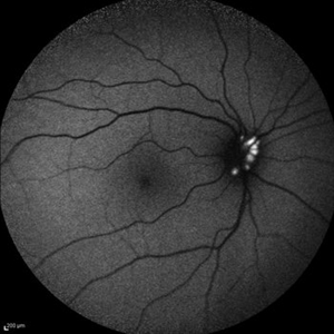
Fundus autofluorescence image of the right eye of a 24-year-old woman with optic disc drusen and VA 20/20.
Photographer: Solmaz Shahmohammadi, Negah Eye Center, Tehran
Imaging device: Heidelberg Spectralis
Condition/keywords: fundus autofluorescence (FAF), optic disc drusen
-
 Optic Disc Coloboma
Apr 25 2017 by Nimrod Dar
9 year-old patient, noticed a gradual deterioration in her visual acuity at her LE (6/15). On her examination, a double optic disc can be seen. OCT scan revealed an intra retinal fluid and macular schisis.
Photographer: Nimrod Dr, MD
Condition/keywords: coloboma of the optic nerve
-
 Optic disc pit 2
-
 Coloboma of Disc & Choroid
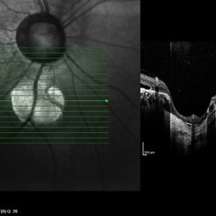
Oct 6 2012 by Hamid Ahmadieh, MD
OCT image of a 25-year-old woman with a small coloboma of choroid associated with coloboma of disc.
Photographer: Hamid Ahmadieh, MD, Ophthalmic Research Center, Labbafinejad Medical Center, Shahid Beheshti University of Medical Sciences
Imaging device: Heidelberg Spectralis
Condition/keywords: coloboma of choroid, coloboma of optic disc, optical coherence tomography (OCT)
 Loading…