File number: 14334
Comments
-
 Raj K. Maturi, MD (December 1 2014)
Hi Bryan,
Thank you for taking the time to comment. I appreciate it.
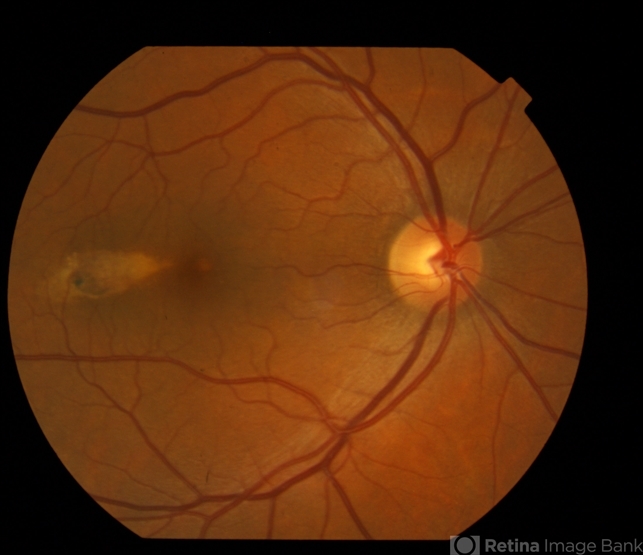
This was an isolated lesion. Agree that FAP is a concern, and in fact, the primary differential diagnosis in cases of Torpodo. I've copied a report form Ocular Surgery News (2013) that describes this well (see below link):
http://www.healio.com/ophthalmology/retina-vitreous/news/print/osn-retina/%7B02e1e3b0-e003-4f0b-86d9-a73d1a79c04a%7D/fundus-lesion-targets-the-central-macula?page=1 -
 Bryan K. Rutledge, MD (December 1 2014)
Looks more like pigmented ocular fundus lesion (rather than nevus), similar to what I have seen in patients with FAP. If other pigmented lesions, consider testing.
Sign in to comment.
 Initializing download.
-
 By Raj K. Maturi, MD
Midwest Eye Institute & Retina Partners Midwest - Uploaded on Feb 26, 2014.
- Last modified by Caroline Bozell on Feb 26, 2014.
- Rating
- Appears in
- Miscellaneous
- Condition/keywords
- fundus photograph, torpedo maculopathy, choroidal nevus
- Photographer
- Charlotte Harris COA Midwest Eye Institute Indianapolis, Indiana
- Imaging device
-
Fundus camera
TOPCON 50EX - Description
- Color fundus of a 47-year-old female with torpedo maculopathy- atypical choroidal nevus.
")
")
")


