Search results (4766 results)
-
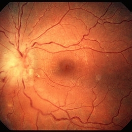
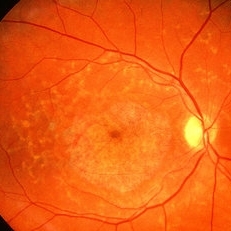
 Lacquer Cracks
Oct 13 2012 by Geoffrey G. Emerson, MD, PhD, FASRS
Lacquer cracks
Condition/keywords: lacquer cracks, myopic macular degeneration
-
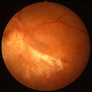
---thumb.jpg/image-square;max$300,300.ImageHandler) Proliferative Diabetic Retinopathy (PDR) & Traction Retinal Detachment
Proliferative Diabetic Retinopathy (PDR) & Traction Retinal Detachment
Feb 13 2013 by From the Collections of Thomas M. Aaberg, MD and Thomas M. Aaberg Jr., MD
Florid NV with early macular TRD.
Condition/keywords: neovascularization (NV), tractional retinal detachment
-
 Ozurdex implant
Aug 23 2012 by Daniel A. Adelberg, MD, FASRS
Anterior Segment photograph of a 50 year old with Uveitis and Cystoid Macular Edema status post Intravitreal injection of an Ozurdex dexamethasone implant
Photographer: Robert Ramsey, Southwestern Eye Center, Mesa Arizona
Condition/keywords: Ozurdex implant
-
 Familial Dominant Drusen
Nov 22 2015 by Mallika Goyal, MD
Bilateral drusen over the entire retinal mid-periphery and periphery of a 29-year-old male with no visual complaints. Macular centre is normal though there are some drusen in the temporal macula.
Photographer: Mallika Goyal, MD, Apollo Health City, Jubilee Hills, Hyderabad, India
Condition/keywords: familial drusen
-
 Toxoplasma Neuroretinitis (Jensen`s Disease)
Feb 25 2013 by Henry J. Kaplan, MD
Toxoplasma neuroretinitis in the left eye of a patient with macular star formation, retinitis adjacent to the optic nerve head with disc swelling.
Condition/keywords: Jensen disease, ocular toxoplasmosis, toxoplasmosis
-
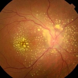 Uveitis Posterior
Jul 19 2019 by JEFFERSON R SOUSA, Tecg.º (Biomedical Systems Technology)
A 23-year-old male patient attended the clinic with low vision of the right eye. In the evaluation it presented important fundoscopical alterations like retinal exudations in the posterior pole and nasal retina, aspects of macular star. It was proven that it was a posterior uveitis.
Photographer: JEFFERSON R SOUSA - Study Center and Ophthalmological Research Dr. Andre M V Gomes, Institute Dr. Suel Abujamra São Paulo-Brazil
Imaging device: Topcon TRC-50 DX, Imaginet 4.0, angle de 50 graus. Flash 50w-s
Condition/keywords: uveitis
-
---thumb.JPG/image-square;max$300,300.ImageHandler) Disciform Scar
Disciform Scar
Jul 13 2013 by Jason S. Calhoun
Poor central vision in the left eye due to macular degeneration. Disciform scar.
Photographer: Jason S. Calhoun, Department of Ophthalmology, Mayo Clinic Jacksonville, Florida
Imaging device: TOPCON TRC 50-EX
Condition/keywords: disciform scar, macular degeneration
-
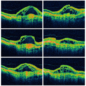 Wet Macular Degeneration OCT
Oct 13 2012 by Geoffrey G. Emerson, MD, PhD, FASRS
Condition/keywords: optical coherence tomography (OCT)
-
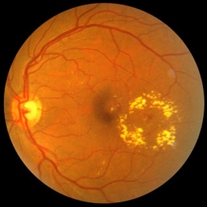
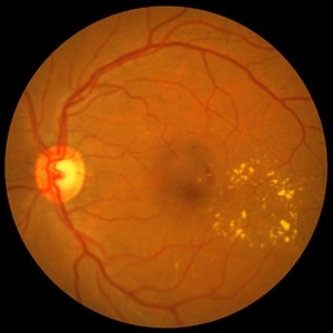 NPDR with CSME
Oct 8 2012 by Jeffrey G. Gross, MD, FASRS
NPDR with CSME with circinate ring of lipid s/p laser.
Condition/keywords: circinate ring, laser, macular edema, nonproliferative diabetic retinopathy
-
 Macular Choroidal Osteoma
Aug 17 2012 by Jonathan L. Prenner, MD
Macular choroidal osteoma in a 29-year-old woman
Condition/keywords: macular choroidal osteoma
-
 Fundus Flavimaculatus
May 2 2013 by Henry J. Kaplan, MD
Yellow pisciform flecks at the level of RPE accompanied by Bull's eye, Right eye; #1.
Condition/keywords: bull's eye maculopathy, fundus flavimaculatus, Stargardt disease
-
 PDR with Traction RD of Macular
Oct 8 2012 by Jeffrey G. Gross, MD, FASRS
PRD, with traction RD of macular, pre-op, 20/200.
Condition/keywords: 20/200, macular, pre-op, tractional retinal detachment
-
---thumb.jpg/image-square;max$300,300.ImageHandler) Central Retinal Artery Occlusion Sparing Macula
Central Retinal Artery Occlusion Sparing Macula
Oct 18 2012 by Larry Halperin, MD
Central retinal artery occlusion sparing macula
Condition/keywords: central retinal artery occlusion (CRAO), macula
-
 Geographic Atrophy, Fundus photograph
Aug 23 2012 by Gerardo Garcia-Aguirre, MD
Fundus photograph of an 85-year-old patient with age related macular degeneration and geographic atrophy. A large area with well-defined borders is observed, in which the choroidal vasculature is visualized.
Photographer: Noemí Hernández, Asociación para Evitar la Ceguera en México
Imaging device: Zeiss FF4
Condition/keywords: geographic atrophy
-
 NPDR with CSME
Oct 8 2012 by Jeffrey G. Gross, MD, FASRS
NPDR with CSME with circinate ring of lipid.
Condition/keywords: circinate ring, macular edema, nonproliferative diabetic retinopathy
-
 Tractional Retinal Detachment
Sep 27 2012 by Virgilio Morales-Canton, MD
OCT image of a 42-year-old male patient with a localized traction of the superior macula secondary to proliferative diabetic retinopathy.
Imaging device: Cirrus
Condition/keywords: tractional retinal detachment
-
---thumb.jpg/image-square;max$300,300.ImageHandler) Geographic atrophy
Geographic atrophy
Aug 29 2012 by Young Hee Yoon, MD, PhD
OCT image of an 78-year-old woman. Her best-corrected visual acuity was counting fingers at 30cm.
Photographer: Ji Hee Kim, Asan Medical Center
Imaging device: Heidelberg spectralis
Condition/keywords: dry age-related macular degeneration (dry AMD), geographic atrophy
-
 Macula off Rhegmatogenous Retinal Detachment
Aug 28 2012 by Sharon Fekrat, MD FACS FASRS
62 year old man with a rhegmatogenous retinal detachment involving the foveal center in his left eye as depicted on this Zeiss Stratus OCT image.
Photographer: Michael P. Kelly, FOPS Director, Duke Eye Labs, Duke University Eye Center, Durham, NC
Imaging device: Zeiss Stratus
-
 Myelinated Nerve Fibers
Sep 17 2012 by Michael P. Kelly, FOPS
Retinal fundus photograph of a macular hole.
Photographer: Michael P. Kelly, FOPS Director, Duke Eye Labs, Duke University Hospital, Duke Eye Center
Imaging device: Topcon
Condition/keywords: macular hole, myelinated nerve fibers
-
---thumb.jpg/image-square;max$300,300.ImageHandler) C3F8 gas bubble after retinal detachment surgery
C3F8 gas bubble after retinal detachment surgery
Feb 1 2013 by Sharon Fekrat, MD FACS FASRS
63 year old man s/p encircling scleral buckle and 23g pars plana vitrectomy for a macula off phakic rhegmatogenous retinal detachment. This fundus photograph shows the effect of the encircling buckle and the residual C3F8 intravitreal gas bubble in the right eye.
Photographer: Tiffanie Keaton, Duke Eye Imaging, Duke University Eye Center, Durham, NC
Imaging device: Optos
Condition/keywords: intravitreal gas bubble, vitrectomy
-
 Familial Dominant Drusen
Nov 22 2015 by Mallika Goyal, MD
Bilateral drusen over the entire retinal mid-periphery and periphery of a 29-year-old male with no visual complaints. Macular centre is normal though there are some drusen in the temporal macula.
Photographer: Mallika Goyal, MD, Apollo Health City, Jubilee Hills, Hyderabad, India
Condition/keywords: familial drusen
-
---thumb.jpg/image-square;max$300,300.ImageHandler) Reticular Pattern Dystrophy
Reticular Pattern Dystrophy
Aug 7 2013 by From the Collections of Thomas M. Aaberg, MD and Thomas M. Aaberg Jr., MD
Color fundus photograph reveals typical reticular - type pattern dystrophy, OS.
Condition/keywords: pattern macular dystrophy, reticular dystrophy
-
 Traumatic Macular Hole with Retinal Detachment and PVR - montage
Sep 27 2012 by Pauline T Merrill, MD, FASRS
Fundus photo montage of a 13-year-old boy s/p soccer ball injury 1 month previously.
Photographer: Karen Parque, Illinois Retina Associates, Chicago, IL
Condition/keywords: proliferative vitreoretinopathy (PVR), traumatic macular hole
-
 Central Retinal Artery Occlusion & Cilioretinal Artery Sparing
Dec 22 2012 by Hamid Ahmadieh, MD
Early phase FA image of the right eye of a 34-year-old man with sudden drop of vision due to CRAO. The macula is involved despite cilioretinal artery sparing .
Photographer: Zohre Salimi; Labbafinejad Medical Center, Shahid Beheshti University of Medical Sciences , Tehran
Imaging device: Heidelberg HRA
Condition/keywords: central retinal artery occlusion (CRAO), cilioretinal sparing
-
---thumb.jpg/image-square;max$300,300.ImageHandler) Tamoxifen Retinopathy- OCT
Tamoxifen Retinopathy- OCT
Aug 30 2012 by Young Hee Yoon, MD, PhD
OCT image of an 58-year-old woman with a bilateral tamoxifen maculopathy. She had taken tamoxifen for 24 months due to breast cancer. In spite of discontinuation 2 years ago, her macula remained unchanged. Her best-corrected visual acuity was 20/50 in the right and 20/100 in the left.
Photographer: Soon Tae Kim, Asan Medical Center
Imaging device: Heidelberg Spectralis
Condition/keywords: drug toxicity
 Loading…