Search results (4766 results)
-
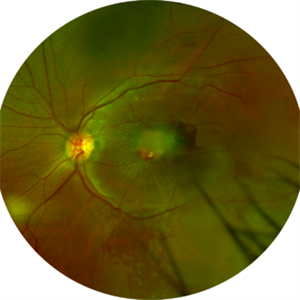
 Acute Macular Neuroretinopathy
Dec 11 2019 by Lauren Whaley
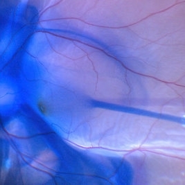
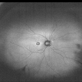
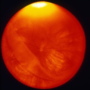
34-year-old female patient presented with changes in vision after recent upper respiratory infection. Referring doctor originally thought it was a blood pressure issue. She noticed a "C" shape in her vision. Infrared image was captured showing exactly what patient was describing! Doctor confirmed with this image that it was AMN.
Photographer: Lauren R. Whaley, COA
Imaging device: Heidelberg Spectralis
Condition/keywords: 30 degrees, acute macular neuroretinopathy, Heidelburg Spectralis, left eye, macula, near infrared autofluorescence (NIRAF)
-
 Brilliant Blue Dye Injection to Stain ILM in a Macular Hole with Retinal Detachment
Feb 4 2022 by Manish Nagpal, MD, FRCS (UK), FASRS
Intraoperative still of a Brilliant blue dye injection being done to stain the ILM.
Photographer: Manish Nagpal, Director, Retina Foundation, Ahmedabad
Imaging device: Sony PMW -10 MD surgical camera
Condition/keywords: full thickness macular hole, macula, retina
-
 Central Areolar Choroidal Dystrophy
Apr 14 2013 by Edwin H. Ryan, MD
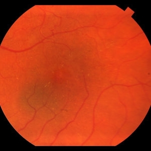
Fundus photograph of a 52-year-old woman with CACD. 5/200 OD, 20/50 OS.
Condition/keywords: central areolar choroidal dystrophy (CACD), geographic atrophy, macula
-
 Central Areolar Choroidal Dystrophy
Apr 14 2013 by Edwin H. Ryan, MD
Fundus photograph of a 52-year-old woman with CACD. 5/200 OD, 20/50 OS.
Condition/keywords: central areolar choroidal dystrophy (CACD), geographic atrophy, macula
-
 Central Areolar Choroidal Dystrophy
Apr 14 2013 by Edwin H. Ryan, MD
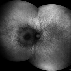
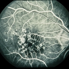
Mid-FA of a 52-year-old woman with CACD. 5/200 OD, 20/50 OS.
Condition/keywords: central areolar choroidal dystrophy (CACD), geographic atrophy, macula
-
 Central Areolar Choroidal Dystrophy
Apr 14 2013 by Edwin H. Ryan, MD
Late FA of a 52-year-old woman with CACD. 5/200 OD, 20/50 OS.
Condition/keywords: central areolar choroidal dystrophy (CACD), geographic atrophy, macula
-
 Central Areolar Choroidal Dystrophy
Apr 14 2013 by Edwin H. Ryan, MD
Late FA of a 52-year-old woman with CACD. 5/200 OD, 20/50 OS.
Condition/keywords: central areolar choroidal dystrophy (CACD), geographic atrophy, macula
-
 Central Areolar Choroidal Dystrophy
Apr 14 2013 by Edwin H. Ryan, MD
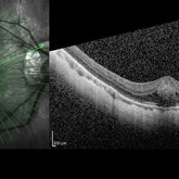
SD-OCT of a 52-year-old woman with CACD. 5/200 OD, 20/50 OS.
Condition/keywords: central areolar choroidal dystrophy (CACD), geographic atrophy, macula
-
 Central Areolar Choroidal Dystrophy
Apr 14 2013 by Edwin H. Ryan, MD
SD-OCT of a 52-year-old woman with CACD. 5/200 OD, 20/50 OS. Submacular fluid noted.
Condition/keywords: central areolar choroidal dystrophy (CACD), geographic atrophy, macula
-
 Central Bouquet Hemorrhage
May 31 2025 by Moazzam Parvez
OCT image of a 26 year old gentleman of right eye macula with a central foveolar cotton ball like lesion . Inward traction by Müller cells over CB causes upward displacement of foveal cones without major disturbance of ellipsoid zone (EZ) and ELM . Cotton ball sign is characterized by small, fuzzy subfoveal hyperreflective area between the inner segment ellipsoid zone (EZ) and the interdigitation zone (IZ) .
Photographer: Dr Moazzam Parvez , Netralayam , Kolkata
Imaging device: Heidelberg Spectralis
Condition/keywords: Central bouquet haemorrhage, macula, myopia
-
---thumb.jpg/image-square;max$300,300.ImageHandler) Central Retinal Artery Occlusion Sparing Macula
Central Retinal Artery Occlusion Sparing Macula
Oct 18 2012 by Larry Halperin, MD
Central retinal artery occlusion sparing macula
Condition/keywords: central retinal artery occlusion (CRAO), macula
-
 Chloroquine maculopathy
Jun 22 2022 by JORGE SOBERANES
Fundus autofluorescence of a bull´s eye maculopathy of a 55-year-old woman treated for ten years with choloquine.
Photographer: Jorge I. Soberanes MD, Asociación para Evitar la Ceguera en México.
Imaging device: Zeiss Clarus 700 (Green autofluorescence)
Condition/keywords: bull's eye maculopathy, chloroquine, fundus autofluorescence (FAF), macula, maculopathy
-
 Choroidal Nevus
May 27 2016 by Olivia Rainey
Color fundus image of a small choroidal nevus near the macula.
Photographer: Olivia Rainey
Imaging device: Topcon50dx
Condition/keywords: 20 degrees, choroidal nevus, color fundus photograph, color photo, macula
-
 Choroidal Rupture Across Macula
Oct 23 2012 by Larry Halperin, MD
Choroidal rupture across macula
Condition/keywords: choroidal rupture, macula
-
 Combined Hamartoma
Feb 29 2016 by Andrea Arriola-Lopez, MD MSc
40 year-old man with diminished VA since 6 month ago. Fundus examination revealed macular folds, yellow-whitish elevated lesion at the fovea and a subretinal hemorrhage.
Photographer: Andrea Elizabeth Arriola-Lopez MD, MSc
Imaging device: OPTOS Dakota
Condition/keywords: combined hamartoma, macula, subretinal hemorrhage
-
 Cone Dystrophy
Mar 22 2015 by Andrea Arriola-Lopez, MD MSc
Fundus autofluorescence of an 31-year-old male with cone dystrophy.
Photographer: Andrea Elizabeth Arriola López, MSc. Asociación para Evitar la Ceguera, I.A.P. México D.F.
Imaging device: OPTOS, Dakota.
Condition/keywords: autofluorescence imaging, cone dystrophy, fundus autofluorescence (FAF), macula, macula lesion
-
---thumb.jpg/image-square;max$300,300.ImageHandler) Cone Dystrophy
Cone Dystrophy
Feb 20 2013 by From the Collections of Thomas M. Aaberg, MD and Thomas M. Aaberg Jr., MD
High mag color photo of the macula of OD in a patient with cone dystrophy; VA=20/80.
Condition/keywords: color photo, cone dystrophy, macula
-
---thumb.jpg/image-square;max$300,300.ImageHandler) Cone Dystrophy
Cone Dystrophy
Feb 20 2013 by From the Collections of Thomas M. Aaberg, MD and Thomas M. Aaberg Jr., MD
Color photo of the fundus of OD in a patient with cone dystrophy; VA=20/80.
Condition/keywords: color photo, cone dystrophy, macula
-
---thumb.jpg/image-square;max$300,300.ImageHandler) Cone Dystrophy
Cone Dystrophy
Feb 20 2013 by From the Collections of Thomas M. Aaberg, MD and Thomas M. Aaberg Jr., MD
Green filter photo of the macula of an eye with cone dystrophy.
Condition/keywords: color photo, cone dystrophy, macula
-
---thumb.jpg/image-square;max$300,300.ImageHandler) Cone Dystrophy
Cone Dystrophy
Feb 20 2013 by From the Collections of Thomas M. Aaberg, MD and Thomas M. Aaberg Jr., MD
Green filter photo of the macula of an eye with cone dystrophy.
Condition/keywords: cone dystrophy, green filter, macula
-
 Congenital Hypertrophy of the Retinal Pigment Epithelium
Nov 11 2019 by Jessica Norkus
Bilateral Optos ultra wide field imaging of a 31-year-old female patient with CHRPE lesions. Lesions in OD were suspicious of Gardner Syndrome due to familial history of cancerous polyps in colon. Patient underwent colonoscopy and was deemed clear.
Photographer: Jessica Norkus, COA, Retina Specialists of Michigan
Imaging device: Optos Ultra Wide Field Camera
Condition/keywords: bear tracks, bilateral, color fundus photograph, color photo, congenital hypertrophy of the retinal pigment epithelium (CHRPE), fundus autofluorescence (FAF), fundus photograph, lacunae, macula, optic disc, Optos, pseudocolor, ultra-wide field imaging
-
---thumb.jpg/image-square;max$300,300.ImageHandler) Flecked Retina
Flecked Retina
-
 Foveal Detachment in Dome-Shaped Macula
May 6 2017 by Mitzy E Torres Soriano, MD
Vertical scans of optical coherence tomography showing a foveal detachment at the top of dome-shaped macula in a 48-year-old female with high myopia.
Photographer: Mitzy E. Torres Soriano
Condition/keywords: detachment, high myopia, macula
-
 Giant Retinal Tear With Retinal Detachment, Macula On
Oct 2 2013 by Jerald A. Bovino, MD
This patient has a giant retinal tear with a retinal detachment. The macula is still attached.
Condition/keywords: macula, retinal tear
-
 Grouped Albinotic Spots in Macula
Mar 4 2014 by David Callanan, MD
39-year-old female, 20/15 OU; normal color, ERG, EOG, thresholds, VF.
Condition/keywords: albinism, macula
 Loading…