Search results (4766 results)
-
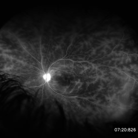
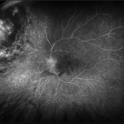
 Wide-Field-OCT-montage
Jan 8 2018 by Netan Choudhry, MD, FRCS(C) FASRS
This is an SD-OCT montage image of a 55 year old male with optic neuropathy representing a wide-field OCT spanning 130 degrees.
Photographer: John Golding, Vitreous Retina Macula Specialists of Toronto
Imaging device: Heidelberg Spectralis OCT system
Condition/keywords: wide angle imaging
-
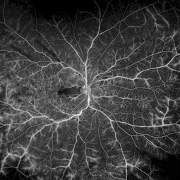
 Whole Eye OCT
Jan 4 2019 by Netan Choudhry, MD, FRCS(C) FASRS
Swept-Source OCT montage of a 45-year-old male with Alports disease and posterior subcapsular cataract.
Photographer: John Golding BA, Vitreous Retina Macula Specialists of Toronto
Imaging device: Topcon DRI Triton
Condition/keywords: Alports disease, optical coherence tomography (OCT), swept source
-
 Foreign Body SS
Feb 14 2015 by Shlomit Schaal, MD, PhD, MHCM
A four millimeter metal foreign body removed surgically using foreign body forceps. The macula is protected by PFCL heavy fluid. There is a traumatic retinal detachment inferiorly (top of photo, surgeon's view).
Photographer: Shlomit Schaal, MD, PhD
Condition/keywords: intraocular foreign body
-
 Acute Posterior Multifocal Placoid Pigment Epitheliopathy
Feb 20 2024 by Soobien Lee
A 20-year-old caucasian female with viral prodrome and vision loss OS>OD secondary to Acute Posterior Multifocal Placoid Pigment Epitheliopathy (APPME). OCT of the left macula shows bacillary layer detachment.
Photographer: Kim Seay, Elman Retina Group
Condition/keywords: acute posterior multifocal placoid pigment epitheliopathy (APMPPE), bacilliary layer detachment, OCT, Uveitis, white dot syndrome
-
 Cat Scratch
Feb 15 2017 by Hua Gao, MD, PhD, FASRS
A female patient of 57-year-old presented with neuroretinitis due to cat-scratch disease with positive Bartonella henselae antibodies. Two weeks after symptom onset she developed exudates in a "macular star" pattern.
Condition/keywords: cat scratch retinitis
-
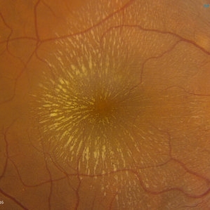
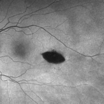 Torpedo Maculopathy
Feb 20 2024 by Soobien Lee
Optos fundus autofluorescence photograph of a 35-year-old asymptomatic female with no ocular or medical history with stable and chronic appearing torpedo-shaped macula lesion in the left eye.
Photographer: Peter Sotirakos, Elman Retina Group
Imaging device: Optos Ultra-Widefield Autoflurescence Imaging
Condition/keywords: autofluorescence imaging, genetics, macula, maculopathy, Optos, torpedo maculopathy
-
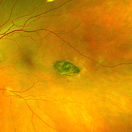 Torpedo Maculopathy
Feb 20 2024 by Soobien Lee
Optos color fundus photograph of a 35-year-old asymptomatic female with no ocular or medical history with stable and chronic appearing torpedo-shaped macula lesion in the left eye.
Photographer: Peter Sotirakos, Elman Retina Group
Imaging device: Optos Ultra-Widefield Imaging
Condition/keywords: macula, Optos, torpedo maculopathy
-
 Central Retinal Artery Occlusion & Cilioretinal Artery Sparing
Dec 22 2012 by Hamid Ahmadieh, MD
Early phase FA image of the right eye of a 34-year-old man with sudden drop of vision due to CRAO. The macula is involved despite cilioretinal artery sparing .
Photographer: Zohre Salimi; Labbafinejad Medical Center, Shahid Beheshti University of Medical Sciences , Tehran
Imaging device: Heidelberg HRA
Condition/keywords: central retinal artery occlusion (CRAO), cilioretinal sparing
-
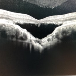 Choroidal Excavation
Jun 2 2019 by Nelson Chamma Capelanes, MD
SD-OCT of a 32-year-old woman showing a subfoveal choroidal excavation associated with chronic central serous chorioretinopathy.
Photographer: Nelson Chamma Capelanes, Promacula, Brazil
Imaging device: Heidelberg Spectralis SD-OCT
Condition/keywords: choroidal excavation, chronic central serous chorioretinopathy (CSCR), pachychoroid
-
 Retinal Detachment with PVR (s/ SPR, PPV, MPV, 360 Retinectomy, PFO, PI, FAx, SO)
Aug 22 2019 by Merrick Avila
Ultra-wide field pseudocolor fundus photograph of a 64-year-old female with a treated retinal detachment with proliferative vitreoretinopathy. Patient has a history of complex retinal detachments that have been treated multiple times. On exam 8-22-19, there were large macular holes with LP vision. There was a long discussion about guarded nature of her condition and goals or trial for repair including globe sparing prevention of phthisis.
Photographer: Merrick Avila
Imaging device: Optos
Condition/keywords: diabetic retinopathy, hemorrhage, Optos, proliferative vitreoretinopathy (PVR), retinectomy, silicone oil
-
 Retinal Vasculitis in Behcet's OS
Jun 29 2018 by Gareth Lema, MD, PhD
IVFA at 7 minutes showing retinal vasculitis, cystoid macular edema, and disc staining.
Photographer: Ross Eye Institute, University at Buffalo Jacobs School of Medicine, Buffalo. NY
Imaging device: Optos
Condition/keywords: Behcet's Disease, cystoid macular edema (CME), disc staining, retinal vasculitis
-
 Tractional Retinal Detachment
Sep 27 2012 by Virgilio Morales-Canton, MD
OCT image of a 42-year-old male patient with a localized traction of the superior macula secondary to proliferative diabetic retinopathy.
Imaging device: Cirrus
Condition/keywords: tractional retinal detachment
-
 Acute Macular Neuroretinopathy
Dec 11 2019 by Lauren Whaley
34-year-old female patient presented with changes in vision after recent upper respiratory infection. Referring doctor originally thought it was a blood pressure issue. She noticed a "C" shape in her vision. Infrared image was captured showing exactly what patient was describing! Doctor confirmed with this image that it was AMN.
Photographer: Lauren R. Whaley, COA
Imaging device: Heidelberg Spectralis
Condition/keywords: 30 degrees, acute macular neuroretinopathy, Heidelburg Spectralis, left eye, macula, near infrared autofluorescence (NIRAF)
-
---thumb.jpg/image-square;max$300,300.ImageHandler) Central Retinal Vein Occlusion
Central Retinal Vein Occlusion
Oct 30 2012 by Lihteh Wu, MD
35-year-old hypertensive man with an acute CRVO. Notice the peripapillary cotton wool spots, superficial flame shaped hemorrhages and deeper dot and blot hemorrhages in all 4 quadrants. This is the typical blood and thunder appearance of a CRVO.
Condition/keywords: central retinal vein occlusion (CRVO), cotton wool spots
-
 Choroidal Fracture
Oct 27 2024 by César Adrián Gómez Valdivia, MD
Fundus photograph of a traumatic choroidal fracture & extra-macular sub-retinal hemorrhage.
Photographer: @eyemissu2
Imaging device: TOPCON TRC-50DX
Condition/keywords: Choroidal Fracture
-
 CRVO
CRVO
Apr 22 2017 by Gabriel Costa Andrade, PhD
Panoramic retinography (Optos® California) of the right eye of a 48-year-old female patient with a history of low-vision in the right eye 2 months ago. At the exam presented visual acuity of 20/200 in the right eye and 20/20 in the left eye. Angiography shows diffuse perivascular leakage associated with areas of hypoperfusion in macula and periphery.
Photographer: Gabriel Andrade
Imaging device: Optos® California
Condition/keywords: central retinal vein occlusion (CRVO)
-
 Cuticular and soft drusen
Jun 14 2021 by Gerardo Garcia-Aguirre, MD
Fundus photograph (left) and Retro mode infrared image (right) of an eye with soft and cuticular drusen. Drusen are highlighted and better visualized with retro mode imaging.
Photographer: Gerardo Garcia-Aguirre
Imaging device: Nidek Mirante
Condition/keywords: drusen, dry age-related macular degeneration (dry AMD)
-
 Displaced & folded macula
Oct 10 2022 by Ricardo Leitão Guerra
Tractional retinal detachment due to sickle cell retinopathy leading to a displaced and folded appearance of the macula in this 36-yo male. Subretinal bands are also noticed crossing the macula towards inferior retinal detachment area.
Photographer: Ricardo Leitão Guerra
Imaging device: Clarus 700 - Zeiss
Condition/keywords: folds, sickle cell retinopathy, subretinal bands, tractional retinal detachment
-
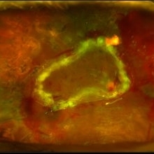
 Epiretinal Membrane/Macular Pucker With Combined Hamartoma of Retina and RPE
Jul 8 2015 by Emmanuel Chang, MD PhD FACS FASRS
10-year-old with history of progressive severe distortion in the left eye over the past year.
Photographer: Retina and Vitreous of Texas
Imaging device: Heidelberg Autofluorescence
Condition/keywords: combined hamartoma, epiretinal membrane (ERM), retinal pigment epithelium (RPE) hamartoma
-
 Green Goblin Detachment
Jan 13 2022 by Netan Choudhry, MD, FRCS(C) FASRS
Tractional retinal detachment with macular hole in a 76-year-old female.
Photographer: John Golding BA, Vitreous Retina Macula Specialists of Toronto, OCTane Imaging Lab
Imaging device: Multicolor fundus photo taken on the Spectralis OCT2 (Heidelberg Engineering GmbH).
Condition/keywords: macular hole, Multispectral imaging, tractional retinal detachment
-
 Hypertensive Retinopathy
Feb 25 2013 by Suber S. Huang, MD, MBA, FASRS
32-year-old African American male with Grade IV hypertensive retinopathy and acute renal failure. Vision OD 20/70, OS 20/25. Creatine 7.1. BP: 250/150.
Photographer: Geoffrey Pankhurst, University Hospitals, Eye Institute/Dept. Ophthalmology and Visual Sciences Case Western Reserve University Cleveland, OH
Imaging device: Topcon TRC 50x
Condition/keywords: acute renal failure, disc edema, exudate, hypertension, hypertensive retinopathy, ischemia, macular edema, macular ischemia, optic disc edema
-
 Lady in a dress
Feb 9 2023 by Shelby Helton
Fluorescein Angiography on a 67-year-old male with significant RPE changes secondary to a severe subretinal hemorrhage that required a vitrectomy with subretinal TPA in 2013.
Photographer: Shelby Helton
Imaging device: Heidelberg Spectralis
Condition/keywords: wet age-related macular degeneration (wet AMD)
-
 Myopic Traction Maculopathy
Mar 17 2025 by Drew Mitchell
HD 1 line 100x 9 mm scan of a right eye with MTM at stage 3c. Macular Schisis Detachment.
Photographer: Drew Mitchell OCT-C
Imaging device: Zeiss Cirrus 5000
Condition/keywords: full thickness macular hole, Macular hole, myopic foveoschisis, myopic macular schisis, myopic traction maculopathy, PVD
-
 Proliferative Diabetic Retinopathy
Jul 15 2022 by Gabriel Costa Andrade, PhD
Fundus angiography of an 22-year-old man with proliferative diabetic retinopathy and macular ischemia.
Photographer: Dr Gabriel Andrade
Condition/keywords: Diabetes
-
 Radiation Retinopathy; BRVO with Macular Edema
Apr 26 2023 by Denica Rodriguez
Ultra-wide field fluorescein angiography of a 61 year old male with radiation retinopathy following brachytherapy for choroidal melanoma of his left eye. Following treatment, patient developed a branch retinal vein occlusion both ischemic and non-ischemic. Anti-VEGF injections were recommended. The fine needle biopsy showed a class 2 uveal melanoma. Patient also has diabetic retinopathy affecting both eyes. Patient's vision at the time the image was taken was Dcc 20/80-1.
Photographer: Denica Rodriguez COA, ST
Imaging device: Optos California
Condition/keywords: branch retinal vein occlusion (BRVO), Choroidal melanoma, diabetic retinopathy, FA, fluorescein angiogram (FA), I-125 brachytherapy, macular edema, melanoma, Optos, radiation retinopathy, Retina, ultra-wide field imaging
 Loading…