Search results (38 results)
-
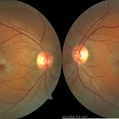
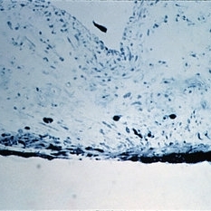
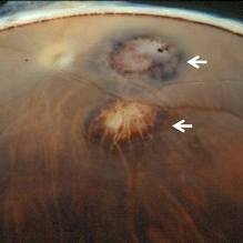
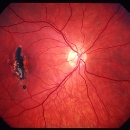 Choroidal rupture
Jan 11 2013 by Alex P. Hunyor, MD
Choroidal rupture, right eye. Note marked RPE hyperplasia
Condition/keywords: choroidal rupture
-
 Retinal hyperplasia
Feb 19 2018 by JEFFERSON R SOUSA, Tecg.º (Biomedical Systems Technology)
Female patient, 28 years in monitoring to control a hyperpigmented lesion in the temporal retina of the right eye.
Photographer: Photographer JEFFERSON ROCHA DE SOUSA, Clinic Dr. Marco Antonio Albhy Oftalmology, Institute Dr. Suel Abujamra São Paulo-Brazil
Imaging device: Retinografo Topcin TRC-NW6S. Mosaic, Flash 25.
Condition/keywords: hyperplasia, hyperplastic retinal pigment epithelium (RPE)
-
 Relentless Placoid Chorioretinitis
Jan 22 2021 by Renata Garcia Franco, Md
20-year-old male with reduction of vision in both eyes, scotoma and metamorphopsia. Widespread multiple chorioretinal lesions with RPE hyperplasia, which appear from posterior pole to peripheral retina and inactive choroidal neovascular membrane.
Photographer: Fatima Hernandez, Instituto de la Retina del Bajio SC
Imaging device: Zeiss
Condition/keywords: chorioretinitis
-
 Relentless Placoid Chorioretinitis
Jan 22 2021 by Renata Garcia Franco, Md
20-year-old male with reduction of vision in both eyes, scotoma and metamorphopsia. Widespread multiple chorioretinal lesions with RPE hyperplasia, which appear from posterior pole to peripheral retina.
Photographer: Fatima Hernandez, Instituto de la Retina del Bajio SC
Imaging device: Zeiss
Condition/keywords: chorioretinitis
-
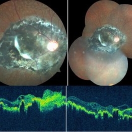
 PEHCR (Peripheral Exudative Hemorrhagic Chorioretinopathy)
May 12 2023 by Niloofar Piri, MD
Ultrawide fundus photograph of the left eye demonstrating extensive peripheral hemorrhagic exudative detachment in a 79 yo Caucasian female with prior history of non-exudative AMD. Recent diagnosis of Acute myeloid leukemia with low platelet count which might have contributed to the above presentatuon. Please note the temporal subretinal hemorrhage as well as RPE atrophy and hyperplasia in the macula.
Photographer: Rocio Bentivegna, MD, Saint Louis University; Jessica Maddox, COA, Saint Louis University
Condition/keywords: peripheral exudative hemorrhagic chorioretinopathy (PEHCR)
-
 Chorioretinitis Sclopetaria
May 4 2021 by Priya Rasipuram Chandrasekaran, MBBS, DO, DNB, FRCS
This fundus photo and montage shows pigmentary changes with fibroglial proliferation of the disc and macula in a 36-year-old male following injury with an iron chain. This is usually following a high velocity non-penetrating missile or blast injury categorized as coup injury and can be both direct or indirect. The layers affected are the highly inelastic Bruch’s membrane with choriocapillaris and retinal pigment epithelium in contrast to the highly elastic retina and sclera. The high impact injury causes full thickness defect in the retina, Bruch’s membrane and choroid leading to retraction of the retina and choroid, leaving the intact bare sclera behind. Pathology included defects in the Bruch’s membrane and choroid, and extensive photoreceptor loss with hyperplasia of retinal pigment epithelium. Over the weeks, loose fibrous tissue gets replaced by dense connective tissue leading to scarring between retina and choroid as seen in our patient. The background shows fundus albipunctatus.
Condition/keywords: chorioretinitis sclopetaria
-
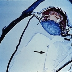 Slide 8-4
Mar 4 2019 by Lancaster Course in Ophthalmology
Persistence and hyperplasia of the primary vitreous (PHPV). The dense fibrovascular tissue molds the posterior surface of the lens and has drawn the ciliary processes and peripheral retina toward the center of the mass. A section of the hyaloid artery is present in the vitreous cavity (arrow). (A.F.l.P. No. 744398)
Condition/keywords: ciliary, fibrovascular tissue, hyaloid artery, persistent hyperplastic primary vitreous (PHPV)
-
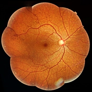 Congenital Hypertrophy of Retinal Pigment Epithelium
Sep 7 2019 by Hashim Ali Khan, OD, FAAO
Color fundus montage of a 22-year-old man with congenital hypertrophy of retinal pigment epithelium.
Condition/keywords: bear tracks, congenital hypertrophy of the retinal pigment epithelium (CHRPE), RPE hyperplasia
-
 Congenital Toxoplasmosis
Feb 2 2021 by Niloofar Piri, MD
43-year-old female with large oval chorioretinal scar in posterior pole with heavy RPE hyperplasia and history of hydrocephalus s/p VP shunt since birth. Findings are consistent with congenital toxoplasmosis.
Condition/keywords: congenital toxoplasmosis
-
 Idiopathic Parafoveal Telengiectasia - Type 2
Nov 27 2020 by Priya Rasipuram Chandrasekaran, MBBS, DO, DNB, FRCS
This is the fundus photo of a 61-year-old male presenting with bilateral gradual loss of vision since 6 months. Bilateral fundus photo shows parafoveal greying with early crystals and retinal pigment hyperplasia around a dilated venule suggestive of type 2 idiopathic parafoveal telangiectasia.
Condition/keywords: parafoveal telangiectasia
-
 Slide 9-73
Feb 26 2019 by Lancaster Course in Ophthalmology
Inferior retinal dialysis with a localized area of long-standing retinal detachment and demarcation line (upper left). There is total atrophy of the photoreceptor cell layer (upper right). The demarcation line (lower views) is an area of RPE hypertrophy and hyperplasia with nodular basement membrane production and retinal adhesion.
Condition/keywords: photoreceptor cell, retinal dialysis, retinal pigment epithelium (RPE) hypertrophy
-
 Slide 8-11
Mar 4 2019 by Lancaster Course in Ophthalmology
Radial perivascular lattice degeneration. The lesion is located at or posterior to the equator and is associated with a major vessel. As with a typical lattice there is discontinuity of the internal limiting membrane, a loss of inner retinal layers, an overlying vitreous degeneration, vitreoretinal adhesion at the margin, sclerosis of vessels, and retinal pigment epithelial hypertrophy, hyperplasia, and migration into the retina. (E.P. No. 31493)
Condition/keywords: hyperplasia, lattice degeneration, lesion, retinal pigment epithelium, vitreoretinal adhesion, vitreoretinal degeneration
-
 Slide 5-4
Feb 20 2019 by Lancaster Course in Ophthalmology
Marked downward proliferation of acanthotic epithelium (pseudoepitheliomatous hyperplasia) at the edge of a fungal ulcer.
Condition/keywords: acanthosis, fungal ulcer
-
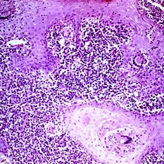
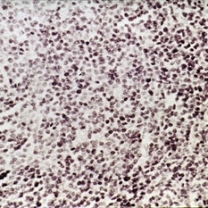 Slide 6-57
Mar 20 2019 by Lancaster Course in Ophthalmology
Benign reactive lymphoid hyperplasia, showing monotonous infiltrate of mature lymphocytes with occasional mitotic figure (H&E x252).
Condition/keywords: hyperplasia, lymphocytes
-
 Slide 12-23
Feb 27 2019 by Lancaster Course in Ophthalmology
Pigmentary glaucoma. Necrosis of the iris pigment epithelium, macrophages filled with pigment in the iris stroma, and atrophy and hyperplasia of the iris dilator muscle are present (H&E x101).
Condition/keywords: glaucoma, hyperplasia, retinal necrosis
-
 Sympathetic Ophthalmia
May 24 2023 by Niloofar Piri, MD
Montage fundus photograph of the left eye with end stage Sympathetic Ophthalmia, demonstrating optic nerve pallor, severe arterial attenuation, extensive chorioretinal atrophy (sclera is exposed in most areas), and peripheral RPE hyperplasia. Patient is a 50 yo Asian female with history of multiple vitrectomies due to retinal detachment and loss of vision who developed Sympathetic Ophthalmia in the other eye. This picture is 20 years after the disease process started, with end stage picture and HM vision.
Photographer: Sean Kelso, Saint louis university
Condition/keywords: sympathetic ophthalmia
-
 Slide 9-51
Feb 26 2019 by Lancaster Course in Ophthalmology
Reticular degenerative retinoschisis. In this case the process extends posteriorly from the equatorial area to the midperiphery. The gross view (upper left) also shows a band of dark pigmentation due to peripheral retinal pigment epithelial (RPE) hypertrophy, a few areas of paving-stone degeneration within the area of RPE hypertrophy, and peripheral retinal thinning with focal areas of RPE hyperplasia and migration into the retina.
Condition/keywords: hyperplasia, hypertrophy, retinal pigment epithelium, retinoschisis
-
 Slide 2-31
Feb 19 2019 by Lancaster Course in Ophthalmology
Extensive lymphocytic infiltration of the choroid and episcleral tissues in benign lymphoid hyperplasia. Note the serous macular detachment.
Condition/keywords: choroid, episcleral tissues, lymphocytic infiltration, lymphoid hyperplasia, macular detachment
-
 Slide 9-35
Feb 26 2019 by Lancaster Course in Ophthalmology
"Black sunburst" sign of sickle-cell retinopathy. These are localized lesions characterized by hypertrophy, hyperplasia, and migration of the retinal pigment epithelium into the retina in a perivascular location. This latter has given the lesion its spiculate ophthalmoscopic and gross appearance.
Condition/keywords: hyperplasia, hypertrophy, sickle cell, spiculate ophthalmoscopic
-
 Slide 7-29
Feb 25 2019 by Lancaster Course in Ophthalmology
Benign lymphoid hyperplasia of the conjunctiva may resemble a normal lymph node.
Condition/keywords: conjunctiva, hyperplasia, lymph node
-
 Slide 9-42
Feb 26 2019 by Lancaster Course in Ophthalmology
Bassen-Kornzweig syndrome. There is thinning of the outer nuclear layer in the parafoveal area (upper view). In the midperiphery of the retina there is more marked degeneration of the photoreceptor cells, hyperplasia, and migration of the retinal pigment epithelium into the retina in a perivascular location.
Condition/keywords: Bassen-Kornzweig syndrome, hyperplasia, photoreceptor cell, retinal pigment epithelium
-
 Slide 9-97
Feb 26 2019 by Lancaster Course in Ophthalmology
Disciform macular lesion with marked hyperplasia of RPE (A.F.I.P. No. 797119).
Condition/keywords: disciform macular lesion, hyperplasia, retinal pigment epithelium
-
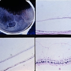
 Slide 9-103
Feb 26 2019 by Lancaster Course in Ophthalmology
Acute leukemia with focal areas of RPE defects (lower left), and areas of double-row (upper right) and nodular (lower right) RPE hyperplasia.
Condition/keywords: acute leukemia, retinal pigment epithelium
-
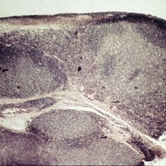
 Toxoplasmic Chorioretinitis
May 18 2020 by McGill University Health Centre
Toxoplasmic chorioretinitis is caused by parasitic infection from Toxoplasma gondii. Two forms are recognized: congenital and acquired. Congenital toxoplasmic chorioretinitis occurs because the infection is transplacental: T. gondii is among infections that cause TORCH syndrome. Acquired toxoplasmic chorioretinitis is produced by parasite ingestion, usually from raw or undercooked food. After parasitemia, the parasite directly invades the photoreceptors in the retina. In this enucleation specimen, chronic and subacute lesions coexist. In the active lesion located in the macula (arrow), the retina is necrotic, and reactive RPE cell hyperplasia surrounds the lesion. The chronic lesion (arrowhead) demonstrates atrophy of the retina and RPE in the center; in the periphery, RPE proliferation is present.
Condition/keywords: toxoplasmosis chorioretinitis
-
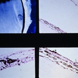
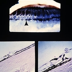 Slide 9-61
Feb 26 2019 by Lancaster Course in Ophthalmology
Hyperplasia of RPE at ora serrata. Fine-stippled, dark pigmentation at the ora serrata is due to pigment epithelial hyperplasia with migration internally (arrow). Histopathologic sections (lower views) show the strands of hyperplastic epithelium internal to the retina and pars plana.
Condition/keywords: ora serrata, retinal pigment epithelium
 Loading…