 Initializing download.
-
 By Niloofar Piri, MD
SSM Health Group, St Louis University
Co-author(s): Osasu Adah, MD Saint Louis University; Jacob Grodsky, MD Saint Louis University; Sean Kelso, Saint Louis University - Uploaded on May 24, 2023.
- Last modified by Joshua Friedman on May 25, 2023.
- Rating
- Appears in
- Miscellaneous
- Condition/keywords
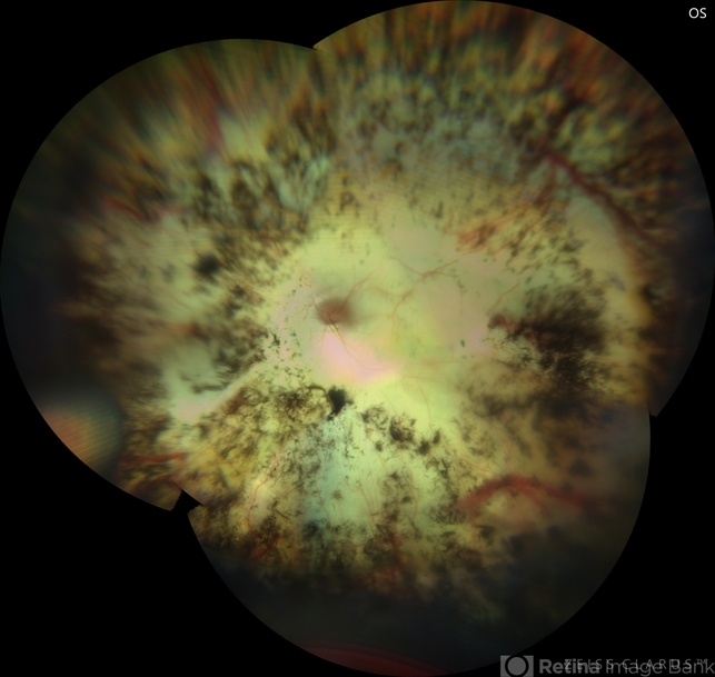
- sympathetic ophthalmia
- Photographer
- Sean Kelso, Saint louis university
- Imaging device
- Fundus camera
- Description
- Montage fundus photograph of the left eye with end stage Sympathetic Ophthalmia, demonstrating optic nerve pallor, severe arterial attenuation, extensive chorioretinal atrophy (sclera is exposed in most areas), and peripheral RPE hyperplasia. Patient is a 50 yo Asian female with history of multiple vitrectomies due to retinal detachment and loss of vision who developed Sympathetic Ophthalmia in the other eye. This picture is 20 years after the disease process started, with end stage picture and HM vision.


