 Initializing download.
-
 By Priya Rasipuram Chandrasekaran, MBBS, DO, DNB, FRCS
Lotus eye hospital
Co-author(s): Lotus eye hospital, Salem, India - Uploaded on Nov 27, 2020.
- Last modified by Caroline Bozell on Dec 1, 2020.
- Rating
- Appears in
- Miscellaneous
- Condition/keywords
- parafoveal telangiectasia
- Imaging device
- Fundus camera
- Description
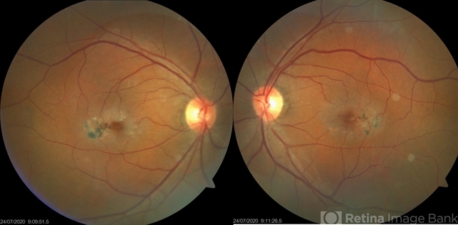
- This is the fundus photo of a 61-year-old male presenting with bilateral gradual loss of vision since 6 months. Bilateral fundus photo shows parafoveal greying with early crystals and retinal pigment hyperplasia around a dilated venule suggestive of type 2 idiopathic parafoveal telangiectasia.

---thumb.jpg/image-square;max$79,0.ImageHandler "Parafoveal Retinal Telangiectasis")


