Search results (387 results)
-
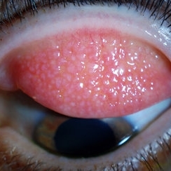 Giant Papillary Conjunctivitis, Left Upper Eyelid
Jul 22 2013 by Jason S. Calhoun
Contact lens wearer in for exam. Has rough feeling underneath both eyelids. Patient thought it was through SCL wear. Patient VA was 20/20. right eye, 20/30, left eye. Underneath the left upper eyelid, you can see papillary inflammation and redness.
Photographer: Jason S. Calhoun, Department of Ophthalmology, Mayo Clinic Jacksonville, Florida
Imaging device: TOPCON D-90 SL NIKON CAMERA
Condition/keywords: giant papillary conjunctivitis
-
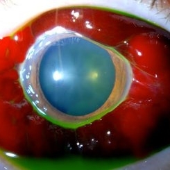
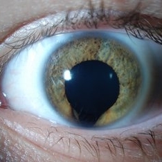 Keyhole Pupil Coloboma
Jul 13 2013 by Jason S. Calhoun
14-year-old male presents with decreased vision in the left eye. Dx with iris and retinal coloboma in the left eye. Patient VA was 20/20, right eye, 20/100 left eye with pinhole improvement 20\50. Patient was fitted for SCL in the left eye.
Photographer: Jason S. Calhoun, Department of Ophthalmology, Mayo Clinic Jacksonville, Florida
Imaging device: TOPCON D-90 SL NIKON CAMERA
Condition/keywords: deformed pupil
-
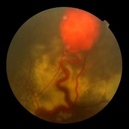
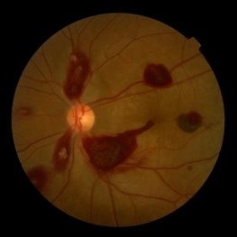 Ocular Manifestation of Acute Leukemia
Sep 8 2012 by Hamid Ahmadieh, MD
Color fundus photograph of a 26-year-old man with acute leukemia.
Photographer: Hamid Ahmadieh, MD, Ophthalmic Research Center, Labbafinejad Medical Center, Shahid Beheshti University of Medical Sciences , Tehran
Imaging device: Topcon Fundus Camera
Condition/keywords: acute leukemia, white centered retinal hemorrhage (Roth Spot)
-
 Coloboma, In Stereo
Oct 1 2012 by Michael P. Kelly, FOPS
This is a stereo retinal fundus photograph of a coloboma, with the optic nerve centered, using a Zeiss FF3C retinal fundus camera.
Photographer: Michael P. Kelly, FOPS Director, Duke Eye Labs, Duke University Hospital, Duke Eye Center, Durham, NC
Condition/keywords: coloboma, fundus photograph, stereo pair
-
---thumb.jpg/image-square;max$300,300.ImageHandler) Bergmeister's Papilla
Bergmeister's Papilla
Mar 22 2014 by Hamid Ahmadieh, MD
Color fundus photograph of the right eye of a 50-year-old man with Bergmeister's papilla.
Photographer: Naghmeh Nozhat, Negah Eye Center, Tehran
Imaging device: Topcon Fundus Camera
Condition/keywords: Bergmeister's Papillae, color photo
-
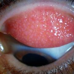
 Giant Papillary Conjunctivitis
Dec 13 2013 by Jason S. Calhoun
Patient wears soft contact lenses complained of irritation when the SCL would move. Inverted eyelid in both eyes and there was papillary +2 underneath the eyelid.
Photographer: Jason S. Calhoun, Ophthalmic Photographer, Department of Ophthalmology, Mayo Clinic Jacksonville
Imaging device: TOPCON D-90 SL NIKON CAMERA
Condition/keywords: giant papillary conjunctivitis
-
 Giant Papillary Conjunctivitis, Left Upper Eyelid
Jul 22 2013 by Jason S. Calhoun
Contact lens wearer, in for exam. Has rough feeling underneath both eyelids. Patient thought it was through SCL wear. Patient VA was 20/20. right eye, 20/30, left eye. Underneath the left upper eyelid, you can see papillary inflammation and redness.
Photographer: Jason S. Calhoun, Department of Ophthalmology, Mayo Clinic Jacksonville, Florida
Imaging device: TOPCON D-90 SL NIKON CAMERA
Condition/keywords: giant papillary conjunctivitis
-
 Rhegmatogenous Retinal Detachment (Widefield/Optos)
Dec 16 2016 by Courtney Crawford, MD, FACS
65-year-old-male with curtain/veil over vision for two days.
Photographer: Ryann Nafe
Imaging device: Optos Fundus Camera
-
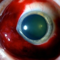
 Gas Bubble in Anterior Chamber
Jul 14 2013 by Jason S. Calhoun
Gas bubble injected in anterior chamber to control IOP.
Photographer: Jason S. Calhoun, Department of Ophthalmology, Mayo Clinic Jacksonville, Florida
Imaging device: TOPCON D-90 SL NIKON CAMERA
Condition/keywords: gas bubble
-
 Retinal Angiomatous Proliferation in Age-Related Macular Degeneration with Subretinal Neovascularization
Sep 24 2012 by James B. Soque, CRA, OCT-C, COA, FOPS
75-year-old white male with classic SRN with RAP. Lesion OD is active, and patient is receiving anti-VEGF treatment. Mid phase FA, 50 Deg, Mag 2x.
Photographer: James Soque, CRA, COA, Island Retina, Shirley, NY, USA
Imaging device: Topcon TRC 50 DX, OIS 5.0 MP Color, FA Camera, OIS Software
Condition/keywords: age-related macular degeneration (AMD), fundus autofluorescence (FAF), leakage, retinal angiomatous proliferation (RAP), subretinal neovascularization (SRNV)
-
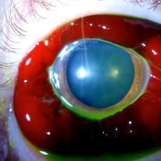
 Sub-Conjunctival Hemorrhage (Chemosis)
Jul 13 2013 by Jason S. Calhoun
Chemosis or swelling of the conjunctiva with sub-conjunctival hemorrhage.
Photographer: Jason S. Calhoun, Department of Ophthalmology, Mayo Clinic Jacksonville, Florida
Imaging device: TOPCON D-90 SL NIKON CAMERA
Condition/keywords: chemosis
-
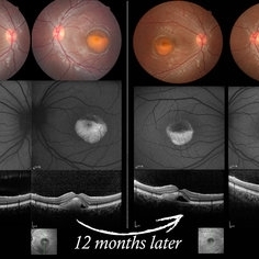 Vitelliform Macular Dystrophy or Best Disease
Dec 16 2016 by Young Hee Yoon, MD, PhD
Bilateral fundus photographs and autofluorescence images of 15-year-old girl who was diagnosed as vitelliform macular dystrophy or Best disease. Vitelliform macular lesion showed morphologic change during one year.
Photographer: Hyejin Jo, Sunghyun Kim, Heoni Hong, Minjung Chae, Mihwa Shin, Asan medical center, Seoul
Imaging device: Topcon TRC-500X fundus camera, Heidelberg HRA 2 autofluorescence, Heldelberg Spectralis OCT
Condition/keywords: Best disease, pseudohypopyon, scrambled-egg, vitelliform macular dystrophy
-
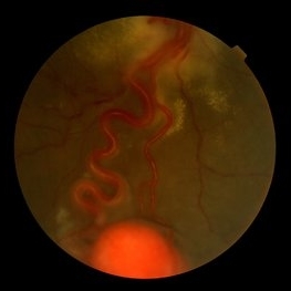
 Central Retinal Artery Occlusion & Cilioretinal Artery Sparing
Dec 22 2012 by Hamid Ahmadieh, MD
Color fundus photograph of the right eye of a 34-year-old man with sudden drop of vision due to CRAO. Please notice cherry red spot despite cilioretinal artery sparing .
Photographer: Zohre Salimi; Labbafinejad Medical Center, Shahid Beheshti University of Medical Sciences, Tehran
Imaging device: Topcon Fundus Camera
Condition/keywords: central retinal artery occlusion (CRAO), cherry red spot, cilioretinal sparing
-
 Eyelid Cyst
Jul 14 2013 by Jason S. Calhoun
Upper underneath eyelid cyst.
Photographer: Jason S. Calhoun, Department of Ophthalmology, Mayo Clinic Jacksonville, Florida
Imaging device: TOPCON D-90 SL NIKON CAMERA
Condition/keywords: cystic lesion
-
 Sub-Conjunctival Hemorrhage (Chemosis)
Jul 13 2013 by Jason S. Calhoun
Chemosis or swelling of the conjunctiva with sub-conjunctival hemorrhage.
Photographer: Jason S. Calhoun, Department of Ophthalmology, Mayo Clinic Jacksonville, Florida
Imaging device: TOPCON D-90 SL NIKON CAMERA
Condition/keywords: chemosis
-
 Giant Retinal Tear
Oct 9 2012 by Audina M. Berrocal, MD FASRS
Teenager with high myopia and a GRT
Photographer: Ditte Hess CRA, BPEI
Imaging device: Fundus Camera
Condition/keywords: high myopia, retinal degeneration, retinal tear
-
 Von Hippel-Lindau
Sep 3 2012 by Hamid Ahmadieh, MD
Color fundus photograph of a 35-year-old woman with retinal angiomatosis.
Photographer: Hamid Ahmadieh, MD, Ophthalmic Research Center, Labbafinejad Medical Center, Shahid Beheshti University of Medical Sciences , Tehran
Imaging device: Topcon Fundus Camera
Condition/keywords: retinal angiomatous proliferation (RAP), Von Hippel-Lindau
-
 Giant Papillary Conjunctivitis
Dec 13 2013 by Jason S. Calhoun
Patient wears soft contact lenses complained of irritation when the SCL would move. Inverted eyelid in both eyes and there was papillary +2 underneath the eyelid.
Photographer: Jason S. Calhoun, Ophthalmic Photographer, Department of Ophthalmology, Mayo Clinic Jacksonville
Imaging device: TOPCON D-90 SL NIKON CAMERA
Condition/keywords: giant papillary conjunctivitis
-
 Fibrovascular Retinal Pigment Epithelial Detachment - Color Fundus
Jul 16 2014 by James B. Soque, CRA, OCT-C, COA, FOPS
69-year-old white female with Hx of 10 anti-VEFG treatment injections of right eye, VA 20/200, now stable, off drug for 10 months.
Photographer: James B Soque, CRA COA
Imaging device: Topcon TRC 50 DX with MERGE software, 5 MP dig camera
Condition/keywords: color fundus photograph, fibrovascular pigment epithelial detachment (PED), pigment epithelial atrophy, retina
-
 Von Hippel-Lindau
Sep 3 2012 by Hamid Ahmadieh, MD
Color fundus photograph of a 35-year-old woman with retinal angiomatosis.
Photographer: Hamid Ahmadieh, MD, Ophthalmic Research Center, Labbafinejad Medical Center, Shahid Beheshti University of Medical Sciences
Imaging device: Topcon Fundus Camera
Condition/keywords: retinal angiomatous proliferation (RAP), Von Hippel-Lindau
-
 Diabetic Retinopathy, CSME, Color Fundus Photo
Mar 18 2015 by James B. Soque, CRA, OCT-C, COA, FOPS
A 58-year-old diabetic male with a longstanding history of diabetic eye disease. Left eye color fundus photo shows extensive CSME, Clinically Significant Macular Edema, with deposits of hard exudates at fixation. There is extensive scattering of hard exudates and sheathing of the vessels.
Photographer: James B Soque, CRA COA
Imaging device: Topcon TRC 50 DX, OIS 5 MP Camera, MERGE software
Condition/keywords: background diabetic retinopathy (BDR), creamy yellow exudates, diabetes, exudates over the posterior pole, neovascularization of the disc (NVD), vessel sheathing
-
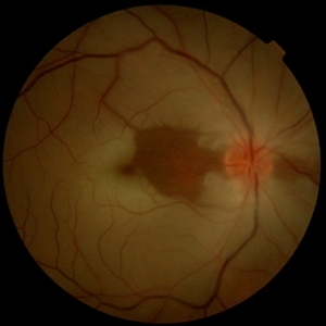
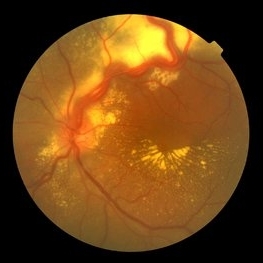
 Papilledema
Sep 8 2012 by Hamid Ahmadieh, MD
Color fundus photograph of the right eye of a 55-year-old woman with an intracranial malignant tumor.
Photographer: Hamid Ahmadieh, MD, Ophthalmic Research Center, Labbafinejad Medical Center, Shahid Beheshti University of Medical Sciences, Tehran, Iran
Imaging device: Topcon Fundus Camera
Condition/keywords: papilledema
-
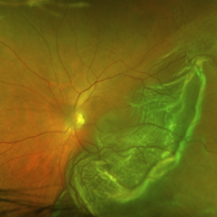
 Von Hippel-Lindau
Sep 5 2012 by Hamid Ahmadieh, MD
Color fundus photograph of a 32-year-old man with retinal angiomatosis.
Photographer: Hamid Ahmadieh, MD, Ophthalmic Research Center, Labbafinejad Medical Center, Shahid Beheshti University of Medical Sciences
Imaging device: Topcon Fundus Camera
Condition/keywords: retinal angiomatous proliferation (RAP), Von Hippel-Lindau
-
 Epiretinal Membrane
Sep 14 2012 by Michael P. Kelly, FOPS
Epiretinal membrane imaged using a high magnification retinal fundus camera and red free illumination.
Photographer: Michael P. Kelly, FOPS, Director, Duke Eye Center Labs, Duke Universtiy Hospital
Condition/keywords: epiretinal membrane (ERM), high magnification, monochromatism, red-free
-
 Sub-Conjunctival Hemorrhage (Chemosis)
Jul 13 2013 by Jason S. Calhoun
Chemosis or swelling of the conjunctiva with Sub conjunctival hemorrhage.
Photographer: Jason S. Calhoun, Department of Ophthalmology, Mayo Clinic Jacksonville, Florida
Imaging device: TOPCON D-90 SL NIKON CAMERA
Condition/keywords: chemosis
 Loading…