Search results (387 results)
-
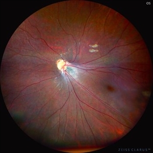
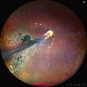 Comets in the Eye (Retinopathy of Prematurity)
Apr 8 2025 by KANWALJEET HARJOT MADAN, M.S. (Ophthalmology), FAICO (Vitreous - Retina)
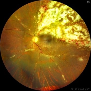
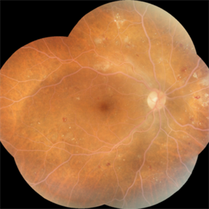
This is the fundus picture of right eye (RE) of a 4 years female child presented with outward deviation of right eye. Her parents also complained of diminution of vision in both eyes. On examination, her best corrected vision in RE was hand movements close to face and was 20/80 in LE. Posterior segment exam revealed presence of macular scar in RE and presence of dry retinal fold with dragging of retinal vessels. LE fundus revealed presence of nasal drag of optic disc. Parents gave history of untreated ROP as an infant. Retinopathy of Prematurity (ROP) is a Vaso proliferative disorder of Retina occurring in premature infants. Advances in neonatal care and ROP treatment has led these babies to live longer with this disease.
Photographer: Dr. Kanwaljeet Harjot Madan, Thind Eye Hospital, Jalandhar City (Punjab) INDIA.
Imaging device: Zeiss Fundus Camera
Condition/keywords: Retinopathy of Prematurity, Vaso proliferative disorder
-
 Comets in the Eye (Retinopathy of Prematurity)
Apr 8 2025 by KANWALJEET HARJOT MADAN, M.S. (Ophthalmology), FAICO (Vitreous - Retina)
This is the fundus picture of right eye (RE) of a 4 years female child presented with outward deviation of right eye. Her parents also complained of diminution of vision in both eyes. On examination, her best corrected vision in RE was hand movements close to face and was 20/80 in LE. Posterior segment exam revealed presence of macular scar in RE and presence of dry retinal fold with dragging of retinal vessels. LE fundus revealed presence of nasal drag of optic disc. Parents gave history of untreated ROP as an infant. Retinopathy of Prematurity (ROP) is a Vaso proliferative disorder of Retina occurring in premature infants. Advances in neonatal care and ROP treatment has led these babies to live longer with this disease.
Photographer: Dr. Kanwaljeet Harjot Madan, Thind Eye Hospital, Jalandhar City (Punjab) INDIA.
Imaging device: Zeiss Fundus Camera
Condition/keywords: Retinopathy of Prematurity
-
 Diabetic Vitreous Hemorrhage
Feb 3 2025 by Hollie Sanders
61 year-old male with a history of type two DM. Per MD, Sub-Retinal, Pre-Retinal, and Vitreous Hemorrhages. Denies any treatment in the past. Treatment initiated in clinic.
Photographer: Hollie Sanders, Tennessee Retina, Nashville, Tennessee
Imaging device: OPTOS Silverstone Fundus camera
Condition/keywords: diabetic vitreous hemorrhage
-
 Iris Nevus
Jan 28 2025 by Korey Starkey
Slit-lamp image of an 89-year-old patient with an iris nevus. Nevus appeared stable on exam, will continue to monitor.
Photographer: Korey Starkey
Imaging device: Slit lamp camera
Condition/keywords: ectropion uveae, iris nevus, slit lamp photo
-
 Iris Melanoma
Jan 28 2025 by Korey Starkey
Slit-lamp image of 90-year-old patient with iris melanoma and new hemorrhage affecting the right eye. Patient re-presented after nearly 1 year, now seeking treatment. Given iris location of tumor, multiple clock hours of iris involved, and increase in size of the known malignant transformation; safest approach was enucleation.
Photographer: Korey Starkey
Imaging device: Slit lamp camera
Condition/keywords: anterior chamber, hemorrhage, iris melanoma, slit lamp photo
-
 Epicapsular Stars
Jan 28 2025 by Korey Starkey
Epicapsular stars and cataract noted in natural lens of 68-year-old patient.
Photographer: Korey Starkey
Imaging device: Slit lamp camera
Condition/keywords: cataract, chicken tracks, epicapsular stars, slit lamp photography
-
 Vitreous Prolapse
Jan 28 2025 by Korey Starkey
Slit lamp image of a 62-year-old patient presented at first visit with vitreous prolapse due to mechanical complications from IOL placement. IOP was being managed with drops, vision was 20/20, patient opted for surgery due to constant haze in vision.
Photographer: Korey Starkey
Imaging device: Slit lamp camera
Condition/keywords: slit lamp photo, vitreous prolapse
-
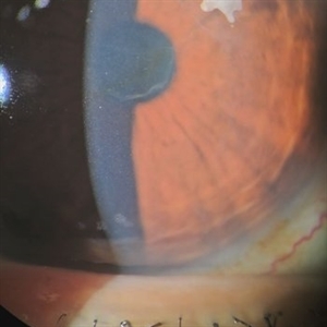
 Coloboma in a Unicameral Eye
Dec 20 2024 by Virginia Gebhart
59 year old female with choroidal coloboma extending into iris. Pt had PC IOL placed in 2016, removed in Aug due to suspected UGH syndrome. Lens haptics were oriented vertically causing haptic to chafe iris superiorly. Most likely etiology was loss of inferior zonules from coloboma. Pt remains aphakic.
Photographer: Virginia Gebhart, Retina Consultants of Carolina
Imaging device: Optos California
Condition/keywords: choroidal coloboma, coloboma
-
 Pseudoxanthoma Elasticum
Dec 3 2024 by Dr Bilal Mir
This is a fundus picture showing angiod streaks, CNVM, comet lesion.
Photographer: Dr Bilal Ahmed mir MS ophthalmology
Imaging device: Zeiss fundus camera
Condition/keywords: Angiod streaks in Pseudoxanthoma elasticum, Pseudoxanthoma elasticum
-
 Oval Pigmented Vitreous Cyst
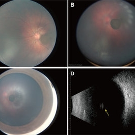
Nov 27 2024 by Xinyu Zhao
An 8-month-old infant was found to have a brown object in the left vitreous during a fundus screening. A wide-field digital retinal camera (RetCam) revealed a pigmented, non-transparent, freely floating, oval cystic lesion in the vitreous, measuring 2 disc diameters (Figures A-D). The cyst appeared cloudy when focused on the retina (Figure A) but was clearly defined in the vitreous (Figure B). Ultrasound showed a well-defined hyperreflective structure with a hyporeflective lumen (Figure D, indicated by the yellow arrow). A diagnosis of a vitreous pigment cyst, rare in infants, was made. Long-term follow-up is necessary to monitor changes affecting the infant’s vision.
Photographer: Xinyu Zhao, Shenzhen Eye Hospital, Shenzhen, China
Imaging device: RetCam
Condition/keywords: infant, vitreous cyst
-
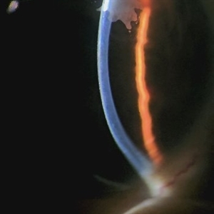
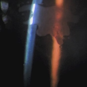
 PFO Bubbles
Oct 24 2024 by Korey Starkey
Slit lamp photograph of a 23 year old female with PFO bubbles inferiorly in the AC. Discussed surgical intervention to remove PFO from AC and vitreous cavity in future.
Photographer: Korey Starkey
Imaging device: Slit lamp camera
Condition/keywords: anterior chamber, PFO, slit lamp photography, submacular perfluorocarbon liquid (PFO)
-
 Ink Blot Epithelial Ingrowth Post-LASIK Refractive Surgery
Jun 29 2024 by Luai Abu-Ismail, MD
Anterior segment photo of a 45-year-old female patient presented 12-year post-LASIK surgery.
Photographer: Dr. Luai Abu-Ismail, Ophthalmology Department, Islamic Hospital.
Imaging device: Slit lamp biomicroscope photo taken by Smart phone camera.
Condition/keywords: cornea, corneal scars and opacities, flap, LASIK
-
 Ink Blot Epithelial Ingrowth Post-LASIK Refractive Surgery
Jun 29 2024 by Luai Abu-Ismail, MD
Anterior segment photo of a 45-year-old female patient presented 12-year post-LASIK surgery.
Photographer: Dr. Luai Abu-Ismail, Ophthalmology Department, Islamic Hospital.
Imaging device: Slit lamp biomicroscope photo taken by Smart phone camera.
Condition/keywords: complication, cornea, corneal scars and opacities, epithelial ingrowth, LASIK, LASIK FLAP, refractive surgery
-
 Gigantic Curved Glass Intraocular Foreign Body
Jun 6 2024 by Veer Singh, MS, FVRS, FMRF, FICO (Retina)
Gigantic Curved Glass Intraocular Foreign Body measuring 15x10 mm size
Photographer: Dr. Veer Singh
Imaging device: Ikegami 4k Microscope Camera
Condition/keywords: Curved Glass, Gigantic, IOFB
-
 Scleral Suspension CMT-Flex SFIOL Implantation
Apr 16 2024 by Veer Singh, MS, FVRS, FMRF, FICO (Retina)
Implantation of the Sutureless Scleral Suspension CMT-Flex SFIOL along with 25G Vitrectomy
Photographer: Dr. Veer Singh
Imaging device: Ikegami 4k Microscope Camera
Condition/keywords: CMT-Flex SFIOL, Scleral Suspension, Trailing Haptic
-
 Cheese Pizza Pie Appearance in CMV Retinitis
Mar 30 2024 by KANWALJEET HARJOT MADAN, M.S. (Ophthalmology), FAICO (Vitreous - Retina)
This is Fundus Photograph of left eye of 53 year male depicting an area of Retinal Necrosis with few Retinal Haemorrhages suggestive of CMV Retinitis. Areas of Perivascular Exudation also seen. On investigations, the patient was found to be HIV positive. He was started on Anti Retro Viral treatment after physician opinion.
Photographer: Dr. Kanwaljeet Harjot Madan, Thind Eye Hospital, Jalandhar City (Punjab) INDIA.
Imaging device: Zeiss Fundus Camera
Condition/keywords: AIDS, cytomegalovirus (CMV), retinitis
-
 Blistered Retina
Jan 27 2024 by prathibha hande, MS DNB
Fundus photo of a 32 year old male presenting with blurred vision. Undiagnosed renal hypertension. Blood pressure at the time of presentation 210/120 mmhg.
Photographer: Mr Prathap K
Imaging device: Mirante SLO fundus camera
Condition/keywords: hypertensive choroidopathy
-
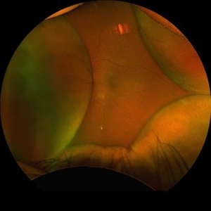
 Sublimiting navicular hemorrhage
Jan 25 2024 by Itzel Romero Ramírez
A 62-year-old male with a history of visual loss in the right eye lasting 3 months, ultra-wide-field fundus imaging was performed on a California optos camera, where the image of sublimiting navicular hemorrhage in the right eye was evident.
Photographer: Itzel Romero Ramírez, Hospital Militar de Especialidades Oftalmológicas, Ciudad de México
Imaging device: Ultra wide field fundus camera California optos camera
Condition/keywords: hemorragia, retina, sublimitante
-
 Sublimiting navicular hemorrhage
Jan 25 2024 by Itzel Romero Ramírez
A 62-year-old man with a history of visual loss in the right eye of 3 months' duration underwent ultra-wide-field fundus imaging with an optos California camera, which revealed sublimiting navicular hemorrhage in the right eye.
Photographer: Itzel Romero Ramírez, Hospital Militar de Especialidades Oftalmológicas, Mexico City
Imaging device: Ultra wide field fundus camera California optos camera
Condition/keywords: hemorrhage, retina, sublimitante
-
 Gore Tex Suture
Nov 1 2023 by Virginia Gebhart
Gore Tex suture of scleral fixated lens in 66 year-old male, 1 day post-op
Photographer: Virginia Gebhart
Imaging device: slit lamp camera
Condition/keywords: Gore Tex Suture, post-op, Scleral fixated IOL
-
 Massive Sub-Retinal Haemorrhage involving Macula : Intra-operative still image
Oct 26 2023 by Veer Singh, MS, FVRS, FMRF, FICO (Retina)
Massive Sub-Retinal Haemorrhage involving Macula : Intra-operative still image
Photographer: Dr. Veer Singh
Imaging device: Ikegami 4k Microscope Camera
Condition/keywords: Haemorrhage, macula, Massive, Sub-Retinal
-
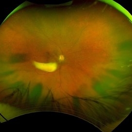
 Choroidal Detachment
Aug 14 2023 by Omar Toncel Churio
Fundus photograph of a woman patient with a choroidal detachment.
Photographer: Omar Toncel Churio, Hospital Militar de Especialidades Oftalmológicas, Ciudad de México
Imaging device: Optos California Retinal Camera
Condition/keywords: choroid, detachment, retina
-
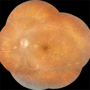 Lipemia retinalis with severe NPDR- Left Eye
Aug 1 2023 by Maneesh M Bapaye, MD, MBA
A 52 years old poorly controlled diabetic male pt. HbA1c of 16.2%, Sr. Cholesterol 460, Sr. Triglycerides 575, VLDL 115
Photographer: Dr.Maneesh Bapaye
Imaging device: Zeiss fundus camera
Condition/keywords: clinically significant macular edema (CSME), lipemia retinalis, nonproliferative diabetic retinopathy
-
 Lipemia Retinalis -Right eye
Aug 1 2023 by Maneesh M Bapaye, MD, MBA
A 52 years old poorly controlled diabetic male pt. HbA1c of 16.2%, Sr. Cholesterol 460, Sr. Triglycerides 575, VLDL 115
Photographer: Dr.Maneesh Bapaye
Imaging device: Zeiss fundus camera
Condition/keywords: clinically significant macular edema (CSME), lipemia retinalis, nonproliferative diabetic retinopathy
 Loading…