Search results (14 results)
-
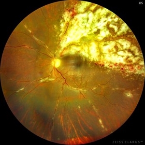 Cheese Pizza Pie Appearance in CMV Retinitis
Mar 30 2024 by KANWALJEET HARJOT MADAN, M.S. (Ophthalmology), FAICO (Vitreous - Retina)
This is Fundus Photograph of left eye of 53 year male depicting an area of Retinal Necrosis with few Retinal Haemorrhages suggestive of CMV Retinitis. Areas of Perivascular Exudation also seen. On investigations, the patient was found to be HIV positive. He was started on Anti Retro Viral treatment after physician opinion.
Photographer: Dr. Kanwaljeet Harjot Madan, Thind Eye Hospital, Jalandhar City (Punjab) INDIA.
Imaging device: Zeiss Fundus Camera
Condition/keywords: AIDS, cytomegalovirus (CMV), retinitis
-
 Blistered Retina
Jan 27 2024 by prathibha hande, MS DNB
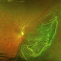
Fundus photo of a 32 year old male presenting with blurred vision. Undiagnosed renal hypertension. Blood pressure at the time of presentation 210/120 mmhg.
Photographer: Mr Prathap K
Imaging device: Mirante SLO fundus camera
Condition/keywords: hypertensive choroidopathy
-
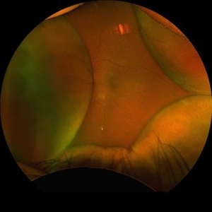 Choroidal Detachment
Aug 14 2023 by Omar Toncel Churio
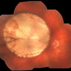
Fundus photograph of a woman patient with a choroidal detachment.
Photographer: Omar Toncel Churio, Hospital Militar de Especialidades Oftalmológicas, Ciudad de México
Imaging device: Optos California Retinal Camera
Condition/keywords: choroid, detachment, retina
-
 Lupus Retinopathy
Mar 14 2021 by Marco Antonio Sauza
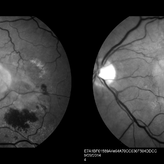
Fluorescein angiography photo of and 13-year-old female with ischemic retinopathy with LES.
Photographer: Marco Sauza
Imaging device: Zeiss fundus camera
Condition/keywords: systemic lupus erythematosus (SLE) retinopathy
-
 Intravitreal Cysticercosis With Full Thickness Macular Hole
Apr 30 2018 by Vishal Agrawal, MD, FRCS,FACS,FASRS
Fundus montage picture of a 40-year-old man presenting with decreased vision in the right eye for the past 2 months. Live intravitreal cysticercosis can be seen lying on the retina. Zooming the image reveals the full thickness macular hole. The scolex invaginates with the light of the camera causing double image of the cyst because of movement .
Photographer: Vishal Agrawal MD,FRCS
Imaging device: Zeiss 524
Condition/keywords: cysticercosis, full thickness macular hole
-
 Bullous RD With Dislocated Lens
Apr 3 2018 by Navneet Mehrotra, DNB
Dislocated clear lens and associated retinal detachment in a young patient with Marfan's syndrome.
Photographer: Navneet Mehrotra
Imaging device: Sony 3 chip camera
Condition/keywords: dislocated crystalline lens, Marfan's syndrome
-
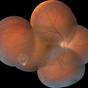
 Retinal Detachment
Feb 17 2018 by JEFFERSON R SOUSA, Tecg.º (Biomedical Systems Technology)
A 42-year-old patient complained of low vision in the left eye. In retinal mapping and background color photography, extensive retinal detachment was observed.
Photographer: JEFFERSON R SOUSA - Study Center and Ophthalmological Research Dr. Andre M V Gomes, Institute Dr. Suel Abujamra São Paulo-Brazil
Imaging device: Fundus camera Topcon TRC-50 DX, Imaginet 5.0, angle de 50 graus. Flash 36 / Mosaic with 10 images.
-
 Retinal Detachment
Feb 8 2018 by JEFFERSON R SOUSA, Tecg.º (Biomedical Systems Technology)
The male patient attended the clinic with low vision. In the retinal and retinal mapping examination, important fudoscopical alterations were observed. Full retinal detachment with Giant rupture in upper temporal arch.
Photographer: JEFFERSON R SOUSA - Study Center and Ophthalmological Research Dr. Andre M V Gomes, Institute Dr. Suel Abujamra São Paulo-Brazil
Imaging device: Fundus camera Topcon TRC-50 DX, Imaginet 5.0, campo de 50 graus. Flash 36 / Mosaic with 16 images.
Condition/keywords: retinal in rupture
-
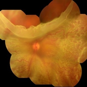
 Coloboma
Jan 23 2018 by JEFFERSON R SOUSA, Tecg.º (Biomedical Systems Technology)
Male patient, 22 years old, with low vision since infancy. In retinal and retinal mapping examinations, important alterations were observed in the formation of retinochoroidal structures suggestive of coloboma.
Photographer: JEFFERSON R SOUSA - Study Center and Ophthalmological Research Dr. Andre M V Gomes, Dr. Suel Abujamra Institute São Paulo-Brazil
Imaging device: Acquisition of the image in the Camera background Topcon TRC-50 Dx - IA, Keystone field photo of 50 Degrees. Composition automatic of Imaginet with manual adjustment
Condition/keywords: coloboma, coloboma of choroid
-
 Rhegmatogenous Retinal Detachment (Widefield/Optos)
Dec 16 2016 by Courtney Crawford, MD, FACS
65-year-old-male with curtain/veil over vision for two days.
Photographer: Ryann Nafe
Imaging device: Optos Fundus Camera
-
 Hemi Vein Occlusion, Fluorescein Angiogram, Montage
Dec 17 2015 by James B. Soque, CRA, OCT-C, COA, FOPS
74-year-old woman, with recurrent superior hemi vein occlusion, montage image of fluorescein angiogram left eye. Currently receiving Lucentis injections OS.
Photographer: James Soque, CRA COA
Imaging device: Topcon RC 50 DX Fundus Camera with MERGE Winstation Software for Fluorescen Angiography
Condition/keywords: montage, occlusion of retinal vein, superior arcade
-
 Neovascular ARMD With Subretinal Hemorrhage, Red-Free Photos - Stereo
Nov 26 2014 by James B. Soque, CRA, OCT-C, COA, FOPS
Stereo FC, RF and FA of a 77-year-old white female with visual acuity CC 20/200-3, with left eye neovascular ARMD, drusen, and subretinal hemorrhage with hard exudates temporally. Peripheral retina reveals cobblestone degeneration.
Photographer: James Soque, CRA, COA, Island Retina, Shirley, NY
Imaging device: Topcon TRC 50 EX, with MERGE software and OIS 5 MP digital Camera
Condition/keywords: neovascular age-related macular degeneration (AMD), red-free, stereo pair
-
 Optic Atrophy and Attenuated Retinal Vessels Following Endophthalmitis
Jul 12 2014 by Philip J. Polkinghorne, MD
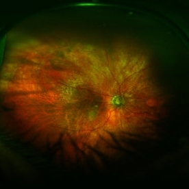
This elderly lady underwent a vitrectomy for post-surgical endophthalmitis. The infection was successfully treated but the functional outcome was poor because of optic atrophy and attenuated retinal vessels.
Photographer: Alex Fraser
Imaging device: Optos Camera
Condition/keywords: attenuated vessels, endophthalmitis, optic atrophy, post-vitrectomy
-
 Picture With 20D and Cell Phone Camera
Mar 29 2014 by Bhartendu Kumar Varma, DNB
Picture with 20D and cell phone camera.
Photographer: Bhartendu Varma, Delhi
Imaging device: Cell phone camera
Condition/keywords: cell phone camera
 Loading…