Search results (387 results)
-
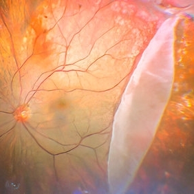
 Ocular Manifestation of Acute Leukemia
Sep 8 2012 by Hamid Ahmadieh, MD
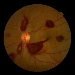
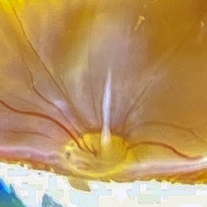
Color fundus photograph of a 26-year-old man with acute leukemia.
Photographer: Hamid Ahmadieh, MD, Ophthalmic Research Center, Labbafinejad Medical Center, Shahid Beheshti University of Medical Sciences , Tehran
Imaging device: Topcon Fundus Camera
Condition/keywords: acute leukemia, white centered retinal hemorrhage (Roth Spot)
-
 IOFB Over Disc BRAO Post Hyaloid Removal
Feb 25 2017 by Manish Nagpal, MD, FRCS (UK), FASRS
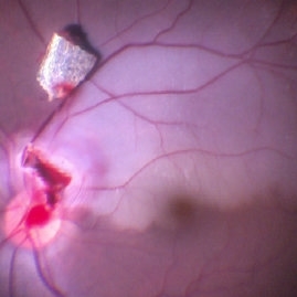
Intraoperative photo of a foreign body piercing the inferotemporal margin of disc revealing a infero temporal BRAO immediately after hyaloid removal. The foreign boy has just been removed from the disc and is freely lying on the retina.
Photographer: MANISH NAGPAL
Imaging device: STILL CAPTURED FROM A 3CHIP HD camera attached to microscope
Condition/keywords: intraocular foreign body
-
 Blistered Retina
Jan 27 2024 by prathibha hande, MS DNB
Fundus photo of a 32 year old male presenting with blurred vision. Undiagnosed renal hypertension. Blood pressure at the time of presentation 210/120 mmhg.
Photographer: Mr Prathap K
Imaging device: Mirante SLO fundus camera
Condition/keywords: hypertensive choroidopathy
-
 Capillary Hemangioma
Dec 14 2016 by Young Hee Yoon, MD, PhD
Wide fundus photo of a 35-year-old man with huge capillary hemagioma in the right eye. He is diagnosed with Von Hippel-Lindau disease. His best-corrected visual acuity was 20/50.
Photographer: Yu Jin Jang and Hun Eui Hong, Asan Medical Center
Imaging device: Wide fundus camera
Condition/keywords: retinal capillary hemangioma, Von Hippel-Lindau
-
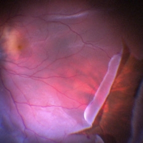
 Epiretinal Membrane
Sep 14 2012 by Michael P. Kelly, FOPS
Epiretinal membrane imaged using a high magnification retinal fundus camera and red free illumination.
Photographer: Michael P. Kelly, FOPS, Director, Duke Eye Center Labs, Duke Universtiy Hospital
Condition/keywords: epiretinal membrane (ERM), high magnification, monochromatism, red-free
-
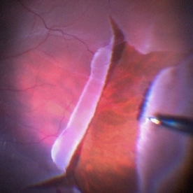
 ERM With Retinal Detachment
May 25 2017 by Manish Nagpal, MD, FRCS (UK), FASRS
Per operative photo prior to ERM removal in a case of retinal detachment with ERM.
Photographer: MANISH NAGPAL
Imaging device: SONY 3 CHIP HD CAMERA
Condition/keywords: epiretinal membrane (ERM), internal limiting membrane (ILM) peeling
-
 Intraoperative Photo Taken During Vitrectomy
Jan 26 2017 by Manish Nagpal, MD, FRCS (UK), FASRS
Intraoperative photo while doing vitectomy near a horseshoe tear to clear the adherent vitreous enhanced by peripheral scleral indentation while using chandelier light.
Photographer: Manish Nagpal
Imaging device: Still captured from a 3 chip HD camera on microscope
Condition/keywords: cutter, scleral indentation, vitrectomy, vitreous
-
 Large Retinal Tear
Mar 24 2017 by Manish Nagpal, MD, FRCS (UK), FASRS
Intraoperative photo of a large retinal tear with everted edges.
Photographer: manish nagpal
Imaging device: Still captured from 3 Chip HD camera on microscope
Condition/keywords: retinal tear
-
 Large Retinal Tear
Mar 24 2017 by Manish Nagpal, MD, FRCS (UK), FASRS
Intraoperative photo of a large retinal tear with everted edges.
Photographer: Manish Nagpal
Imaging device: Still captured from a 3 chip HD camera on microscope
Condition/keywords: retinal tear
-
 Lupus Retinopathy
Mar 14 2021 by Marco Antonio Sauza
Fluorescein angiography photo of and 13-year-old female with ischemic retinopathy with LES.
Photographer: Marco Sauza
Imaging device: Zeiss fundus camera
Condition/keywords: systemic lupus erythematosus (SLE) retinopathy
-
 Rubeosis
Jul 14 2013 by Jason S. Calhoun
Slit lamp photo shows active rubeosis in the left eye.
Photographer: Jason S. Calhoun, Department of Ophthalmology, Mayo Clinic Jacksonville, Florida
Imaging device: TOPCON D-90 SL NIKON CAMERA
Condition/keywords: rubeosis
-
 Triamcinolone Stained Hyaloid
Feb 11 2017 by Manish Nagpal, MD, FRCS (UK), FASRS
Intraoperative photo of triamcinolone stained hyaloid being being lifted with vacuum generated by the cutter to detach it.
Photographer: Manish Nagpal
Imaging device: Still captured from a 3 chip HD camera on microscope
Condition/keywords: hyaloid, triamcinolone
-
 Acute Retina Necrosis-Active
Dec 30 2015 by Nader Moinfar, MD, MPH, FACS, FASRS
Healthy 55-year-old patient presenting with subacute decline in vision, vitritis, periphlebits, and necrotizing retinitis.
Imaging device: Optos Wide Field Camera
Condition/keywords: acute retinal necrosis
-
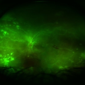
 Acute syphilitic posterior placoid chorioretinitis
Apr 24 2022 by Aniruddha K Agarwal, MD
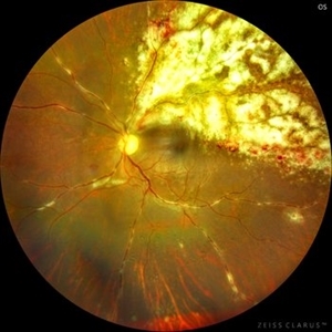
Green-light fundus autofluorescence (FAF) of the right eye from a 55-year-old man with risk factors for sexually trasnmitted diseases who presented to the retina clinic for a central scotoma. Funduscopy revealed a placoid lesion in the posterior pole. FAF highlights a hyperautofluorescent placoid lesion involving the macula with granular hyperfluorescence. The patient tested positive for syphilis and received intravenous penicillin treatment.
Photographer: Esther CIANCAS, MD, PhD, Gema CRESPO-RODRÍGUEZ, RN
Imaging device: Zeiss Clarus fundus camera
Condition/keywords: chorioretinitis, IUSG, syphilis, uveitis
-
 Bergmeister Papilla
Feb 20 2020 by Nisarg Joshi, MD
Gross pathology photo of a Bergmeister Papilla. It is a remnant of incompletely resorbed hyaloid vasculature from ocular development. This glial tissue is seen emminating from the optic nerve, which also shows glaucomatous cupping. The eye was enucleated due to a choroidal melanoma.
Photographer: Nisarg Joshi, MD, Geisinger Medical Center
Imaging device: Digital camera
Condition/keywords: Bergmeister's Papillae, hyaloid artery, persistent fetal vasculature (PFV)
-
 Central retinal artery occlusion
Nov 30 2022 by Ethan K Sobol, MD
A central retinal artery occlusion with cilioretinal artery sparing, imaged using a Volk Panretinal 2.2 and an iPhone camera in the emergency department.
Photographer: Jared Raabe, MD, Emory University Hospital
Imaging device: IPhone 13 Pro
Condition/keywords: central retinal artery occlusion (CRAO)
-
 Cheese Pizza Pie Appearance in CMV Retinitis
Mar 30 2024 by KANWALJEET HARJOT MADAN, M.S. (Ophthalmology), FAICO (Vitreous - Retina)
This is Fundus Photograph of left eye of 53 year male depicting an area of Retinal Necrosis with few Retinal Haemorrhages suggestive of CMV Retinitis. Areas of Perivascular Exudation also seen. On investigations, the patient was found to be HIV positive. He was started on Anti Retro Viral treatment after physician opinion.
Photographer: Dr. Kanwaljeet Harjot Madan, Thind Eye Hospital, Jalandhar City (Punjab) INDIA.
Imaging device: Zeiss Fundus Camera
Condition/keywords: AIDS, cytomegalovirus (CMV), retinitis
-
 Corneal Abnormal Blood Vessels
Jul 14 2013 by Jason S. Calhoun
Corneal neovascularization, abnormal blood vessels growing on the epithelium.
Photographer: Jason S. Calhoun, Department of Ophthalmology, Mayo Clinic Jacksonville, Florida
Imaging device: TOPCON D-90 SL NIKON CAMERA
Condition/keywords: cornea
-
 Diabetic Retinopathy, CSME, Exudates, NVD, Color Fundus Photo, Montage
Mar 18 2015 by James B. Soque, CRA, OCT-C, COA, FOPS
A 58-year-old diabetic male with a longstanding history of diabetic eye disease. Left eye color fundus photo shows extensive CSME, Clinically Significant Macular Edema, with deposits of hard exudates at fixation. There is extensive scattering of hard exudates and sheathing of the vessels.
Photographer: James B Soque, CRA COA
Imaging device: Topcon TRC 50 DX, OIS 5 MP Camera, MERGE software
Condition/keywords: background diabetic retinopathy (BDR), creamy yellow exudates, diabetes, exudates over the posterior pole, neovascularization of the disc (NVD), vessel sheathing
-
---thumb.JPG/image-square;max$300,300.ImageHandler) DSEK
DSEK
Jul 14 2013 by Jason S. Calhoun
Descemet's stripping endothelial keratoplasty, air bubble in anterior chamber.
Photographer: Jason S. Calhoun, Department of Ophthalmology, Mayo Clinic Jacksonville, Florida
Imaging device: TOPCON D-90 SL NIKON CAMERA
Condition/keywords: cornea
-
 Epicapsular Stars
Jan 28 2025 by Korey Starkey
Epicapsular stars and cataract noted in natural lens of 68-year-old patient.
Photographer: Korey Starkey
Imaging device: Slit lamp camera
Condition/keywords: cataract, chicken tracks, epicapsular stars, slit lamp photography
-
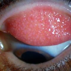 Giant Papillary Conjunctivitis, Left Upper Eyelid
Jul 22 2013 by Jason S. Calhoun
Contact lens wearer, in for exam. Has rough feeling underneath both eyelids. Patient thought it was through SCL wear. Patient VA was 20/20. right eye, 20/30, left eye. Underneath the left upper eyelid, you can see papillary inflammation and redness.
Photographer: Jason S. Calhoun, Department of Ophthalmology, Mayo Clinic Jacksonville, Florida
Imaging device: TOPCON D-90 SL NIKON CAMERA
Condition/keywords: giant papillary conjunctivitis
-
 Giant Retinal Tear
Jan 11 2022 by Manish Nagpal, MD, FRCS (UK), FASRS
Intraoperative photo a temporal giant retinal tear with everted flap and some laser marks noted on the bare choroid from previous barrage attempt elsewhere.
Photographer: Manish Nagpal, Retina Foundation, Ahmedabad, India
Imaging device: Sony PMW -10 MD surgical camera
Condition/keywords: giant retinal tear
-
 Gigantic Curved Glass Intraocular Foreign Body
Jun 6 2024 by Veer Singh, MS, FVRS, FMRF, FICO (Retina)
Gigantic Curved Glass Intraocular Foreign Body measuring 15x10 mm size
Photographer: Dr. Veer Singh
Imaging device: Ikegami 4k Microscope Camera
Condition/keywords: Curved Glass, Gigantic, IOFB
-
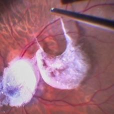
 ILM Peeling in Progress
Feb 4 2022 by Manish Nagpal, MD, FRCS (UK), FASRS
Intraoperative shot of ILM peeling in progress using forceps.
Photographer: Manish Nagpal, Director, Retina Foundation, Ahmedabad
Imaging device: Sony PMW -10 MD surgical camera
Condition/keywords: ILM flap, ILM staining, internal limiting membrane (ILM) peeling, macular hole, retina, retina surgery
 Loading…