Search results (885 results)
-
---thumb.jpg/image-square;max$300,300.ImageHandler) Presumed Ocular Histoplasmosis Syndrome
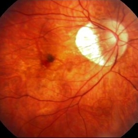
Presumed Ocular Histoplasmosis Syndrome
Feb 26 2013 by Henry J. Kaplan, MD
Color fundus photograph of the right eye of a patient with POHS shows typical punched out scars and peripapillary atrophy.
Condition/keywords: presumed ocular histoplasmosis syndrome (POHS)
-
---thumb.jpg/image-square;max$300,300.ImageHandler) Myopic fundus
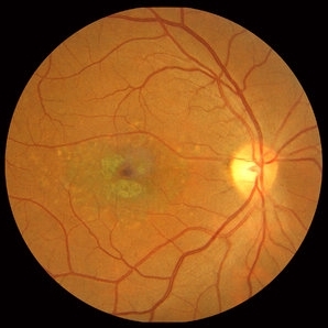
Myopic fundus
Jan 11 2013 by Hyung-Woo Kwak, MD
Myopic fundus reveals yellow-colored lacquer cracks and peripapillary atrophy. There was visible choroidal vessel due to thin retina.
Photographer: Misook Lee, Kyung Hee Univsersity Hospital, Seoul
Imaging device: Zeiss f 450 plus
Condition/keywords: myopic fundus
-
---thumb.jpg/image-square;max$300,300.ImageHandler) Geographic atrophy
Geographic atrophy
Aug 29 2012 by Young Hee Yoon, MD, PhD
OCT image of an 78-year-old woman. Her best-corrected visual acuity was counting fingers at 30cm.
Photographer: Ji Hee Kim, Asan Medical Center
Imaging device: Heidelberg spectralis
Condition/keywords: dry age-related macular degeneration (dry AMD), geographic atrophy
-
 Geographic Atrophy, Fundus photograph
Aug 23 2012 by Gerardo Garcia-Aguirre, MD
Fundus photograph of an 85-year-old patient with age related macular degeneration and geographic atrophy. A large area with well-defined borders is observed, in which the choroidal vasculature is visualized.
Photographer: Noemí Hernández, Asociación para Evitar la Ceguera en México
Imaging device: Zeiss FF4
Condition/keywords: geographic atrophy
-
---thumb.jpg/image-square;max$300,300.ImageHandler) Inferior Sector Iris Atrophy With Depigmentation
Inferior Sector Iris Atrophy With Depigmentation
Aug 1 2013 by From the Collections of Thomas M. Aaberg, MD and Thomas M. Aaberg Jr., MD
Inferior sector iris atrophy with depigmentation.
Condition/keywords: depigmentation, inferior sector iris atrophy
-
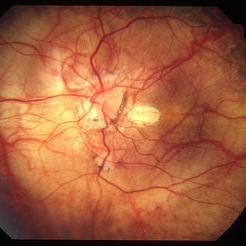
 Myopic Choroidal Neovascular Membrane
Mar 25 2013 by Ratimir Lazic, MD, PhD
Color fundus photography of a 33-year-old female. In macular area subretinal hemorrhage can be seen. Area of atrophy temporal from PNO. Myopic changes of posterior pole and mid periphery can be noticed. The patient has been treated with 2 consecutive ranibizumab intravitreal injections. BCVA at baseline was 0,05 (Snellen lines) and 0,3 (Snellen lines) 2 months after.
Photographer: Marko Lukic, MD
Imaging device: Zeis Visucam Lite 2
Condition/keywords: high myopia, myopic choroidal neovascularization (CNV), ranibizumab
-
 Stargardt macular dystrophy slide 1
Oct 22 2012 by Ronald C. Gentile, MD
16-year-boy with difficulty in school seeing the black board. The macula area of the right eye had areas with a beaten bronze appearance and atrophy. Small pisci-form flecks can be seen surrounding the fovea.
Photographer: The New York Eye & Ear Infirmary Department of Medical Imaging
Condition/keywords: small pisci-form flecks, Stargardt disease
-
---thumb.jpg/image-square;max$300,300.ImageHandler) Peripapillary Atrophy
Peripapillary Atrophy
Feb 13 2013 by From the Collections of Thomas M. Aaberg, MD and Thomas M. Aaberg Jr., MD
Papilledema, intra-retinal hemorrhage, periopticneuritis.
Condition/keywords: intraretinal hemorrhage, papilledema, periopticneuritis, peripapillary atrophy
-
 Peripapillary Atrophy With High Myopia
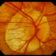
Feb 4 2015 by H. Michael Lambert, MD
Peripapillary atrophy and central macular degeneration seen in high myopia.
Condition/keywords: high myopia, peripapillary atrophy
-
 Fibrovascular Retinal Pigment Epithelial Detachment - Color Fundus
Jul 16 2014 by James B. Soque, CRA, OCT-C, COA, FOPS
69-year-old white female with Hx of 10 anti-VEFG treatment injections of right eye, VA 20/200, now stable, off drug for 10 months.
Photographer: James B Soque, CRA COA
Imaging device: Topcon TRC 50 DX with MERGE software, 5 MP dig camera
Condition/keywords: color fundus photograph, fibrovascular pigment epithelial detachment (PED), pigment epithelial atrophy, retina
-
 Geographic Atrophy
Aug 29 2012 by Young Hee Yoon, MD, PhD
Fundus photograph of an 78-year-old woman. Her best-corrected visual acuity was counting fingers at 30cm.
Photographer: Kyoung Woon Kim, Asan Medical Center
Imaging device: Canon CR-DGI/Version 5.1.2
Condition/keywords: dry age-related macular degeneration (dry AMD), geographic atrophy
-
 Myopic macular degeneration
Jan 11 2013 by Alex P. Hunyor, MD
Myopic macular degeneration, left eye - extensive chorioretinal atrophy.
Condition/keywords: myopic degeneration, myopic fundus, myopic macular degeneration
-
---thumb.JPG/image-square;max$300,300.ImageHandler) TID (Trans Illumination Defect)
TID (Trans Illumination Defect)
Jul 8 2013 by Jason S. Calhoun
74-year-old patient who VA 20/70 OD, 20/50 OS. Complaints of blurred vision. Iris atrophy, both eyes. ERM, right eye, Patient to have cataract surgery to improve distance vision.
Photographer: Jason S. Calhoun, Department of Ophthalmology, Mayo Clinic Jacksonville, Florida
Condition/keywords: translucency of iris
-
Stargardts Disease in Fundus Autofluorescence
Sep 12 2012 by Michael P. Kelly, FOPS
Fundus autofluorescence of a patient with Stargardts disease. Note the central area of hypo-autofluorescence indicating atrophy surrounded by smaller areas of hyper-autofluorescence. Note also the much smaller, and in greater number, pinpoints of hyper-autofluorescence extending from the vascular arcades into the mid-periphery.
Photographer: Michael P. Kelly, FOPS, Director, Duke Eye Labs, Duke University Hospital, Duke Eye Center
Imaging device: Heidelberg Spectralis
Condition/keywords: fundus autofluorescence (FAF), Stargardt disease
-
 Ocular Albinism Carrier
Feb 13 2013 by From the Collections of Thomas M. Aaberg, MD and Thomas M. Aaberg Jr., MD
RPE atrophy, degeneration, thinned arterioles.
Condition/keywords: degeneration, ocular albinism, retinal pigment epithelium atrophy, thinned arterioles
-
 Stargardt macular dystrophy slide 1
Oct 22 2012 by Ronald C. Gentile, MD
45-year-old man with progressive central vision loss since the age of 10. Small flecks can be seen surrounding the central area of atrophy.
Photographer: The New York Eye & Ear Infirmary Department of Medical Imaging
Condition/keywords: Stargardt disease, vision loss
-
 Retinitis Pigmentosa
Apr 6 2022 by Marianna Kavalaraki, MD, Msc
Fundus photography of a 21-year-old man with retinitis pigmentosa. Fundus findings include retinal pigmentary changes in the form of widespread pigment clumpings predominantly in the mid-peripheral fundus, arteriolar attenuation, RPE and retinal atrophy in the posterior pole.
Photographer: Marianna Kavalaraki, General Hospital of Nikaia Piraeus, Department of Ophthalmology
Imaging device: Canon CF-60DSi Digital Fundus Camera
Condition/keywords: retinitis pigmentosa
-
 Gyrate Atrophy of Choroid and Retina
Apr 19 2014 by Mallika Goyal, MD
Right eye fundus of a 45-year-old male patient with advanced gyrate atrophy of the choroid and retina with macular sparing. Optic nerve head is healthy.
Photographer: Mallika Goyal, MD, Apollo Health City, Hyderabad, India
Condition/keywords: choroid, gyrate atrophy
-
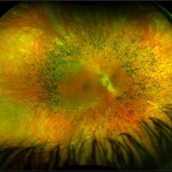 Retinitis Pigmentosa
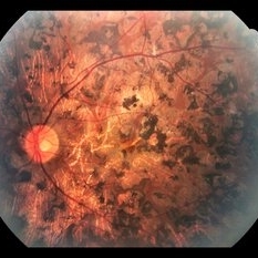
May 26 2017 by Olivia Rainey
Ultra-wide-field pseudocolor image of the right eye of an 39-year-old female with Retinitis Pigmentosa. She had slightly atypical appearance due to asymmetry: sectoral atrophy in left eye, compared to 360 degree bone spicule formation in right eye. Ddx: Pigmentary degeneration vs infection vs X-linked RP carrier due to asymmetry. Recommended genetic testing through My Retina Tracker, as well as visual field and ERG testing. Patient's vision was sc20/100 PH 20/70 in the right eye and sc20/80 PH 20/40 in the left.
Photographer: Olivia Rainey
Imaging device: Optos California
Condition/keywords: bone spicule, fundus photograph, Optos, peripheral bone spicules, pseudocolor, retinitis pigmentosa, ultra-wide field imaging
-
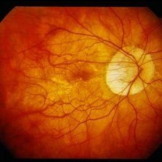
 High Myopia
May 2 2013 by Henry J. Kaplan, MD
Chorioretinal atrophy in high myopia and tilted disc.
Condition/keywords: high myopia, tilted disc
-
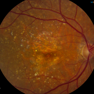 AMD with Calcific Drusen and Geographic Atrophy
Apr 19 2013 by Brandon G. Busbee, MD
AMD with calcific drusen and geographic atrophy.
Photographer: Alecia Camp, CRA - Tennessee Retina - Nashville, TN
Imaging device: Topcon TRC 50-EX
Condition/keywords: geographic atrophy
-
 Stargardt macular dystrophy slide 2
Oct 22 2012 by Ronald C. Gentile, MD
The central areas of atrophy were symmetric in both eyes with increased visibility of the choroidal vessels within the atrophic macula.
Photographer: The New York Eye & Ear Infirmary Department of Medical Imaging
Condition/keywords: Stargardt disease
-
---thumb.jpg/image-square;max$300,300.ImageHandler) Presumed Ocular Histoplasmosis Syndrome
Presumed Ocular Histoplasmosis Syndrome
Feb 26 2013 by Henry J. Kaplan, MD
POHS; left eye: large punched out pigmented scars and peripapillary atrophy.
Condition/keywords: presumed ocular histoplasmosis syndrome (POHS)
-
 Peripapillary Atrophy With High Myopia
Feb 4 2015 by H. Michael Lambert, MD
Peripapillary atrophy and central macular degeneration seen in high myopia.
Condition/keywords: high myopia, peripapillary atrophy
-
 Asymptomatic Lesion
Nov 9 2012 by Norman Byer
This is the same lesion as seen in the previous slide pair. Here the scleral indentation is carried more posterior revealing a tiny, round, full thickness retinal hole. This is not a tear produced by traction even though vitreous is always attached to these flaps. You will note that the hole is round and is separated by a slight distance from the flap itself. It is probably the result of long continued atrophy and devitalization of the retina. A posterior vitreous was not detached. This lesion has not changed its appearance for more than a year of observation, but the age of the hole is actually unknown.
Condition/keywords: asymptomatic, atrophy, full thickness retinal hole, posterior scleral indentation, retinal hole, round hole
 Loading…