Search results (15 results)
-
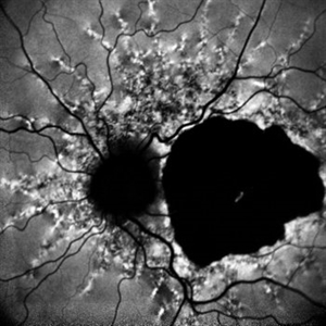
 Presumed Congenital Toxoplasmosis
Aug 16 2025 by Vishal Agrawal, MD, FRCS,FACS,FASRS
Fundus picture of 7 a year-old boy with esotropia. OCT showed complete atrophy & disorganization of the overlying RPE and neurosensory retina.
Photographer: Dr Ayushi Gupta
Imaging device: Clarus 700
Condition/keywords: coloboma of macula, toxoplasmosis
-
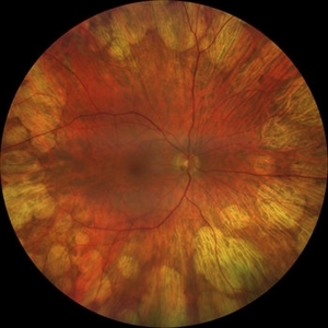
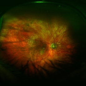
 Gyrate Atrophy
Apr 12 2023 by Ahmed Abbas Hashmi, OD
Left eye fundus of a 53-year-old male patient with advanced gyrate atrophy of the choroid and retina with macular sparing. Optic nerve head is healthy.
Photographer: Ahmed Abbas Hashmi
Imaging device: Topcon TRC-NW8F
Condition/keywords: chorioretinal atrophy
-
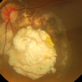
 PEHCR (Peripheral Exudative Hemorrhagic Chorioretinopathy)
May 12 2023 by Niloofar Piri, MD
Ultrawide fundus photograph of the left eye demonstrating extensive peripheral hemorrhagic exudative detachment in a 79 yo Caucasian female with prior history of non-exudative AMD. Recent diagnosis of Acute myeloid leukemia with low platelet count which might have contributed to the above presentatuon. Please note the temporal subretinal hemorrhage as well as RPE atrophy and hyperplasia in the macula.
Photographer: Rocio Bentivegna, MD, Saint Louis University; Jessica Maddox, COA, Saint Louis University
Condition/keywords: peripheral exudative hemorrhagic chorioretinopathy (PEHCR)
-
 Angioid Streaks
Dec 14 2022 by Pramod Kumar Suman, MBBS, MD
Fundus autofluorescence photograph of a 65-year-old male with numerous narrow, irregular streaks radiating in a circumferential pattern within the posterior pole with macular atrophy.
Photographer: Pramod Kumar Suman, Retina Foundation, Ahmedabad
Imaging device: Mirante
Condition/keywords: angioid streaks
-
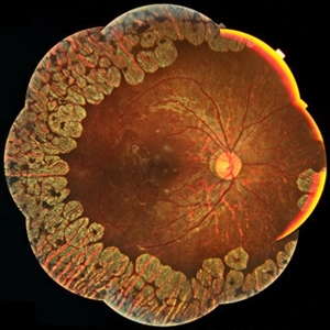
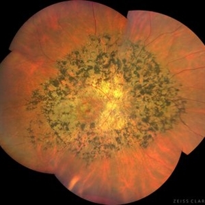 Tapetoretinal Degeneration
Sep 7 2022 by JEFFERSON R SOUSA, Tecg.º (Biomedical Systems Technology)
Patient 52 years old, Male, progressive loss of vision since the age of 20. Retinography showed mobilization of pigments in osteoblasts, extensive area of atrophy of the pigmentary epithelium and choroid. On fluorescein angiography, typical changes following the characteristic patterns of paracentra retinal retinitis pigmentosa. Autofluorescent fundus with a sectorial autohypofluorescence pattern in the regions of atrophies.
Photographer: JEFFERSON ROCHA DE SOUSA - Retinal Department at Instituto Dr. Suel Abujamra Sao Paulo-Brazil
Imaging device: Clarus 700 - Zeiss, composite of four 135 degree images.
Condition/keywords: pericentral retinitis pigmentosa, tapeoretinal degeneration
-
 Thioridazine-toxicity
Apr 30 2022 by Niloofar Piri, MD
61 yo male with PMH of longstanding schizophrenia since 20s with secondary intellectual disability presented with decreased vision following a recent stroke. He was found to have bilateral chorio-retinal atrophy involving posterior pole with scalloped edges and coin shaped atrophic area at margins extending into mid-periphery, diagnosis most concerning for intermediate stage thioridazine toxicity given the history. Mother could find handwritten prescriptions from 1990s when he was on Thioridazine 800 mg daily for unknown period of time. Patient had better vision in the left eye which was affected by recent stroke and prompted him to seek medical care. Fundus photograph of the right eye is demonstrated here.
Photographer: Jacob Grodsky, MD
Condition/keywords: drug toxicity, thioridazine toxicity, toxic retinopathy
-
 Paravenous-Pigmented-Retinochoroidal-Atrophy
Dec 17 2021 by Aditya S Kelkar, MS, FRCS, FASRS,FRCOphth
Right-eye Fundus Photo of a 30-year-old male.
Imaging device: Clarus 500
Condition/keywords: pigmented paravenous chorioretinal atrophy (PPCRA), retinochoroidopathy
-
 Gyrate Atrophy
Oct 30 2020 by JEFFERSON R SOUSA, Tecg.º (Biomedical Systems Technology)
Female patient, 28-year-old, with low vision in both eyes since childhood. In routine examination, important changes were observed with atrophic, symmetrical and bilateral aspects with apparently preservation of the central retina.
Condition/keywords: gyrate atrophy
-
 Gyrate Atrophy
Sep 23 2020 by Hashim Ali Khan, OD, FAAO
Widefield color fundus image of a young male with gyrate atrophy.
Imaging device: Optomap
Condition/keywords: gyrate atrophy
-
 Didanosine Toxicity
Jan 27 2020 by Nimesh A. Patel, MD, FASRS
Patient with history of HIV treated with didanosine. Developed gyrate like peripheral retinal atrophy with central sparing. Vision is 20/25
Imaging device: Clarus
Condition/keywords: AIDS, didanosine, HIV
-
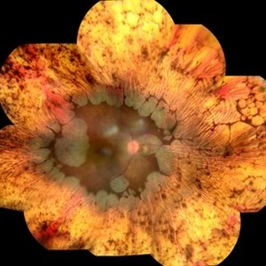
 Gyrate Atrophy
Jan 6 2019 by Hashim Ali Khan, OD, FAAO
Montage of Multiple Fundus Photographs from the right eye of a 25-year-old woman with gyrate atrophy.
Photographer: Ahmed Abbass
Imaging device: Topcon TRC-NW8F
Condition/keywords: gyrate atrophy, hereditary retinal dystrophy, retinal dystrophy
-
 Intraocular Foreign Body
Feb 7 2019 by Somnath Chakraborty, MD
Left eye fundus photo montage of a 45-year-old male showing a large iron foreign body, impacted inferior to the infero-temporal branch vessels with a large patch of surrounding chorio-retinal atrophy, secondary to resolving Commotio retinae
Photographer: Saptarshi Mehta
Condition/keywords: commotio retinae, intraocular foreign body, trauma
-
 Retinoblastoma Regressed
Dec 31 2015 by P. Mahesh Shanmugam, MBBS, DO, FRCSEd, PhD, FAICO
Regressed Retinoblastoma S/P chemotherapy and multiple sessions of TTT. Central calcific residue with surrounding chorio-retinal atrophy is well noted.
Condition/keywords: retinoblastoma
-
 Optic Atrophy and Attenuated Retinal Vessels Following Endophthalmitis
Jul 12 2014 by Philip J. Polkinghorne, MD
This elderly lady underwent a vitrectomy for post-surgical endophthalmitis. The infection was successfully treated but the functional outcome was poor because of optic atrophy and attenuated retinal vessels.
Photographer: Alex Fraser
Imaging device: Optos Camera
Condition/keywords: attenuated vessels, endophthalmitis, optic atrophy, post-vitrectomy
-
 ARMD With Geographic Atrophy, Peripheral Degeneration
Dec 6 2013 by James B. Soque, CRA, OCT-C, COA, FOPS
92-year-old white female with exudative macular degeneration, geographic atrophy, and peripheral retinal degeneration.
Photographer: James Soque, CRA COA, Island Retina, Shirley, New York
Imaging device: Topcon TRC 50DX with OIS 10.6.45
Condition/keywords: fundus photograph, geographic atrophy
 Loading…