Search results (885 results)
-
 AMD with Calcific Drusen and Geographic Atrophy
Apr 19 2013 by Brandon G. Busbee, MD
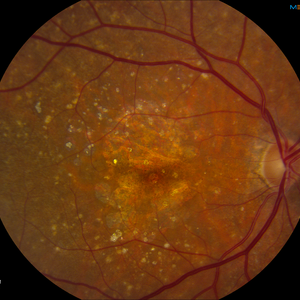
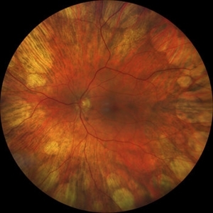
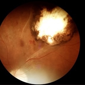
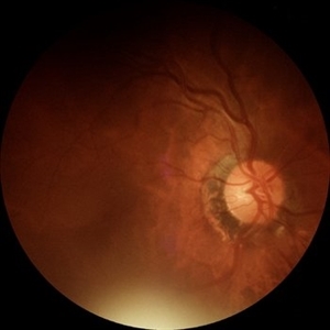
AMD with calcific drusen and geographic atrophy.
Photographer: Alecia Camp, CRA - Tennessee Retina - Nashville, TN
Imaging device: Topcon TRC 50-EX
Condition/keywords: geographic atrophy
-
 Pigmented Paravenous Retinochoroidal Atrophy (PPRCA)
Jun 30 2025 by Maria Letícia Costa Holanda
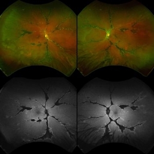
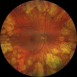
Fundoscopy of a 42-year-old asymptomatic man with pigmented paravenous chorioretinal atrophy. Pigmented paravenous retinochoroidal atrophy (PPRCA) is a rare disorder of unknown etiology. The disease is characterized by pigment accumulation along the distribution of retinal veins. The findings are usually incidental with minimal effect on vision.
Photographer: Guilherme da Cruz Reis, CLINOS Eye Hospital - Feira de Santana (BA),Brazil
Condition/keywords: pigmented paravenous chorioretinal atrophy (PPCRA)
-
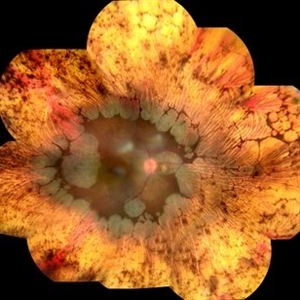
 Retinoblastoma Regressed
Dec 31 2015 by P. Mahesh Shanmugam, MBBS, DO, FRCSEd, PhD, FAICO
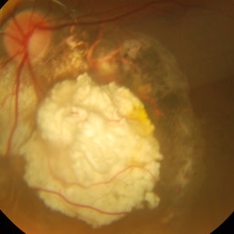
Regressed Retinoblastoma S/P chemotherapy and multiple sessions of TTT. Central calcific residue with surrounding chorio-retinal atrophy is well noted.
Condition/keywords: retinoblastoma
-
 Rod Cone dystrophy
Nov 29 2022 by Niloofar Piri, MD
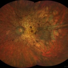
Fundus photograph of the left eye in a 58 yo male with rod cone dystrophy. He presented with night blindness and peripheral vision loss since youth and recent decrease in central vision for the past 10 years. Notice waxy pallor of the nerve, severe arterial narrowing and chorioretinal atrophy mainly around the arcades as well as posterior pole along with RPE hyperplastic changes and atrophy. RPE atrophy in midperiphery has coin shaped appearance. FAF has characteristic appearance (uploaded separately) He has one pathogenic variants of both CEP290 and PRPH2 genes.
Photographer: Sean Kelso, Saint Louis University
Condition/keywords: hereditary retinal deg, hereditary retinal dystrophy, Rod cone dystrophy
-
 Didanosine Toxicity
Jan 27 2020 by Nimesh A. Patel, MD, FASRS
Patient with history of HIV treated with didanosine. Developed gyrate like peripheral retinal atrophy with central sparing. Vision is 20/25
Imaging device: Clarus
Condition/keywords: AIDS, didanosine, HIV
-
 Didanosine Toxicity
Jan 27 2020 by Nimesh A. Patel, MD, FASRS
Patient with history of HIV treated with didanosine. Developed gyrate like peripheral retinal atrophy with central sparing. Vision is 20/25
Imaging device: Clarus
Condition/keywords: AIDS, didanosine, HIV
-
 Diffuse Chorioretinal Atrophy
Feb 21 2024 by Virginia Gebhart
61 year male with myopic degeneration and diffuse chorioretinal atrophy. BCVA 20/200.
Photographer: Virginia Gebhart
Imaging device: Topcon TRC 50DX
Condition/keywords: chorioretinal atrophy, myopic degeneration
-
 Geographic Atrophy
Oct 13 2012 by Geoffrey G. Emerson, MD, PhD, FASRS
Geographic atrophy
Condition/keywords: advanced geographic atrophy, choroid, dry age-related macular degeneration (dry AMD)
-
 Geographic Atrophy 2nd to Central areolar choroidal dystrophy
Nov 24 2012 by Roy Schwartz, MD
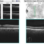
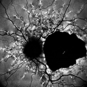
Fundus autofluorescence and SD-OCT of a 70-year-old woman with geographic atrophy sec. to Central areolar choroidal dystrophy
Condition/keywords: central areolar choroidal dystrophy (CACD), geographic atrophy
-
 Gyrate Atrophy
Oct 30 2020 by JEFFERSON R SOUSA, Tecg.º (Biomedical Systems Technology)
Female patient, 28-year-old, with low vision in both eyes since childhood. In routine examination, important changes were observed with atrophic, symmetrical and bilateral aspects with apparently preservation of the central retina.
Condition/keywords: gyrate atrophy
-
 Myopic retinopathy
Dec 27 2021 by Eduardo Javier Pinuer Alvarado
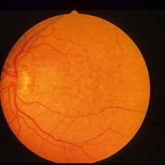
Fundus photograph of an 50-year-old man with myopic retinopathy, posterior staphyloma, myopic chorioretinal atrophy and tilted and oblique disc.
Photographer: Eduardo Pinuer A, Universidad Austral de Chile.
Imaging device: CR-2 AF Digital Non-Mydriatic Retinal Camera, Canon.
Condition/keywords: myopic chorioretinal atropthy, myopic retinopathy, posterior staphyloma, retinopathy
-
 PEHCR (Peripheral Exudative Hemorrhagic Chorioretinopathy)
May 12 2023 by Niloofar Piri, MD
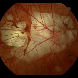
Ultrawide fundus photograph of the left eye demonstrating extensive peripheral hemorrhagic exudative detachment in a 79 yo Caucasian female with prior history of non-exudative AMD. Recent diagnosis of Acute myeloid leukemia with low platelet count which might have contributed to the above presentatuon. Please note the temporal subretinal hemorrhage as well as RPE atrophy and hyperplasia in the macula.
Photographer: Rocio Bentivegna, MD, Saint Louis University; Jessica Maddox, COA, Saint Louis University
Condition/keywords: peripheral exudative hemorrhagic chorioretinopathy (PEHCR)
-
 Pigmented Paravenous Chorioretinal Atrophy (PPCRA)
Jun 27 2025 by Maria Letícia Costa Holanda
Fundoscopy of a 42-year-old asymptomatic man with pigmented paravenous chorioretinal atrophy. Pigmented paravenous retinochoroidal atrophy (PPRCA) is a rare disorder of unknown etiology. The disease is characterized by pigment accumulation along the distribution of retinal veins. The findings are usually incidental with minimal effect on vision.
Photographer: Guilherme da Cruz Reis, CLINOS Eye Hospital - Feira de Santana (BA),Brazil
Condition/keywords: pigmented paravenous chorioretinal atrophy (PPCRA)
-
 Pigmented Paravenous Retinochoroidal Atrophy (PPRCA)
Jun 27 2025 by Maria Letícia Costa Holanda
Fundoscopy of a 42-year-old asymptomatic man with pigmented paravenous chorioretinal atrophy. Pigmented paravenous retinochoroidal atrophy (PPRCA) is a rare disorder of unknown etiology. The disease is characterized by pigment accumulation along the distribution of retinal veins. The findings are usually incidental with minimal effect on vision.
Photographer: Guilherme da Cruz Reis, CLINOS Eye Hospital - Feira de Santana (BA),Brazil
Condition/keywords: pigmented paravenous chorioretinal atrophy (PPCRA)
-
 Presumed Congenital Toxoplasmosis
Aug 16 2025 by Vishal Agrawal, MD, FRCS,FACS,FASRS
Fundus picture of 7 a year-old boy with esotropia. OCT showed complete atrophy & disorganization of the overlying RPE and neurosensory retina.
Photographer: Dr Ayushi Gupta
Imaging device: Clarus 700
Condition/keywords: coloboma of macula, toxoplasmosis
-
 Retinocoroiditis Inactiva Por Toxoplasmosis
Apr 28 2025 by Paulina Araujo
Fundus photography demonstrates a 2-disc-diameter chorioretinal scar in the superior temporal arcade, consistent with inactive toxoplasmic retinochoroiditis. The lesion exhibits pigmented borders and central atrophy, with adjacent splinter hemorrhages and vascular sheathing. No vitreous inflammation or active satellite lesions are present.
Photographer: Paulina D.Araujo Martínez, Asociación para Evitar la Ceguera en México I.A.P., Hospital Dr Luis Sánchez Bulnes.
Condition/keywords: toxoplasmosis chorioretinitis
-
 Thioridazine-toxicity
Apr 30 2022 by Niloofar Piri, MD
61 yo male with PMH of longstanding schizophrenia since 20s with secondary intellectual disability presented with decreased vision following a recent stroke. He was found to have bilateral chorio-retinal atrophy involving posterior pole with scalloped edges and coin shaped atrophic area at margins extending into mid-periphery, diagnosis most concerning for intermediate stage thioridazine toxicity given the history. Mother could find handwritten prescriptions from 1990s when he was on Thioridazine 800 mg daily for unknown period of time. Patient had better vision in the left eye which was affected by recent stroke and prompted him to seek medical care. Fundus photograph of the right eye is demonstrated here.
Photographer: Jacob Grodsky, MD
Condition/keywords: drug toxicity, thioridazine toxicity, toxic retinopathy
-
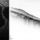
 Acute and Chronic OCT of BRAO
Sep 9 2021 by Aleksandra V. Rachitskaya, MD, FASRS
Acute and chronic OCT findings in BRAO. Acutely, inner retinal hyper-reflectivity is seen. Chronically, retina atrophy ensues.
Condition/keywords: BRAO, OCT
-
---thumb.jpg/image-square;max$300,300.ImageHandler) Age Related Macular Degeneration - Geographic Atrophy
Age Related Macular Degeneration - Geographic Atrophy
May 3 2013 by Suber S. Huang, MD, MBA, FASRS
Geographic Atrophy.
Imaging device: Retina Diseases Imaging Analysis Reading Center
Condition/keywords: advanced geographic atrophy, atrophic scar, atrophic spot, geographic atrophy, macula lesion, pigment epithelial atrophy
-
 Angioid Streaks
Dec 14 2022 by Pramod Kumar Suman, MBBS, MD
Fundus autofluorescence photograph of a 65-year-old male with numerous narrow, irregular streaks radiating in a circumferential pattern within the posterior pole with macular atrophy.
Photographer: Pramod Kumar Suman, Retina Foundation, Ahmedabad
Imaging device: Mirante
Condition/keywords: angioid streaks
-
 ARMD
ARMD
Jan 13 2014 by David Callanan, MD
HM OU marked RPE atrophy, 63-year-old female.
Condition/keywords: age-related macular degeneration (AMD)
-
 Atrophy
Jun 4 2025 by Paulina Araujo
The fundus photograph captures the central 55 degrees of the right eye, revealing alpha and beta peripapillary atrophy.
Photographer: Paulina D.Araujo Martínez, Asociación para Evitar la Ceguera en México I.A.P., Hospital Dr Luis Sánchez Bulnes.
Condition/keywords: atrophy
-
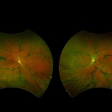
 Choroideremia
Sep 21 2022 by Zach Seim
Ultra-widefield fundus photo of a 74 year old male presenting with severe vision loss beginning at age 55. Patient sought a second opinion with our office and was diagnosed with Choroideremia. Patient denies hearing loss, heart problems, balance issues, polydactyly, kidney problems, and dental problems. Patient reports that nobody in the family had blindness. Choroideremia is an X-linked chorioretinal dystrophy characterized by the diffuse, progressive degeneration of the retinal pigment epithelium (RPE), photoreceptors and choriocapillaris. It is caused by a mutation in the CHM gene.
Photographer: Zach Seim
Imaging device: Optos California
Condition/keywords: choroideremia, hereditary choroidal atrophy, hereditary retinal dystrophy, Optos, pseudocolor, ultra-wide field imaging
-
 Choroideremia
Sep 21 2022 by Zach Seim
Ultra-widefield fundus photo of a 74 year old male presenting with severe vision loss beginning at age 55. Patient sought a second opinion with our office and was diagnosed with Choroideremia. Patient denies hearing loss, heart problems, balance issues, polydactyly, kidney problems, and dental problems. Patient reports that nobody in the family had blindness. Choroideremia is an X-linked chorioretinal dystrophy characterized by the diffuse, progressive degeneration of the retinal pigment epithelium (RPE), photoreceptors and choriocapillaris. It is caused by a mutation in the CHM gene.
Photographer: Zach Seim
Imaging device: Optos California
Condition/keywords: choroideremia, hereditary choroidal atrophy, hereditary retinal dystrophy, left eye, light perception, low vision, Optos, pseudocolor, ultra-wide field imaging
-
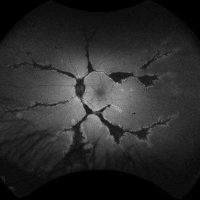
 Choroideremia
Jan 26 2013 by Ratimir Lazic, MD, PhD
FAG image of a 66-year-old male. Diffuse chorioretinal atrophy is present. Large choroidal vessels can be seen due to atrophy of the RPE and choriocapilaris.
Photographer: Marko Lukic, MD
Imaging device: Zeis Visucam Lite 2
Condition/keywords: choroideremia, fundus photograph
 Loading…