Search results (885 results)
-
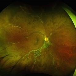
 Gyrate Atrophy
Nov 22 2025 by Gaurav Kamble
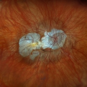
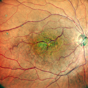
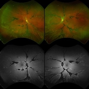
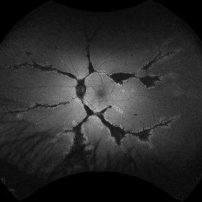
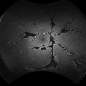
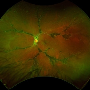
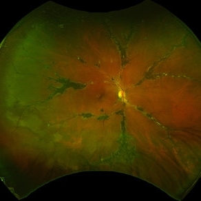
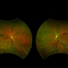
A 12-year-old female presented with progressive blurring of vision for distance and had a known history of convulsions. Ocular examination revealed bilateral proptosis and megalocornea. Fundus evaluation showed well-defined scalloped areas of peripheral chorioretinal degeneration characteristic of gyrate atrophy, along with cystoid macular edema involving the macular region. The overall clinical picture was consistent with gyrate atrophy.
Photographer: Ms. Vishaka Shah , Isha Eye Care Pvt Ltd ,Khadakpada, Kalyan
Imaging device: Optos Imaging Daytona
Condition/keywords: gyrate atrophy
-
 Starstruck by Stargardt
Nov 17 2025 by SHRADDHA RAJ SHRIVASTAVA
Left eye G-FAF image of a 26 year old patient diagnosed with Stargardt Disease, showing hyperautofluorescent flecks of increased lipofuscin accumulation and dark areas of hypoautofluorescence representing retinal pigment epithelium (RPE) atrophy.
Photographer: Dr. Shraddha Raj Shrivastava
Imaging device: Nidek Mirante SLO/OCT (Confocal scanning/Spectral domain OCT)
Condition/keywords: fleck dystrophy, fundus autofluorescence (FAF), hereditary macular dystrophy, heredomacular degeneration, lipofuscin, Stargardt Disease
-
 The Great Disc-guise
Nov 12 2025 by SHRADDHA RAJ SHRIVASTAVA
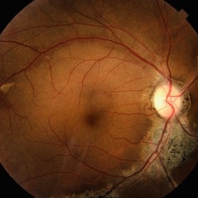
Right eye pseudocolor fundus photo of a 20 year old with Both eyes Pathological Myopia (spherical refractive error of - 18.00 DS in BE), showing a tilted myopic disc with peripapillary atrophy, and extensive posterior staphyloma baring the underlying choroidal vessels and scleral tissue. We can also see a well-defined round chorioretinal atrophic (CRA) patch superonasal to the disc, giving the illusion of double disc on cursory fundus examination.
Photographer: Dr. Shraddha Raj Shrivastava
Imaging device: Nidek Mirante SLO/OCT (Confocal scanning/Spectral domain OCT)
Condition/keywords: chorioretinal atrophy, High Myopia, pathologic myopia, peripapillary atrophy, posterior staphyloma
-
 Retinitis Pigmentosa: Now available in its Pericentral edition
Nov 7 2025 by SHRADDHA RAJ SHRIVASTAVA
Left eye Green-FAF image, of a 50 year old patient, diagnosed with bilateral Pericentral variant of Retinitis Pigmentosa. The disease is characterized by pigmentary changes closer to the macula, and an earlier involvement of central visual acuity as compared to typical RP. We can see prominent, scalloped hypoautofluorescent lesions in the pericentral region, which corresponds to areas of severe RPE atrophy and photoreceptor cell loss. Macula shows preserved background autofluorescence, with darker areas corresponding to no detectable fluorescence due to macular atrophy (loss of melanin/lipofuscin).
Photographer: Dr. Shraddha Raj Shrivastava
Imaging device: Nidek Mirante SLO/OCT (Confocal scanning/Spectral domain OCT)
Condition/keywords: ATYPICAL RETINITIS PIGMENTOSA, fundus autofluorescence (FAF), pericentral retinitis pigmentosa, RP variant
-
 Retinitis Pigmentosa: Now available in its Pericentral edition
Nov 7 2025 by SHRADDHA RAJ SHRIVASTAVA
Right eye fundus photo of a 50 year old patient, diagnosed with bilateral Pericentral variant of Retinitis Pigmentosa. True to the subtype, the pigmentation is closer to fixation. There are bony spicules like pigmentary changes and RPE atrophy seen around the macula and disc (posterior pole), just adjacent to the arcades, while the peripheral fundus appears unaffected. The macula shows severe macular atrophy and scarring. Similar changes were observed in the left eye.
Photographer: Dr. Shraddha Raj Shrivastava
Imaging device: Nidek Mirante SLO/OCT (Confocal scanning/Spectral domain OCT)
Condition/keywords: pericentral retinitis pigmentosa, retinitis pigmentosa (RP) dystrophy, Rod cone dystrophy, RP variant
-
 All That Glows Yellow Isn’t Mellow: Coats' Disease Unveiled
Nov 4 2025 by SHRADDHA RAJ SHRIVASTAVA
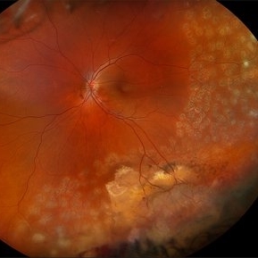
Montage fundus image of an 11 year old boy diagnosed with left eye Coats' disease (stage 3A1), reveals a hyperemic disc and surrounding intra-retinal exudates superior to the disc. There is a single fibroglial nodule at the macula causing submacular fibrosis with exudation. We can see areas of pigmentary changes and RPE atrophy in posterior pole and mid-peripheral retina supero-temporally. There is massive yellowish subretinal exudation in all the quadrants, which are associated with telangiectatic aneurysmal capillary dilation, more prominently seen in the nasal periphery. Supero-nasally we can also see an orange-red elevated vaso-proliferative mass with overlying dilated capillaries, which has likely developed secondary to untreated long standing disease. We can also see associated extrafoveal subtotal exudative retinal detachment in the inferior and nasal quadrants.
Photographer: Dr. Shraddha Raj Shrivastava
Imaging device: Nidek Mirante SLO/OCT (Confocal scanning/Spectral domain OCT)
Condition/keywords: COATS DISEASE, exudative detachment, leukocoria, subretinal exudates, Xanthocoria, yellow exudate
-
 ERMageddon - Wrinkle in the Space-time Fabric of Macula
Oct 29 2025 by SHRADDHA RAJ SHRIVASTAVA
38 year old female with Epiretinal Membrane (ERM) over macula, post laser barrage for multiple symptomatic Horse-shoe Tears (HSTs) and Lattice Degenerations (seen on wide-field image). Posterior pole revealed tilted disc with peripapillary atrophy. There is thick opaque epiretinal membrane obscuring the underlying superior arcade vessels and causing foveal ectopia with distortion of perimacular vasculature. Patient was planned for Right Eye pars plana vitrectomy for ERM peeling.
Photographer: Dr. Shraddha Raj Shrivastava
Imaging device: Nidek Mirante SLO/OCT (Confocal scanning/Spectral domain OCT
Condition/keywords: BARRAGE LASER, ectopic fovea, epiretinal membrane (ERM), horseshoe tear, lattice degeneration, vitreomacular traction (VMT)
-
 ERMageddon - Wrinkle in the Space-time Fabric of Macula
Oct 29 2025 by SHRADDHA RAJ SHRIVASTAVA
38 year old female with Epiretinal Membrane (ERM) over macula, post laser barrage for multiple symptomatic Horse-shoe Tears (HSTs) and Lattice Degenerations. Posterior pole revealed tilted disc with peripapillary atrophy. There is thick opaque epiretinal membrane obscuring the underlying superior arcade vessels and causing foveal ectopia with distortion of perimacular vasculature. Patient was planned for Right Eye pars plana vitrectomy for ERM peeling.
Photographer: Dr. Shraddha Raj Shrivastava
Imaging device: Nidek Mirante SLO/OCT (Confocal scanning/Spectral domain OCT
Condition/keywords: ectopic fovea, epiretinal membrane (ERM), ERM, horseshoe tear, vitreomacular traction (VMT)
-
 Chronic Serous Central Choroidopathy
Oct 15 2025 by Vicente Nicanor Mancilla Guerrero
Color mosaic fundus image of a 60-year-old male patient with CSC recurrences, with the presence of retinal tapestry atrophy with gravitational pattern.
Photographer: Vicente Mancilla G, Ophthalmic Medical Technologist
Imaging device: Canon cx-1
Condition/keywords: chronic central serous chorioretinopathy (CSCR)
-
 Stargardt Disease Autofluorescence
Oct 14 2025 by NIDHI PANWAR, MD FRCS Glasgow FNB FICO
Classical autofluorescence in Stargardt disease with foveal atrophy.
Photographer: Ms Amrutha Shaji Optometrist
Imaging device: Optos
Condition/keywords: Stargardt disease
-
 Dry AMD, Advanced Atrophic with Subfoveal Involvement
Oct 3 2025 by Kimberly Wakester
Optomap RGB of an 76-year-old woman with Dry AMD, Advanced Atrophic with Subfoveal Involvement in the right eye. Longstanding, more likely due to myopic/choroidal atrophy. End-stage disease, would not recommend aggressive intervention. Patient is to continue follow up care and repeat OCT/imaging as directed per doctor. Just a fun mention about the image, if you look at the optic nerve and the vessels it appears to look like an eye.
Photographer: Kimberly Wakester, COA, OCT-C
Imaging device: Optos California
Condition/keywords: Advanced Atrophic with Subfoveal Involvement, dry age-related macular degeneration (dry AMD), Myopic Degeneration
-
 Extensive Macular Atrophy with Pseudodrusen (EMAP)
Aug 31 2025 by Gabriel Costa Andrade, PhD
Autofluorescence image of the right eye of a 53-year-old male with history of rheumatic fever and low vision in OU with diagnosis of Extensive Macular Atrophy with Pseudodrusen (EMAP). The image shows a large area of macular atrophy associated with pseudodrusen. Genetic testing was negative for hereditary retinal diseases.
Photographer: Gabriel Andrade
Condition/keywords: Autoflourescence, macula, Retina
-
 Vasoproliferative Tumor (FEVR) s/p PPV/PRP
Aug 27 2025 by Virginia Gebhart
39 year old female with an amelanotic vascular lesion inferotemporal with CR atrophy inferior edge and likely lipid exudate superior edge. Pt presented with vitreous and sub-hyaloid hemorrhage. Findings from exam, ultrasound, FA all consistent with FEVR, stage 2. PPV with PRP performed, pt vison has improved from CF@2ft at initial visit to 20/100 PH 20/60 at 1 week post-op. Pt's 2 children have been recently examined with identical findings of FEVR
Photographer: Virginia Gebhart, Retina Consultants of Carolina
Imaging device: Optos California
Condition/keywords: familial exudative vitreoretinopathy (FEVR), pan-retinal photocoagulation (PRP), Vasoproliferative Tumor
-
 Presumed Congenital Toxoplasmosis Macular Coloboma
Aug 16 2025 by Vishal Agrawal, MD, FRCS,FACS,FASRS
7-year-old boy presented with esotropia in OD with light perception positive. Fundus reveals a large macular coloboma occupying nearly the entire macula. OCT scan shows complete atrophy and disorganization of the overlying RPE and neurosensory retina. A much smaller lesion was observed in OS with BCVA 20/40.
Photographer: Dr Ayushi Gupta
Imaging device: Clarus 700
Condition/keywords: Coloboma, congenital toxoplasmosis
-
 Presumed Congenital Toxoplasmosis
Aug 16 2025 by Vishal Agrawal, MD, FRCS,FACS,FASRS
Fundus picture of 7 a year-old boy with esotropia. OCT showed complete atrophy & disorganization of the overlying RPE and neurosensory retina.
Photographer: Dr Ayushi Gupta
Imaging device: Clarus 700
Condition/keywords: coloboma of macula, toxoplasmosis
-
 Congenital Retinal Macrovessel
Aug 7 2025 by Cesar Valdez, MD
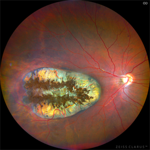
Fundus photograph and fluorescein angiography of a 43-year-old man with typical findings of congenital retinal macrovessel and RPE atrophy due to chronic macular edema.
Photographer: César Valdez, Instituto Mexicano de Oftalmología, IAP. Querétaro, México.
Imaging device: Zeiss Clarus 700
Condition/keywords: congenital retinal macrovessel
-
 Congenital Retinal Macrovessel
Aug 7 2025 by Cesar Valdez, MD
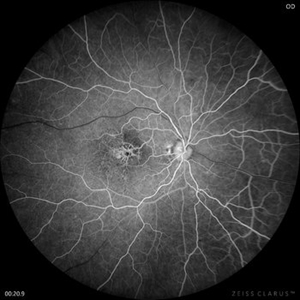
Fundus photograph and fluorescein angiography of a 43-year-old man with typical findings of congenital retinal macrovessel and RPE atrophy due to chronic macular edema.
Photographer: César Valdez, Instituto Mexicano de Oftalmología, IAP. Querétaro, México.
Imaging device: Nidek Mirante
Condition/keywords: Retina
-
 Pigmented Paravenous Retinochoroidal Atrophy (PPRCA)
Jun 30 2025 by Maria Letícia Costa Holanda
Fundoscopy of a 42-year-old asymptomatic man with pigmented paravenous chorioretinal atrophy. Pigmented paravenous retinochoroidal atrophy (PPRCA) is a rare disorder of unknown etiology. The disease is characterized by pigment accumulation along the distribution of retinal veins. The findings are usually incidental with minimal effect on vision.
Photographer: Guilherme da Cruz Reis, CLINOS Eye Hospital - Feira de Santana (BA),Brazil
Condition/keywords: pigmented paravenous chorioretinal atrophy (PPCRA)
-
 Pigmented Paravenous Retinochoroidal Atrophy (PPRCA)
Jun 27 2025 by Maria Letícia Costa Holanda
Fundoscopy of a 42-year-old asymptomatic man with pigmented paravenous chorioretinal atrophy. Pigmented paravenous retinochoroidal atrophy (PPRCA) is a rare disorder of unknown etiology. The disease is characterized by pigment accumulation along the distribution of retinal veins. The findings are usually incidental with minimal effect on vision.
Photographer: Guilherme da Cruz Reis, CLINOS Eye Hospital - Feira de Santana (BA),Brazil
Condition/keywords: Pigmented Paravenous Retinochoroidal Atrophy
-
 Pigmented Paravenous Retinochoroidal Atrophy (PPRCA)
Jun 27 2025 by Maria Letícia Costa Holanda
Fundoscopy of a 42-year-old asymptomatic man with pigmented paravenous chorioretinal atrophy. Pigmented paravenous retinochoroidal atrophy (PPRCA) is a rare disorder of unknown etiology. The disease is characterized by pigment accumulation along the distribution of retinal veins. The findings are usually incidental with minimal effect on vision.
Photographer: Guilherme da Cruz Reis, CLINOS Eye Hospital - Feira de Santana (BA),Brazil
Condition/keywords: pigmented paravenous chorioretinal atrophy (PPCRA)
-
 Pigmented Paravenous Chorioretinal Atrophy (PPCRA)
Jun 27 2025 by Maria Letícia Costa Holanda
Fundoscopy of a 42-year-old asymptomatic man with pigmented paravenous chorioretinal atrophy. Pigmented paravenous retinochoroidal atrophy (PPRCA) is a rare disorder of unknown etiology. The disease is characterized by pigment accumulation along the distribution of retinal veins. The findings are usually incidental with minimal effect on vision.
Photographer: Guilherme da Cruz Reis, CLINOS Eye Hospital - Feira de Santana (BA),Brazil
Condition/keywords: pigmented paravenous chorioretinal atrophy (PPCRA)
-
 Pigmented Paravenous Retinochoroidal Atrophy
Jun 18 2025 by César Adrián Gómez Valdivia, MD
Fundus photograph of a 42YO female patient diagnosed with Pigmented Paravenous Retinochoroidal Atrophy. Findings were bilateral. Image shows hypoautofluorescence in the affected areas due to overall loss of RPE cells and thus lower lipofuscin levels.
Photographer: @eyemissu2
Imaging device: TOPCON TRC-50DX
Condition/keywords: Pigmented Paravenous Retinochoroidal Atrophy
-
 Pigmented Paravenous Retinochoroidal Atrophy
Jun 18 2025 by César Adrián Gómez Valdivia, MD
Fundus photograph Image of a 42YO female patient diagnosed with Pigmented Paravenous Retinochoroidal Atrophy. Findings were bilateral. Image shows hypoautofluorescence in the affected areas due to overall loss of RPE cells and thus lower lipofuscin levels.
Photographer: @eyemissu2
Imaging device: California ICG OPTOS
Condition/keywords: Pigmented Paravenous Retinochoroidal Atrophy
 Loading…