Search results (1033 results)
-
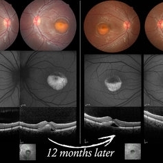
 Bilateral Retinoschisis Retinal Detachment
Sep 15 2012 by Barbara Parolini, MD
Fundus photograph of a case of bilateral retinoschisis and retinal detachment. The border of the external layer breaks and the border of the schisis have been treated with argon laser. An epiretinal membrane formed after the formation of retinal detachment.
Photographer: Dr Rino Frisina, Istituto Clinico S.Anna, Brescia, Italy
Imaging device: optos
Condition/keywords: epiretinal membrane formation, retinoschisis
-
 Plaquenil Toxicity
Apr 30 2013 by Theodore Leng, MD, MS, FASRS
SD-OCT scan from a 44-year-old woman with bilateral plaquenil toxicity. There is damage visible in the outer retina in a perifoveal distribution.
Condition/keywords: hydroxychloroquine toxicity, plaquenil toxicity
-
 Familial Dominant Drusen
Nov 22 2015 by Mallika Goyal, MD
Bilateral drusen over the entire retinal mid-periphery and periphery of a 29-year-old male with no visual complaints. Macular centre is normal though there are some drusen in the temporal macula.
Photographer: Mallika Goyal, MD, Apollo Health City, Jubilee Hills, Hyderabad, India
Condition/keywords: familial drusen
-
 Plaquenil Toxicity
Apr 30 2013 by Theodore Leng, MD, MS, FASRS
SD-OCT scan from a 44-year-old woman with bilateral plaquenil toxicity. There is damage visible in the outer retina in a perifoveal distribution.
Condition/keywords: hydroxychloroquine toxicity, plaquenil toxicity
-
 Normal Nasal Ora Serrata
Nov 9 2012 by Norman Byer
This is the normal nasal ora serrata showing a prominent meridional fold. Such folds are most commonly seen at the lower part of the upper nasal quadrant, and are present in 26% of the population. They are a normal developmental variation and are often bilateral.
Condition/keywords: meridional fold, normal developmental variation, normal nasal ora serrata, upper nasal quadrant
-
 Kearns-Sayre Syndrome
Sep 18 2012 by Michael P. Kelly, FOPS
Retinal fundus photograph of a Kearns-Sayre Syndrome patient.
Photographer: Michael P. Kelly, FOPS Director, Duke Eye Labs, Duke University Hospital, Duke Eye Center
Imaging device: Canon 60UV
Condition/keywords: bilateral pigmentary retinopathy, cardiac conduction abnormalities, chronic progressive ophthalmoplegia, heart-block, Kearns-Sayre Syndrome, ptosis
-
 Familial Dominant Drusen
Nov 22 2015 by Mallika Goyal, MD
Bilateral drusen over the entire retinal mid-periphery and periphery of a 29-year-old male with no visual complaints. Macular centre is normal though there are some drusen in the temporal macula.
Photographer: Mallika Goyal, MD, Apollo Health City, Jubilee Hills, Hyderabad, India
Condition/keywords: familial drusen
-
---thumb.jpg/image-square;max$300,300.ImageHandler) Tamoxifen Retinopathy- OCT
Tamoxifen Retinopathy- OCT
Aug 30 2012 by Young Hee Yoon, MD, PhD
OCT image of an 58-year-old woman with a bilateral tamoxifen maculopathy. She had taken tamoxifen for 24 months due to breast cancer. In spite of discontinuation 2 years ago, her macula remained unchanged. Her best-corrected visual acuity was 20/50 in the right and 20/100 in the left.
Photographer: Soon Tae Kim, Asan Medical Center
Imaging device: Heidelberg Spectralis
Condition/keywords: drug toxicity
-
 Plaquenil Toxicity
Apr 30 2013 by Theodore Leng, MD, MS, FASRS
Fundus autofluorescence from a 44-year-old woman with bilateral plaquenil toxicity. There is an area of hyperautofluorescence that corresponds to areas of outer retinal damage.
Condition/keywords: hydroxychloroquine toxicity, plaquenil toxicity
-
---thumb.JPG/image-square;max$300,300.ImageHandler) endogenous endophthalmitis following meningitis
endogenous endophthalmitis following meningitis
Nov 3 2012 by Mallika Goyal, MD
Right eye of a 19-year-old immunocompetent gentleman with bilateral retinal abscesses and hemorrhages 4 weeks following treatment for meningitis.There was complete resolution with oral levofloxacin alone without any intraocular intervention.
Photographer: Mallika Goyal, MD
Condition/keywords: bilateral retinal abscesses, endogenous endophthalmitis, oral levofloxacin
-
 Vitelliform Macular Dystrophy or Best Disease
Dec 16 2016 by Young Hee Yoon, MD, PhD
Bilateral fundus photographs and autofluorescence images of 15-year-old girl who was diagnosed as vitelliform macular dystrophy or Best disease. Vitelliform macular lesion showed morphologic change during one year.
Photographer: Hyejin Jo, Sunghyun Kim, Heoni Hong, Minjung Chae, Mihwa Shin, Asan medical center, Seoul
Imaging device: Topcon TRC-500X fundus camera, Heidelberg HRA 2 autofluorescence, Heldelberg Spectralis OCT
Condition/keywords: Best disease, pseudohypopyon, scrambled-egg, vitelliform macular dystrophy
-
 Tamoxifen Retinopathy- OCT
Aug 30 2012 by Young Hee Yoon, MD, PhD
OCT image of an 58-year-old woman with a bilateral tamoxifen maculopathy. She had taken tamoxifen for 24 months due to breast cancer. In spite of discontinuation 2 years ago, her macula remained unchanged. Her best-corrected visual acuity was 20/50 in the right and 20/100 in the left.
Photographer: Soon Tae Kim, Asan Medical Center
Imaging device: Zeiss cirrus HD-OCT 4000
Condition/keywords: drug toxicity, toxic maculopathy
-
 Chronic Central Serous Chorioretinopathy re af
Dec 29 2012 by Barbara Parolini, MD
Panoramic autofluorescence fundus photograph of a 56 year old man with chronic central serous chorioretinopathy. BCVA is 20\200.
Photographer: Barbara Parolini, MD
Condition/keywords: bilateral chronic central serous retinopathy
-
 Plaquenil Toxicity
Apr 30 2013 by Theodore Leng, MD, MS, FASRS
Fundus autofluorescence from a 44-year-old woman with bilateral plaquenil toxicity. There is an area of hyperautofluorescence that corresponds to areas of outer retinal damage.
Condition/keywords: hydroxychloroquine toxicity, plaquenil toxicity
-
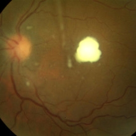
 Yellow Globular Lesion
Nov 9 2012 by Norman Byer
This glistening yellow globular lesion is a so-called pearl of the ora serrata in a 45-year-old man. Notice location in the tooth of the ora, which is a characteristic of this lesion. Histologically pearls are drusen-like structures which form on the inner side of Bruch’s membrane beneath the pigment epithelium. They are seen in about 20% of eyes and are often bilaterally symmetrical. They have no clinical significance but are valuable as landmarks.
Condition/keywords: Bruch's membrane, drusen-like, ora serrata
-
---thumb.jpg/image-square;max$300,300.ImageHandler) Tamoxifen Retinopathy- Fundus photo
Tamoxifen Retinopathy- Fundus photo
Aug 30 2012 by Young Hee Yoon, MD, PhD
Fundus photograph of an 58-year-old woman with a bilateral tamoxifen maculopathy. She had taken tamoxifen for 24 months due to breast cancer. In spite of discontinuation 2 years ago, her macula remained unchanged. Her best-corrected visual acuity was 20/50 in the right and 20/100 in the left.
Photographer: Sung Hyun Kim, Asan Medical Center
Imaging device: Topcon
Condition/keywords: drug toxicity, toxic maculopathy
-
---thumb.JPG/image-square;max$300,300.ImageHandler) Vitreous Snow Balls
Vitreous Snow Balls
Nov 18 2013 by Mallika Goyal, MD
Clumps of inflammatory cells in the inferior vitreous in a patient with bilateral vitreitis.
Photographer: Mallika Goyal, MD, Apollo Health City, Hyderabad
Condition/keywords: vitreous snowballs
-
 Plaquenil Toxicity
Apr 30 2013 by Theodore Leng, MD, MS, FASRS
Right eye 10-2 HVF from a 44-year-old woman with bilateral plaquenil toxicity. A ring scotoma is present.
Condition/keywords: hydroxychloroquine toxicity, plaquenil toxicity
-
 Candida Chorioretinitis
Aug 26 2012 by Andrew N. Antoszyk, MD FASRS
28-year-old female with hyperemesis gravidarum and bilateral retinal and vitreous infiltrates.
Condition/keywords: endogenous endophthalmitis, retinal infiltrates, vitreous infiltrates
-
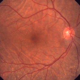 Bilateral Idiopathic Choroidal Folds
Jan 11 2013 by Gerardo Garcia-Aguirre, MD
Fundus photograph of the right eye showing choroidal folds.
Imaging device: Zeiss FF4
Condition/keywords: choroidal folds
-
 Vitelliform Macular Dystrophy
Sep 2 2012 by Hyung-Woo Kwak, MD
The typical appearance is of bilateral, round or oval, yellow, symmetrical, subretinal lesions, typically one-third to one-half optic disc diameter in size.
Imaging device: Zeiss F450 plus
Condition/keywords: Best disease
-
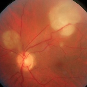 Cryptococcus Endogenous Endophthalmitis
Oct 9 2012 by Alan D. Letson, MD
Elderly man on high dose steroids 20 years duration for RA presents with decreased vision and bilateral chrorioretinal infiltrates and spinal tap produced cryptococcus.
Photographer: Mick Clark
Condition/keywords: cryptococcus, endogenous endophthalmitis, rheumatoid arthritis, steroids
-
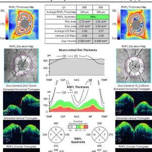 OCT in Patient With IIH Showing Thickened RNFL
Jan 16 2019 by John S. King, MD
18-year-old African American female with increased BMI with a history of headaches, nausea, transient diplopia and vision loss that she notices when getting up from her bed (and goes away after standing upright) for the last two weeks. Went to PCP and was treated for the flu, and after no improvement and visual symptoms known, was sent to ED. MRI did not show any masses and showed empty sella turcia. Vision 20/30 OD and 20/20 OS; no RAPD; IOP 15OU; no anterior segment or vitreous inflammation; discs are elevated with obscuration of the disc margins and some of the smaller vessels; there are no SVPs; there are mild Patton's lines temporally (see Initial Photos). The optic disc cube shows 360 degrees of RNFL thickening (see OCT). Was referred to near-ophthalmologist, Dr. Doyle. She obtained additional work-up, and LP opening pressure was high, and MRV showed bilateral transverse sinus stenosis. Patient showed steady improvement with medical therapy, that included weight loss and oral diamox. On her last visit with Dr. Doyle, vision has remained stable at 20/20-20/25 without an enlarged blindspot; there are SVPs and optic disc edema has resolved (see Post Treatment Photos); she is currently on 1000 mg of diamox and has lost 15 pounds, and no stinting procedure needed.
Imaging device: Cirrus
Condition/keywords: benign idiopatic intracranial hypertension, optic disc edema, papilledema
-
 Pars Planitis - Peripheral Uveitis
Nov 9 2012 by Norman Byer
This 25-year-old man had pars planitis, peripheral uveitis bilaterally. In this eye it produced a small tractional oval tear of the retina and an inferior retinal detachment. The typical creamy yellow exudates of pars planitis can be seen in the lower right very close to the ora serrata.
Condition/keywords: creamy yellow exudates, inferior retinal detachment, pars planitis, peripheral uveitis, tractional retinal tear
-
 Bilateral Idiopathic Choroidal Folds - OCT
Jan 11 2013 by Gerardo Garcia-Aguirre, MD
OCT of the macula showing choroidal folds.
Photographer: Gerardo Garcia-Aguirre, MD
Imaging device: Zeiss Cirrus HD OCT
Condition/keywords: choroidal folds
 Loading…