Search results (1033 results)
-
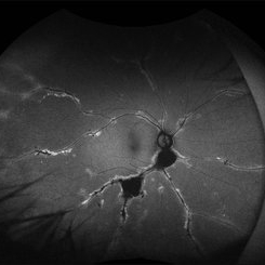
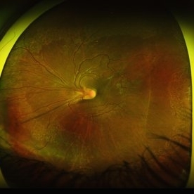
 Purtscher-like Retinopathy in Preeclampsia
Jun 28 2025 by Sriharanathan Poopalaratnam, MD,FRCS
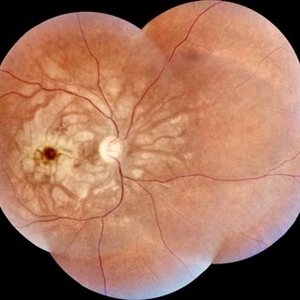
A 24-year-old female, 6 weeks post-emergency cesarean section (LSCS), with a history of pregnancy-induced hypertension (PIH), presented with acute, profound, bilateral painless vision loss of 2 days’ duration
Photographer: Yattiwarra
Condition/keywords: preeclampsia, Purtscher like
-
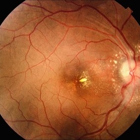
 Serpiginous Choroidopathy
Jun 23 2025 by César Adrián Gómez Valdivia, MD
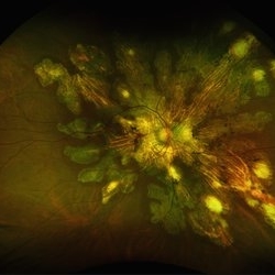
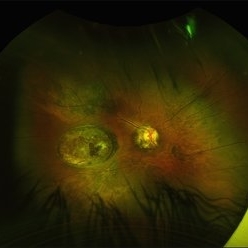
Fundus photograph of a 29 year-old female patient diagnosed with Serpiginous Choroidopathy. Finings were bilateral. The most common complication of SC is choroidal neovascularization affecting up to 35% of patients. Other reported complications are subretinal fibrosis, cystoid macular edema, branch vein occlusion, serous retinal detachment, optic disc neovascularization ,and anterior uveitis.
Photographer: @eyemissu2
Imaging device: TOPCON TRC-50DX
Condition/keywords: serpiginous choroiditis
-
 Serpiginous Choroidopathy
Jun 23 2025 by César Adrián Gómez Valdivia, MD
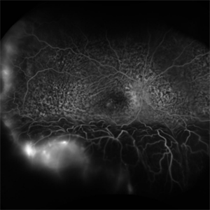
Fundus photograph of a 29 year-old female patient diagnosed with Serpiginous Choroidopathy. Finings were bilateral. The most common complication of SC is choroidal neovascularization affecting up to 35% of patients. Other reported complications are subretinal fibrosis, cystoid macular edema, branch vein occlusion, serous retinal detachment, optic disc neovascularization, and anterior uveitis.
Photographer: @eyemissu2
Imaging device: California ICG OPTOS
Condition/keywords: serpiginous choroiditis
-
 Pigmented Paravenous Retinochoroidal Atrophy
Jun 18 2025 by César Adrián Gómez Valdivia, MD
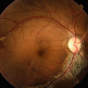
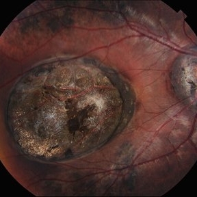
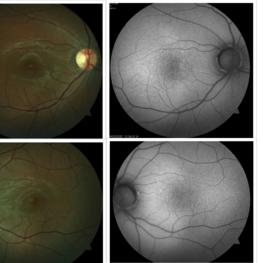
Fundus photograph of a 42YO female patient diagnosed with Pigmented Paravenous Retinochoroidal Atrophy. Findings were bilateral. Image shows hypoautofluorescence in the affected areas due to overall loss of RPE cells and thus lower lipofuscin levels.
Photographer: @eyemissu2
Imaging device: TOPCON TRC-50DX
Condition/keywords: Pigmented Paravenous Retinochoroidal Atrophy
-
 Pigmented Paravenous Retinochoroidal Atrophy
Jun 18 2025 by César Adrián Gómez Valdivia, MD
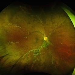
Fundus photograph Image of a 42YO female patient diagnosed with Pigmented Paravenous Retinochoroidal Atrophy. Findings were bilateral. Image shows hypoautofluorescence in the affected areas due to overall loss of RPE cells and thus lower lipofuscin levels.
Photographer: @eyemissu2
Imaging device: California ICG OPTOS
Condition/keywords: Pigmented Paravenous Retinochoroidal Atrophy
-
 Pigmented Paravenous Retinochoroidal Atrophy
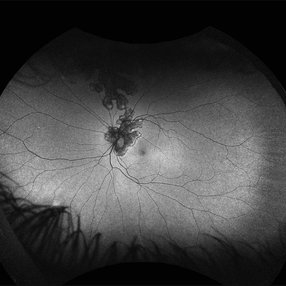
Jun 18 2025 by César Adrián Gómez Valdivia, MD
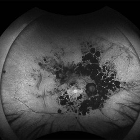
Fundus Autofluorescence Image of a 42YO female patient diagnosed with Pigmented Paravenous Retinochoroidal Atrophy. Findings were bilateral. Image shows hypoautofluorescence in the affected areas due to overall loss of RPE cells and thus lower lipofuscin levels.
Photographer: @eyemissu2
Imaging device: California ICG OPTOS
Condition/keywords: pigmented paravenous chorioretinal atrophy (PPCRA)
-
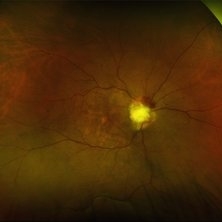
 Serpiginous Choroidopathy
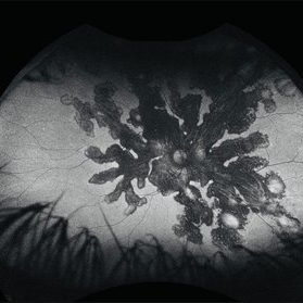
Jun 14 2025 by César Adrián Gómez Valdivia, MD
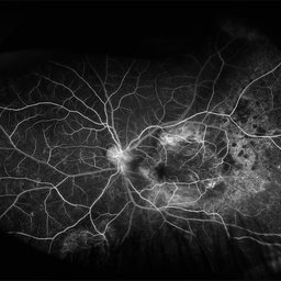
Fundus Autofluorescence of a 29-year-old woman with Serpiginous Choroidopathy. Finings were bilateral.
Photographer: @eyemissu2
Imaging device: California ICG OPTOS
Condition/keywords: Serpiginous Choroidopathy
-
 Serpiginous Choroidopathy
Jun 14 2025 by César Adrián Gómez Valdivia, MD
Fundus Autofluorescence of a 29-year-old woman with Serpiginous Choroidopathy. Finings were bilateral.
Photographer: @eyemissu2
Imaging device: California ICG OPTOS
Condition/keywords: Serpiginous Choroidopathy
-
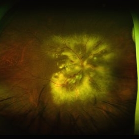
 Idiopathic Multifocal Choroiditis
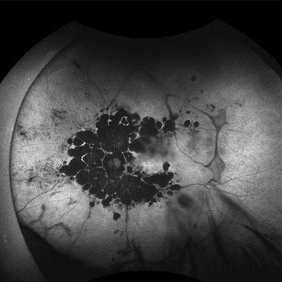
Jun 14 2025 by César Adrián Gómez Valdivia, MD
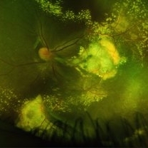
Fundus Autofluorescence of an 46 YO female patient diagnosed with Idiopathic Multifocal Choroiditis. Findings were bilateral.
Photographer: @eyemissu2
Imaging device: California ICG OPTOS
Condition/keywords: choroiditis
-
 Idiopathic Multifocal Choroiditis
Jun 14 2025 by César Adrián Gómez Valdivia, MD
Fundus Autofluorescence of an 46 YO female patient diagnosed with Idiopathic Multifocal Choroiditis. Findings were bilateral.
Photographer: @eyemissu2
Imaging device: California ICG OPTOS
Condition/keywords: choroiditis
-
 Uveal Effusion Syndrome
Jun 13 2025 by Brandon I Fram, MD, BS
75 year-old with bilateral inferior serous detachments, right more than left. Scleral window with biopsy showed scleral thickening with stromal deposits of amorphous glycosaminoglycan-like material.
Imaging device: Fluorescein Angiography
Condition/keywords: exudative retinal detachment, idiopathic uveal effusion syndrome, leopard spots, uveal effusion, uveal effusion syndrome
-
 Macular Coloboma
Jun 5 2025 by César Adrián Gómez Valdivia, MD
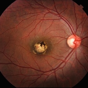
Macular Coloboma found in a 28 year-old male patient, visual acuity was 20/60. Resulting due to fusion failure of the optic fissure, colobomas are commonly found in the infero-nasal quadrant. If the retina is involved, it is reduced to glial tissue with no underlying RPE or choroid. This appears as an area of whitening often with pigment deposition at the junction of the coloboma and normal retina. Findings were bilateral.
Photographer: @eyemissu2
Imaging device: TOPCON TRC-50DX
Condition/keywords: coloboma
-
 Wagon-Wheel Lesion
Jun 5 2025 by César Adrián Gómez Valdivia, MD
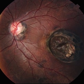
Wagon-wheel lesion found in a 12 YO male patient diagnosed with congenital toxoplasmosis. Findings were bilateral.
Photographer: @eyemissu2
Imaging device: TOPCON TRC-50DX
Condition/keywords: toxoplasmosis chorioretinitis, Wagon-wheel lesion
-
 Wagon-Wheel Lesion
Jun 5 2025 by César Adrián Gómez Valdivia, MD
Wagon-wheel lesion found in a 12 year-old male patient diagnosed with congenital toxoplasmosis. Findings were bilateral.
Photographer: @eyemissu2
Imaging device: California ICG OPTOS
Condition/keywords: toxoplasmosis, Wagon-wheel lesion
-
 Wagon-Wheel Lesion
Jun 5 2025 by César Adrián Gómez Valdivia, MD
Wagon-wheel lesion found in a 12 year-old male patient diagnosed with congenital toxoplasmosis. Findings were bilateral.
Photographer: @eyemissu2
Imaging device: TOPCON TRC-50DX
Condition/keywords: toxoplasmosis, Wagon-wheel lesion
-
 Coats Disease
May 27 2025 by César Adrián Gómez Valdivia, MD
Fluorescein Angiography on an 8 year-old male patient with Coats disease. Vascular leakage causes hard exudates which may be peripheral (near the vascular abnormalities) or midperipheral and central (at the macula. Findings were bilateral.
Photographer: @eyemissu2
Imaging device: California ICG OPTOS
Condition/keywords: Coats disease
-
 Coats Disease
May 27 2025 by César Adrián Gómez Valdivia, MD
Fundus photograph of an 8 year-old male patient with Coats disease. Vascular leakage causes hard exudates which may be peripheral (near the vascular abnormalities) or midperipheral and central (at the macula). Findings were bilateral.
Photographer: @eyemissu2
Imaging device: California ICG OPTOS
Condition/keywords: Coats disease
-
 ROP / Disk Dragging
May 27 2025 by César Adrián Gómez Valdivia, MD
Macular dragging found in a 14 year-old female patient with ROP history. Findings were bilateral.
Photographer: @eyemissu2
Imaging device: California ICG OPTOS
Condition/keywords: Disk Dragging
-
 Infectious Neuroretinitis
May 26 2025 by César Adrián Gómez Valdivia, MD
Neuroretinitis found in a 38 year-oldmale patient with IV drugs abuse history. Findings were bilateral. The lipid-rich component of the exudate is able to penetrate into the outer plexiform layer, creating what is clinically seen as a macular star pattern.
Photographer: @eyemissu2
Imaging device: TOPCON TRC-50DX
Condition/keywords: neuroretinitis
-
 Optic Nerve Metastasis
May 26 2025 by César Adrián Gómez Valdivia, MD
Fundus photograph of a 62 year-old woman with breast cancer history presented to the ER with decreased visual acuity. Optic nerve metastasis were found. Findings were bilateral.
Photographer: @eyemissu2
Imaging device: California ICG OPTOS
Condition/keywords: nerve, Optic, retinal metastasis
-
 Hypertensive Retinopathy
May 26 2025 by César Adrián Gómez Valdivia, MD
Fundus photograph of a 62 year-old woman with history of untreated hypertension and chronic kidney disease. Findings were bilateral.
Photographer: @eyemissu2
Imaging device: OPTOS
Condition/keywords: hypertensive retinopathy
-
 Bilateral Benign Yellow Dot Maculopathy
May 6 2025 by Amol yuvraj ganvir
A 37-year-old female patient presented for a routine eye examination. Her best-corrected visual acuity was 6/6 in both eyes. Fundus examination revealed multiple small yellow dots over the macula in both eyes. FAF imaging demonstrated characteristic hyperautofluorescence corresponding to these dots.
Photographer: Dr. Amol Ganvir, Vitreo-Retina Fellow, Ishwar Eye Centre, Rohtak, Haryana
Imaging device: Visucam-Zeiss
Condition/keywords: Autoflourescence, yellow dots
-
 Stag Horn
Apr 8 2025 by Gustavo Uriel Fonseca Aguirre
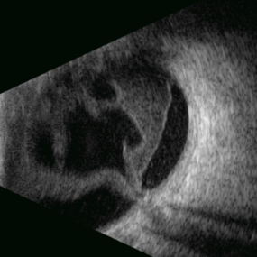
B-mode ultrasound of a young male patient with bilateral panuveitis (currently under investigation) reveals intense vitritis with islands of preserved vitreous and partial posterior hyaloid detachment, creating a characteristic "stag horn" appearance.
Photographer: Gustavo U. Fonseca Aguirre, Hospital Conde de Valenciana, Ciudad de México
Condition/keywords: Panuveitis
-
 Solar Retinopathy
Apr 1 2025 by Isaac Agranoff
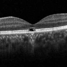
OCT scan of 18-year-old male presenting with 20/40 BCVA OU and bilateral focal outer retinal subfoveal defects. Patient reported long-term history of frequent sungazing, has stopped within past 6-9 months.
Photographer: Isaac Agranoff
Imaging device: Heidelberg Spectralis
Condition/keywords: solar retinopathy
-
 Ciliary Body Metastasis
Mar 26 2025 by Virginia Gebhart
54 year old female referred for iris mass. UBM shows large solid mass originating in the ciliary body and eroding into the anterior chamber under the iris epithelium. Recent CT scans revealed multiple bilateral pulmonary and hepatic nodules. Pt has been scheduled for PET scan and liver biopsy by radiation oncologist.
Photographer: Virginia Gebhart, Retina Consultants of Carolina
Imaging device: Samsung Galaxy
Condition/keywords: choroidal metastasis, ciliary body mass, metastatic cancer
 Loading…