Search results (19 results)
-
 Foveal Hypoplasia / Ocular Albinism
Aug 29 2024 by César Adrián Gómez Valdivia, MD
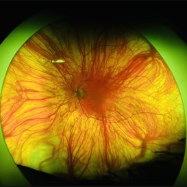
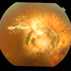
Fundus photograph of a 64-year-old female patient with foveal hypoplasia, ocular albinism and pendular nystagmus. Findings were bilateral. Retinal and choroidal vasculature are exquisitely beautiful.
Photographer: @eyemissu2
Imaging device: California ICG OPTOS
Condition/keywords: foveal hypoplasia, ocular albinism
-
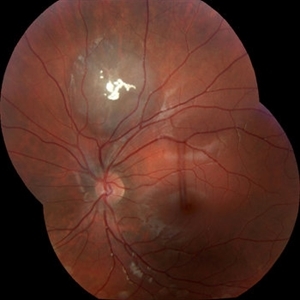
 Pericentral Retinitis Pigmentosa
Sep 6 2024 by Mauricio Bayram-Suverza, MD
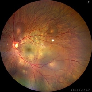
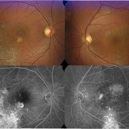
A 65-year-old male patient reports experiencing bilateral blind spots that have gradually intensified over time. Genetic testing was unrevealing. The fundus autofluorescence image shows a hypoautofluorescent ring in the posterior pole, especially nasal to the nerve and along arcades.
Photographer: Mauricio Bayram-Suverza, Casey Eye Institute, OHSU.
Imaging device: Optos California
Condition/keywords: fundus autofluorescence (FAF), inherited retinal disease, nyctalopia, retinal dystrophy, retinitis pigmentosa
-
 Central Serous Chorioretinopathy in Pregnancy (OS)
Apr 28 2024 by Vishal Agrawal, MD, FRCS,FACS,FASRS
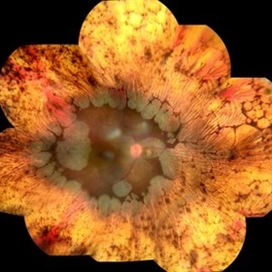
30-year female with sudden loss of vision came for examination. She was in her first trimester of pregnancy. Examination revealed bilateral bullous NSD with subretinal fibrin s/o CSR.
Photographer: Dr Ayushi
Imaging device: Clarus 700
Condition/keywords: Central Serous Chorioretinopathy (CSR), neurosensory detachment of retina, pregnancy
-
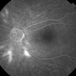
 Venous Loop
Feb 20 2024 by Soobien Lee
A 77-year-old male with a history of bilateral optic neuropathy from bilateral optic nerve sheath meningiomas S/P radiation/proton-beam therapies. Presented with radiation retinopathy OS and a known venous loop OS.
Photographer: Gavin Bragdon, Elman Retina Group
Imaging device: Optos Ultra-Widefield Fluorescein Angiography
Condition/keywords: fluorescein angiogram (FA), Optos, radiation retinopathy, retinal vascular disease, venous loop
-
 Thioridazine-toxicity
Apr 30 2022 by Niloofar Piri, MD
61 yo male with PMH of longstanding schizophrenia since 20s with secondary intellectual disability presented with decreased vision following a recent stroke. He was found to have bilateral chorio-retinal atrophy involving posterior pole with scalloped edges and coin shaped atrophic area at margins extending into mid-periphery, diagnosis most concerning for intermediate stage thioridazine toxicity given the history. Mother could find handwritten prescriptions from 1990s when he was on Thioridazine 800 mg daily for unknown period of time. Patient had better vision in the left eye which was affected by recent stroke and prompted him to seek medical care. Fundus photograph of the right eye is demonstrated here.
Photographer: Jacob Grodsky, MD
Condition/keywords: drug toxicity, thioridazine toxicity, toxic retinopathy
-
 Serous Retinal Detachment in Vogt Koyanagi Harada Patient
Apr 26 2021 by Pablo Baquero Ospina, MD
24-year-old woman with bilateral panuveitis and serous retinal detachment, headache and tinnitus.
Photographer: Pablo Baquero-Ospina, Asociación Para Evitar la Ceguera en México
Imaging device: Heidelberg Spectralis
Condition/keywords: serous retinal detachment, Vogt-Koyanagi-Harada
-
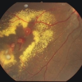
 Gyrate Atrophy
Oct 30 2020 by JEFFERSON R SOUSA, Tecg.º (Biomedical Systems Technology)
Female patient, 28-year-old, with low vision in both eyes since childhood. In routine examination, important changes were observed with atrophic, symmetrical and bilateral aspects with apparently preservation of the central retina.
Condition/keywords: gyrate atrophy
-
 Presumed Tenofovir Induced Toxicity
Nov 7 2019 by Sham Talati, DOMS
46-year-old HIV positive diabetic male with progressive bilateral decrease in vision for last 45 days. Patient had associated liver and kidney disease. Taking Tenofovir for last one year.
Photographer: Sham Talati,Retina Foundation,Ahmedabad
Imaging device: Nidek Mirante
Condition/keywords: autofluorescence imaging, drug toxicity, HIV
-
 Coloboma
Oct 2 2019 by John S. King, MD
27-year-old white female with bilateral, isolated, inferior, chorioretinal colobomas; she has a history of retinal laser anterior to the edge of the coloboma OD secondary to a limited RD. This is the right eye.
Photographer: Shelly Blair
Imaging device: Optos CA
Condition/keywords: coloboma of choroid
-
 Leukemic Retinopathy
Jul 11 2019 by Robert A Lalane, MD
55-year-old male currently undergoing chemotherapy for leukemia. Found to have extensive retinal hemorrhaging throughout various retinal layers bilaterally.
Photographer: Brandy Maxwell, Retina Group of Florida
Condition/keywords: leukemia, retinal hemorrhage
-
 Tumor of Retina (Retinocytoma)
Jan 9 2019 by Janet Brazil
Fundus photograph of a 13-year-old male with a bilateral tumor of the retina, with positive RB gene mutation.
Photographer: Janet Atkinson, Eye Associates of New Mexico
Imaging device: TOPCON TRC-50EX
Condition/keywords: RB gene mutation, tumor
-
 Hemangioma of Retina
Sep 11 2018 by Carolyn Daley
Optos ultra wide field imaging of a 20-year-old with multiple bilateral hemangiomas. Patient was diagnosed with Von Hippel-Lindau Syndrome.
Photographer: Carolyn Daley, Retina Specialists of Michigan
Imaging device: Optos Ultra Wide Field
Condition/keywords: edema, hemangioma, Optos, Von Hippel-Lindau
-
 Susac's Syndrome
Feb 13 2018 by John S. King, MD
Background: 46-year-old WF with CML (stable on Sprycel) saw her PCP for headaches without known cause; Headaches worsened and became confused, disoriented, off balance, and impaired short term memory. Heme-oncology ordered MRI that showed abnormal signal in the cerebellum and other parts of the brain, and LP has elevated protein. LP did show positive tau test, but fortunately, was a false positive for CJD. IV and PO steroids started and symptoms improved. MRI showed much improvement one month since starting steroids. 3 weeks later had a scotoma in right eye and eye doctor did not find anything at that time to cause it. Tinnitus developed (and some intermittent vertigo before that) and ENT referred back to eye doctor, who then referred the patient to Dr. Zocchi. He found a CWS and BRAO OD, and bilateral arteritis. She had some additional work-up for vasculitis. Given the retinal arteritis, cochlear issues, and MRI findings, Dr.Zocchi suspected Susac's Syndrome. She was started on multiple regimens including prednisone, IVIG, azathiprine, and MTX, and has had the best reponse to IVIG (FA shows a recurrence/worsening while adjusting IMT). She is stable and doing well with 20/20 vision in both eyes.
Photographer: Kay Dalby
Imaging device: Topcon
Condition/keywords: retinal vasculitis, Susac's syndrome
-
 Autosomal Recessive Bestrophinopathy - Color Photo OD
Dec 22 2017 by Tony Tsai, MD, FASRS
11-year-old Asian male with 20/40 vision OU, negative family history for ocular conditions, and bilateral atypical vitelliform deposits and subretinal fluid. EOG confirmed abnormally low Arden ratios OU. Genetic testing revealed homozygous recessive mutation in BEST1 gene (p.L140V:c.418C>G). Also known as p.L80V; Ref: Davidson (2009) Am J Hum Genet 85, 581.
Photographer: San Juanita Zazueta
Imaging device: Topcon
Condition/keywords: Best disease
-
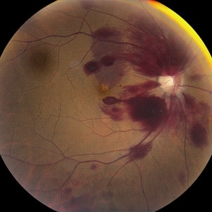
 Bilateral Lebers Miliary Aneurysm in a Female
Sep 5 2017 by Ogugua Ndubuisi Okonkwo, MD, FRCS (Edin), FASRS
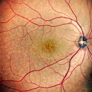
Fundus photograph of the active left eye of a 26-year-old female with bilateral LMA. Shows severe exudation in the nasal retina by leaking aneurysms.
Condition/keywords: aneurysm
-
 Aggressive Posterior Retinopathy of Prematurity
May 8 2017 by Juan Romo-Aguas
Fundus photograph of an 1 month and 21 days female with bilateral aggresive posterior retinopathy of prematurity.
Photographer: Juan C. Romo-Aguas, Asociación Para Evitar la Ceguera en México
Imaging device: Optos Daytona Ultra-widefield Retinal Imaging
Condition/keywords: aggressive posterior retinopathy of prematurity (APROP), retinopathy of prematurity (ROP)
-
 Morning Glory Disc
Apr 22 2016 by Mallika Goyal, MD
Right fundus of a 34-year-old lady with bilateral morning glory disc anomaly with silicon oil in-situ; this eye had rhegmatogenous retinal detachment with multiple peripheral lattice degeneration and was successfully operated. However, there was redetachment within a week of silicon oil removal in absence of any untreated retinal breaks suggesting the abnormal disc as a likely cause of the redetachment.
Photographer: Mallika Goyal, MD, Apollo Health City, Hyderabad, India
Condition/keywords: Morning Glory Syndrome
-
 Chronic Central Serous Chorioretinopathy (CSCR)
Nov 15 2014 by Rita Couceiro, MD, MS
53-year-old black male, with no relevant prior medical history, complained of bilateral blurry vision for the previous 16 years. On examination, visual acuity was 20/50 on the right eye (OD) and 20/100 on the left eye (OS). Anterior segment evaluation was unremarkable. Fundoscopy revealed pigmentary changes near the macular area in both eyes, with a mottling configuration, suggesting chronic CSCR. Fluorescein angiography showed an ink-blot pattern, with leakage superior to the fovea in OD and nasal to the fovea in OS.
Photographer: Telma Gala - Hospital de Santa Maria, Lisbon, Portugal
Condition/keywords: chronic central serous chorioretinopathy (CSCR)
-
 Non-Small-Cell Lung Cancer Choroidal Meta OS Fundus
Apr 30 2013 by Dong Yoon Kim, MD
Fundus photograph of an 64-year-old man with bilateral choroidal metastasis of non-small-cell lung cancer.
Condition/keywords: choroidal metastasis, fundus photograph
 Loading…